


Developmental Malformations
Walker-Warburg syndrome
Apr. 14, 2024
MedLink®, LLC
3525 Del Mar Heights Rd, Ste 304
San Diego, CA 92130-2122
Toll Free (U.S. + Canada): 800-452-2400
US Number: +1-619-640-4660
Support: service@medlink.com
Editor: editor@medlink.com
ISSN: 2831-9125
Toll Free (U.S. + Canada): 800-452-2400
US Number: +1-619-640-4660
Support: service@medlink.com
Editor: editor@medlink.com
ISSN: 2831-9125
Worddefinition
At vero eos et accusamus et iusto odio dignissimos ducimus qui blanditiis praesentium voluptatum deleniti atque corrupti quos dolores et quas.
Pontocerebellar hypoplasias represent a heterogenous group of inherited progressive neurodegenerative disorders with fetal onset and autosomal recessive inheritance. Their common characteristic is prenatal onset hypoplasia or atrophy of the cerebellum and pons as pictured by MRI and a clinical course resulting in progressive microcephaly and severe motor and cognitive impairments. Ten types have been defined (PCH 1 to 11) as well as numerous subtypes. Genetic defects are associated with all types and subtypes have been identified. PCH1, PCH2, PCH4, and PCH6 are the most frequently reported types. The X-linked MicPCH (microcephaly pontocerebellar hypoplasia) due to CASK mutations is etiologically unrelated to the former, because it is not regressive but malformative in its mechanism, but it is discussed here because of similarities in MR presentation. In this updated article, the author provides coverage of these subtypes. Milder expression of the gene defects that cause pontocerebellar hypoplasia may result in a modified picture of cerebellar hypoplasia reminiscent of postnatal onset cerebellar atrophy without attenuation of the pons, and the cerebellar volume approaching the level seen in hereditary cerebellar ataxias. Pontocerebellar hypoplasia also involves supratentorial structures, causing microcephaly, severe intellectual delay, and central motor deficits. A typical aspect of all pontocerebellar hypoplasias is the predominance of supratentorial symptoms (spasticity, dystonia, chorea), rather than cerebellar symptoms. This apparent absence of cerebellar symptoms is due to the modulating rather than initiating role of the cerebellum in control of voluntary motor function. In severe pontocerebellar hypoplasia, cortical functions are impaired together with microcephaly. The severe lack of central control of motor functioning renders the functional role of the cerebellum, which is inherently supportive of intentional posture and movement, less important, even insignificant when cerebral functioning is grossly impaired together with microcephaly. This difference in motor behavior constitutes a major difference with spino-cerebellar ataxias, where typical symptomatology is caused by largely unimpaired supratentorial motor nuclei acting together with impaired cerebellar structures. The author has a special interest in pontocerebellar hypoplasias and has contributed to their classification, their neuropathology, and the identification of the genetic basis of types 1, 2, 4, and 5. Neuroimaging features often are diagnostic, and some cases can be diagnosed prenatally by ultrasound and MRI, including those associated with rare genetic syndrome (73; 46). Some patients are not diagnosed during life but only at autopsy (138).
|
• Pontocerebellar hypoplasias represent a heterogenous group of autosomal recessive progressive disorders originally characterized by fetal onset of pontine and cerebellar growth impairment and microcephaly. Clinical findings are varied but include symptoms of supratentorial involvement, including microcephaly, intellectual impairments and involvement of long tracts. In some subtypes, not enough clinical data are available for an elaborate clinical characterization. Extracranial dysmorphisms and visceral involvement are absent. | |
|
• Less severe cases, corresponding to milder defects of the associated genes, may display less severe cerebellar hypoplasia and normal aspect of the pons, evoking similarity in imaging aspects, but not in symptomatology to the spinocerebellar ataxias (SCA). Phenotypical expression of the neurodegenerative process may at times be initiated postnatally and may not affect brain development and maturation prenatally. | |
|
• Neurologic expression includes ventral spinal horn impairment in type 1 and its subtypes. | |
|
• Type 2 is expressed as dyskinesias and other movement disorders in addition to cerebellar deficits and global developmental delay. | |
|
• Differential diagnosis is large and includes tubulinopathies, CASK deficiency, congenital brainstem disconnection, pontine tegmental cap dysplasia, dystroglycanopathies, and congenital disorders of glycosylation. | |
|
• The group includes 11 distinct types, not including subtypes. | |
|
• Types pontocerebellar hypoplasia type 1 due to SLC25A46 mutations and pontocerebellar hypoplasia type 6 due to RARS mutation involve mitochondrial functions. | |
|
• Arthrogryposis can occur with (PCH1) or without (PCH4) motor neuron involvement, and fetal akinesia sequence also can occur rarely. | |
|
• Some forms are expressed in fetal life by imaging and have a rapid postnatal progressive course leading to early death. | |
|
• The genetic basis is well documented in many forms of pontocerebellar hypoplasia, but new variants are described constantly. |
The first mention of pontocerebellar hypoplasia in a neuropathologic treatise by Brun in 1917 antedates clinical awareness of that entity by 5 to 6 decades (30). Pathologic studies by Brouwer, Koster, and Krause delineated an entity consisting of general hypoplasia of the cerebellar hemispheres, lack of development of primary and secondary folia with relative sparing of vermis, flocculi, and paraflocculi, and ventral pontine hypoplasia or atrophy (29; 77; 79). The main microscopic findings were depletion of internal granule cells, mostly in the cerebellar hemispheres with the vermis and flocculonodular lobes relatively spared, paucity of myelin in the central white matter and within folia, and a peculiar fragmentation of the dentate nuclei into isolated clusters of neurons, remarkably different from the normal festoon-like appearance of the dentate nucleus.
Norman reported the association with spinal anterior horn degeneration (99), Krause (79) and Peiffer and Pfeiffer (105) reported the association with spastic pareses and with chorea. Based on these diverging clinical and pathologic features, Barth proposed an initial subdivision of pontocerebellar hypoplasia in types 1 and 2 (13). Patel and colleagues proposed a further subdivision based on five subtypes (103).
Type 6 (PCH6) was the first for which the causative gene defect was identified (48), followed by PCH2 and PCH4 (31). Presently 10 types are catalogued as such by OMIM, and at least one defective gene is allocated to each.
Widely different etiologies and pathogenic mechanisms underlie the various subtypes. The neuroradiological features of pontocerebellar hypoplasia are helpful for a group diagnosis, but further subtyping is required for molecular-genetic diagnosis, therapeutic advice, and genetic counseling. An excellent review of the genes and genetic subtypes of pontocerebellar hypoplasia was provided by van Dijk and colleagues (145). Most literature on pontocerebellar hypoplasia deals with novel genetic mutations and broadening the spectrum of genotype:phenotype correlations.
OMIM | Gene | Phenotype | Distinguishing characteristic | Inheritance |
1A | VRK1* KIF26B PLA2G6 EXOSC1 | PCH+spinal anterior horn involvement | Prolonged survival, microcephaly. no progression | AR |
1B | EXOSC3 | idem | Spinal ventral horn disease | AR |
1C | EXOSC8 | idem | Ibid | AR |
1D | EXOSC9 | Idem; two genetic variants | PCH | AR |
1? | SLC25A46 | idem | Severe affection, optic atrophy, mitochondrial abnormalities | AR |
2A | TSEN54 | PCH+dystonia/spasticity | Progressive supratentorial brain atrophy. Most common type. | AR |
2B | TSEN2 | idem | Rare, phenotype as 2A | AR |
2C | TSEN34 | idem | Rare, phenotype as 2A | AR |
2D | SEPSECS* | idem | idem | AR |
2F | TSEN15 | idem | Extremely rare, as 2A | AR |
2D | SEPSECS** | mild PCH like | MRI like postnatal onset cerebellar atrophy | AR; splice disrupting |
2E | VPS53** | mild PCH like | MRI like postnatal onset cerebellar atrophy | AR |
2 | INPP4A | PCH, microcephaly, myoclonic epilepsy, developmental delay | Rare | |
2 | PPIL1 | PCH, lissencephaly | Rare | AR |
3 | PCLO | Progressive microcephaly, optic atrophy | Cerebellar hypoplasia | AR |
4,5 | TSEN54 | Olivopontocerebellar atrophy (early lethal type) | Early lethal type with respiratory failure | AR |
6 | RARS2 | Mitochondrial disease | Rapid lethal progress | AR |
7 | TOE1 | PCH | Genital feminization in males | AR |
8 | CHMP1A | PCH | Static course | AR |
9 | AMPD2 | PCH | Underdevelopment middle and basal parts of mesencephalon | AR |
10 | CLP1 | Similar to PCH2A | Founder mutation in Turkish families | AR |
11 | TBC1D23 ATOH1 SLC25A46
| Nondegenerative Developmental delay; hearing impairment
Progressive, infantile lethal; myelopathy; pontocerebellar hypoplasia | Male predominance
Mitochondrial impairment Spinal cord hypoplasia | AR AR AR
|
| ||||
Most ethnic groups are susceptible to these many genetic mutations and clinical expression of pontocerebellar hypoplasia. A family in Sudan was described (08). Both genders are affected equally in most types, but females are selectively involved in PCH7, and males predominate in PCH11 (24).
Type 1: pontocerebellar hypoplasia and spinal ventral horn involvement. This phenotype combines the gross morphologic characteristics of pontocerebellar hypoplasia with ventral horn motor neuron degeneration, similar to spinal muscular atrophy type 1 (Werdnig-Hoffmann disease). It was first reported in 1961 by Norman, who described an infant with respiratory impairment and poor swallowing from birth, adductor spasms, and lack of mental development (99). Death occurred at 6 months. Autopsy findings were cerebellar hypoplasia, whereas degenerative changes and nonspecific neuronal loss were observed in cerebellar dentate and inferior olivary nuclei, and in ventral pontine, bulbar motor, and spinal motor neurons, thalami, and basal ganglia. The inferior olivary nuclei were poorly convoluted. Although there was a reduction of ventral pontine nuclei, the size of the pons was not affected, and the transverse pontine fibers were gliotic but not reduced in numbers. Goutieres and colleagues provided a combined clinical and pathologic definition of this entity in siblings in which the ventral pons was reduced in size and spinal motoneurons were affected similar to Werdnig-Hoffmann disease (54). Patients with type 1 disease may exceptionally present with polyhydramnios and are generally weak and hypotonic from birth, with poor respiration and inspiratory stridor (99; 54; 44; 90; 39; 69; 13). Also, contractures of the limbs may be present at birth, and death may occur in the neonatal period or in infancy in the most severe cases (69). The initial impression of pontocerebellar hypoplasia type 1 as a disease, which is always lethal in the neonatal period and infancy had to be corrected as result of a larger series from Rudnik-Schoeneborn and colleagues, which included cases with survival into childhood and even adolescence (118; 117). Motor nerve conduction velocities were delayed in the reports of Goutieres and colleagues and de Leon and colleagues (54; 44) but normal in the case of Kamoshita and colleagues (69). Autopsy in patients with pontocerebellar hypoplasias and spinal ventral horn disease showed extensive nerve cell loss and gliosis in different systems, including the thalamus, cranial motor nerve nuclei, and pallidum. Findings in the dentate nuclei were destruction and hilar gliosis rather than fragmentation. Brain weights were either normal or diminished. It may be significant that in none of these cases of combined pontocerebellar hypoplasias with spinal anterior horn disease did doubt exist about the regressive nature of the disease, probably because (gliotic) reaction was severe in this phenotype. A series of five patients, including autopsy findings, was reported by Muntoni and colleagues (93), with gene analysis excluding SMN gene mutations. Rudnik-Schoeneborn and colleagues described milder familial and nonfamilial cases with the MR features of pontocerebellar hypoplasia and anterior horn involvement, similar to Werdnig-Hoffmann disease but excluded from the spinal muscular atrophy gene (118).
Rare cases of pontocerebellar hypoplasia type 1B may exhibit micrencephaly with failure of gyration (lissencephalic cortex) and also rhombencephalosynapsis of the cerebellum (125; 133). A founder PPIL1 variant underlies at least some cases of pontocerebellar hypoplasia with micro-lissencephaly, as was show in nine patients from eight unrelated Egyptian families (01).
Muscle biopsy in PCH1 reveals not only a typical pattern of perinatal denervation of striated muscle as in infantile spinal muscular atrophy, but also excessive sarcomeric disorganization and nemaline rods, which may confuse the diagnosis if the initial focus is on the neuromuscular problem rather than the more global CNS developmental disorder (108).
Etiology and pathogenesis of type 1.
Recessive EXOSC3 mutation (type 1B according to OMIM classification). A majority of families with PCH1 in a large study was found to carry biallelic mutations in the EXOSC3 gene (153). The EXOSC3 gene encodes hRp40p, a 3’- 5’ hydrolytic exoribonuclease, which forms a subunit of the exosome complex, which consists of about 10 proteins arranged in a ringlike structure residing in the nucleolus, nucleus, and cytoplasm. The exosome mediates removal of redundant messenger RNA, targeting AU (adenylate/uridylate) rich regions in the untranslated 3’ tail (38; 111; 49). A study of 15 patients from 10 families with EXOSC3-associated PCH1 by Rudnik-Schoeneborn and colleagues put emphasis on familial recurrence, evolving clinical patterns, and variability of imaging data (117). Some important conclusions can be drawn from this study:
(1) Both mild and severe cases may be caused by different, mostly homozygous, bi-allelic mutations in EXOSC3. Survival may range between death in infancy to adolescence. Among other genes in this same family, EXOSC2 causes a neurologic syndrome that includes retinitis pigmentosa, hearing loss, and mild cognitive disability, but not pontocerebellar hypoplasia (49). EXOSC8 mutation, by contrast, results in the same phenotype as EXOSC3 (49). Novel EXOSC9 variants cause PCH1D with spinal motor neuropathy and cerebellar atrophy (121).
Pontocerebellar hypoplasia 1B is a progressive degenerative disease with atrophy of the caudate nucleus as well as of the cerebellum and cerebral cortex; it is distinguished from microcephaly with pontine and cerebellar hypoplasia (MICPCH) in which supratentorial structures are selectively spared (146).
(2) Central motor symptoms (dystonia, spasticity) and peripheral motor symptoms may coexist but may not occur synchronously (eg, spasticity may predominate and later be supplanted by hypotonia as the disease progresses).
(3) Some patients may have a reduced size of the pons on imaging, whereas others show a normal size of the pons.
(4) In some milder cases microcephaly may not occur.
(5) “Major clinical features previously reported in PCH1, including intrauterine abnormalities, postnatal hypoventilation and feeding difficulties, joint contractures, and neonatal death, were rarely observed in mutation-positive infants but were typical among the mutation-negative subjects.” A severe variety of the EXOSC3-related disease, the homozygous missense mutation c.92G -> C, p.G31A mutation, was discovered as a founder mutation in the Roma population in Czechia by Schwabova and associates (127). In what may be regarded as a mild manifestation of EXOSC3 impairment, Zanni and colleagues described a single family from Bangladesh with a mild heterozygous EXOSC3 mutation. The presentation in this family was early-onset spasticity, mild intellectual disability, and cerebellar atrophy. Muscle biopsy in one patient showed neurogenic abnormalities with type grouping of type II fibers (161).
Homozygous mutations in the gene EXOSC3 cause roughly half of all PCH1 cases (153). The EXOSC3 gene encodes hRp40p, a 3’- 5’ hydrolytic exoribonuclease, which forms a subunit of the exosome complex, consisting of about 10 proteins arranged in a ringlike structure residing in the nucleolus, nucleus, and cytoplasm. The exosome interacts with RNAs. Its main function is to act as a quality control instrument for the clearing of redundant and improper RNA transcripts (111).
Recessive EXOSC8 mutation. Boczonadi and colleagues reported three inbred pedigrees (26). The main neurologic findings were onset within the first half year of life of spastic pareses (9 of 11 patients), visual and hearing loss (9 of 11 patients), and respiratory problems. Muscle wasting was present in 8 of 11 patients. Peripheral motor neuron involvement was confirmed by EMG in two patients from the same pedigree. Respiratory chain involvement of complexes I and IV was found in one patient. Most patients died within the first two years of life. MR studies were obtained in eight patients, and vermal hypoplasia presented in four of the eight patients, thin corpus callosum presented in four of the eight patients, and immature myelination presented in two of the eight patients. General cerebellar atrophy, together with cerebral cortical atrophy, was found in one patient.
Recessive SLC25A46 mutation. Wan and colleagues identified mutations of the SLC25A46 gene in severe pontocerebellar hypoplasia in two nonrelated families (152). One male patient and his sister had severe prenatal onset disease with polyhydramnios, bilateral optic atrophy, contractures, and an MRI with features of severe cerebellar hypoplasia and moderate pontine hypoplasia. EMG in the first patient was suggestive of severe axonal sensorimotor neuropathy. Similar findings were present in two siblings of another family who also showed increased serum lactic acid. A defect of mitochondrial splitting, leading to abnormally elongated mitochondria could be reproduced in vitro. No autopsies were performed. Restudying of the original family who served as prototype for type I pontocerebellar hypoplasia with pathologically confirmed spinal anterior horn involvement in the original classification (13) revealed heterozygous deleterious mutations in both alleles (148). This patient also had optic atrophy, which represents a constant finding in all clinically manifest SLC25A46 mutations. Another family presented two siblings, each with a severe neonatal course and death (157). Consistency of the phenotype with variability in clinical severity of PCH1 due to bi-allelic SLC25A46 mutations was reported by Braunisch and colleagues, with reported ages at death varying from the first day of life to 15 months (27). This report confirmed spinal anterior horn loss as well as widespread cerebral involvement, mainly, though not exclusively, affecting the cerebellum, pons, and inferior olivary nuclei. Lactic acid increase was found in some cases, possibly resulting from the mitochondrial fusion defect previously described by Wan and colleagues (152).
A biallelic missense variant in EXOSC1 (p.Ser35Leu) causes PCH1 (132). Type 1D has at least two genetic variants of the EXOSC9 gene (41), and other homozygous variants in EXOSC1 also were described (42).
A pontocerebellar hypoplasia cell line from a patient with PCH1B harboring a homozygous c.395 A>C mutation in EXOSC3 and matched familial controls provides an experimental tool to study the development of PCH1B (134). Imbalanced mRNA isoform expression of long genes essential to neurologic development and function can be recapitulated in vitro in stem cells as well as in vivo from suppression of premature transcription termination and may be relevant in pontocerebellar hypoplasia (80).
The human SLC25A46 mutation is already expressed in fetal life and a rapidly progressive postnatal course leading to death (157). The functions of SLC25A46 have been studied exhaustively in a mouse model with remarkable similarity to the many human expressions of SLC25A46 deficiency (141). The nuclear gene encodes a nuclear mitochondrial protein carrier with a role in mitochondrial dynamics and cristae maintenance. Associated diseases are ataxia, optic atrophy, cerebellar hypoplasia, and abnormalities in the neuromuscular junction leading to Charcot-Marie-Tooth type 2 disease.
Gene-defects mirroring aspects of PCH1. A homozygous mutation causing a stop codon in the vaccinia-related kinase 1 (VRK1) gene was detected in a single family with what appeared to be a milder and atypical type of PCH1 (116). The proband had severe microcephaly from birth, but progressed in motor performance. Results of electromyography, nerve conduction velocity, and SEP studies were consistent with motor and sensory neuropathy. The VRK1 gene encodes a serine/threonine protein kinase involved in cellular proliferation and cell cycle regulation. Vinograd-Byk and colleagues produced a knockdown in the mouse homologue Vrk1 causing impaired proliferation and migration phenotypes (150). Furthermore, VRK1 deficiency leads to downregulation of amyloid-beta precursor protein, known to affect neuronal migration. A new patient with VRK1 mutation studied had cerebral cortical dysplasia rather than atrophy, a finding that fits well with the gene’s role in neuronal migration. In another report, families were described with adult and childhood onset spinal motor neuron disease without cerebellar or pontine abnormalities on imaging (137). In retrospect one might question the logic of classifying the VRK1 mutation with the pontocerebellar hypoplasias.
A mutation in RARS2 was found in a single patient with a clinical diagnosis of PCH1 (96). However, this mutation is more frequently associated with PCH6 (see below). Other novel mutations in PCH1 is in the PLA2G6 gene (51) and the KIF26B gene (156).
A de novo variant of KIF26B, a gene with neurodevelopmental potential, was found in an infant born with arthrogryposis and developmental defects on MRI, which included moderate pontocerebellar hypoplasia. Electromyography and muscle biopsy gave evidence of spinal ventral horn involvement (156).
In the OMIM classification, PCH1 due to VRK1 mutation has become PCH1A; EXOSC3 mutated cases are categorized as type PCH1B. The various genetic-phenotype correlations in PCH1 were reviewed by Ivanov and colleagues (63).
Presentation and course of types 2, 4, and 5. These three types share a common genetic etiology, and will be discussed together.
Type 2: pontocerebellar hypoplasia with chorea or spasticity. Clinical details were first provided by Krause, Biemond, Norman and Urich, and Peiffer and Pfeiffer (79; 22; 100; 105). As part of their mainly neuropathologic studies these authors recorded severe general neurodevelopmental delay, microcephaly, and early death between 5 months and 1.5 years of age. Motor patterns observed were generalized spasticity with neck retraction (100), abnormal spontaneous movements (22), and frank chorea (105). Progressive atrophy of the caudate nucleus and cerebral cortex occurs postnatally in pontocerebellar hypoplasia 2A (146). Splice-disrupting synonymous biallelic variants of SEPSEC5 resulted in HPC2D in two siblings (113).
An extended family comprising five related sibships was discovered in the Dutch genetic isolate of Volendam (18). Study of this family enabled further delineation of a separate neurogenetic entity with the following characteristics: autosomal recessive inheritance, early-onset chorea or dystonia, nearly complete absence of cognitive achievement and voluntary motor control, progressive microcephaly, and lethal course during childhood in the majority of the affected individuals. Exceptional cases of survival into early adulthood are on record. Ultrastructural findings in a cortex biopsy from a patient belonging to the Volendam kindred were consistent with ongoing neuronal degeneration and neuronal death. Neurons were affected by a peculiar focal increase of ribosomes. The consistency of the phenotype was established by the study of 10 pedigrees from Sweden, Germany, and the Netherlands, including, for the first time, MRI features (15). Although the patients were apparently stationary in their performance, several findings indicated a progressive course, especially cerebral cortical atrophy. Early symptoms include impaired swallowing, often necessitating feeding by gavage or gastrostomy, and clonus in the neonatal period, often described as jitteriness. MRI findings are characteristic: the cerebellum shows hypoplasia, often combined with atrophy, with the cerebellar hemispheres more affected than the vermis and the flocculi often relatively spared. The relative sparing of the vermis is typical for pontocerebellar hypoplasia in type 2. The supratentorial parts of the brain are also affected with age-dependent atrophy of the cerebral cortex and delayed myelination without demyelination. The ventricles are usually enlarged, with the frontal parts more affected than the temporal and occipital parts. This may be due in part to atrophy of the caudate nuclei. The corpus callosum is usually thinned, but complete. Clinical and MRI findings have been extended by later studies (52; 135; 33; 95). Patients with pontocerebellar hypoplasia type 2 often present at birth with unrest ("jitteriness"), feeding difficulty, and extended posture. Seizures may occur. Microcephaly in this type is usually progressive. Head circumference is between 0 and -2 standard deviations at birth, but lags continuously, ultimately resulting in severe microcephaly. Funduscopy is normal in all reported PCH2 cases. Cognitive and motor impairments are severe, and unsupported sitting or voluntary hand control is rarely attained. Chorea or dystonia develops in the first year and does not disappear with age. Postural responses are similar to those in tetraplegic cerebral palsy, but plantar responses are flexor. Some patients with pontocerebellar hypoplasia type 2 may become tetraspastic without developing chorea and dyskinesia. Various types of epilepsy and febrile seizures have been reported. No abnormalities are found in internal organs, and patients do not display external dysmorphia. Clinical neurophysiologic measurements (EEG, visual evoked potentials, brainstem acoustic evoked potentials) are nonspecifically altered. Motor nerve conduction velocities are normal. The most distinguishing investigation is MRI of the brain, which shows pontocerebellar hypoplasia with the vermis and flocculi less affected than the hemispheres, occasionally destruction cysts in the cerebellar hemispheres, progressive supratentorial atrophy, decrease in volume of the caudate heads, and no or minimal involvement of white matter. The corpus callosum is often thinned.
Variable atrophy affects the cerebral cortex. Cortical dysplasia is absent. Importantly, some cases of pontocerebellar hypoplasia with proven TSEN-mutations have a normally figured pons whereas their cerebellar gross morphology appears similar to the findings commonly seen in spinocerebellar ataxia. In a review paper, this configuration was labeled “postnatal-onset atrophy like” although clinical symptoms are similar to other patients with PCH2 (95). In addition, spasmodic abdominal pain and gastroesophageal reflux are reported in some patients with PCH2 (66).
Sánchez-Albisua and colleagues were able to study a cohort of patients with PCH2A (the common TSEN 54 p.A307S/p.A307S mutation) (124). Thirty-three patients were included. At follow-up by 11 years 53% had died. Weight, length, and head circumference, mostly in the normal range at birth, became abnormal, especially head circumference (-5.58 SD at 5 years of age). Choreoathetosis was present in 88% (62% with pyramidal signs), and 12% had pure spasticity. Epileptic seizures were manifest in 82% and status epilepticus in 39%. The EEG may be normal in the first few months but evolves into dysrythmic background and superimposed paroxysmal activity over the childhood years (40). Nonepileptic dystonic attacks occurred in 33%. Feeding difficulties were recorded in 100%, sleep disorder in 96%, apneas in 67%, and recurrent infections in 52%. Importantly, gastroesophageal reflux disease was diagnosed in 73%, percutaneous endoscopic gastrostomy in 67%, and a Nissen-fundoplication in 36%. This finding underlines the necessity to screen for reflux in all cases, especially in the case of unexplained “unrest.” All children made some progress, but on a low level, such as fixing and following with the eyes in 76%, attempting to grasp objects (76%), moderate head control (73%), social smile (70%), rolling from prone to supine (58%), and sitting without support (9%).
A novel INPP4A mutation was identified with a phenotype of pontocerebellar hypoplasia, myoclonic epilepsy, microcephaly, and severe developmental delay (71).
Type 3: brainstem and cerebellar hypoplasia. This autosomal recessive disorder is most frequent in middle eastern families and maps to the 7q11.23 locus as a truncating mutation of the PCLO gene or of FGL2 and GSAP genes (78). It can be diagnosed in the neonatal period by Simian creases of the palms, microcephaly, generalized muscular hypertonia, and nonepileptic tonic-clonic movements of the extremities; neuroimaging reveals pontocerebellar hypoplasia with additional brainstem hypoplasia apart from that of the basis pontis (78).
Type 4: olivopontocerebellar hypoplasia. Patients with olivopontocerebellar hypoplasia, also named “PCH type 4,” are born with microcephaly, contractures, polyhydramnios, apneic spells, and display a severe type of clonus or myoclonus so severe that it has been compared to hyperekplexia (36). These babies require respiratory support and die in the neonatal period or early in infancy. The findings of polyhydramnios and congenital contractures are similar to severe type 1 and must be differentiated from this subtype. Type 4 patients are hypertonic rather than hypotonic as in type 2, and they present with impressive clonus or myoclonus immediately after birth. Impaired respiration of medullary origin often leads to death in the first week of life. Spinal cord findings are normal (07; 59; 102; 36; 56; 14).
Comparison of the neuropathology of types 2 and 4 revealed important analogies. In type 4, the inferior olivary nucleus shows no winding at all, whereas winding is reduced in type 2. In both types the vermis and the flocculi are relatively spared. In both types folial development is stunted, but the impairment is more severe in type 4 (14). Mutations have been identified in the TSEN54 subunit of tRNA endonuclease in patients with pontocerebellar hypoplasia type 4 (31; 95). Interestingly, these mutations affect the same gene as type 2A but are more severe (eg, the common p.A307S mutation on one allele and a splice site mutation on the other), confirming the close relationship between these 2 types of pontocerebellar hypoplasia.
Type 5: olivopontocerebellar hypoplasia with fetal onset. Patel and colleagues reported on a family with fetal onset of a type of pontocerebellar hypoplasia in which affection of the vermis predominates (103). Screening of the original family for TSEN mutations revealed compound heterozygosity for the common TSEN54 (p.A307S) mutation plus a novel splice site mutation, a finding proving genetic identity of types 4 and 5 (95b). Mutations in the tRNA splicing endonuclease (TSEN) and its associated RNA kinase cleavage factor polyribonucleotide kinase subunit 1 (CLP1) are the genetic basis of several types of pontocerebellar hypoplasia (Sekulovski and Trowitzsch 2023).
Etiology and pathogenesis of types 2, 4, and 5. Genes have been associated with types 2 and 4, belonging to subunits of the nuclear encoded tRNA endonuclease complex (31). The latter complex consisting of the protein products of TSEN54, TSEN34, TSEN2, and TSEN15 forms a ringlike structure that acts as an enzyme that cleaves an intron from tRNA genes. Only a relatively small number of tRNAs contains an intron. Splicing of this intron is necessary to form functioning aminoacyl-tRNAs as a step in the synthesis of all proteins; splicing is essential to normal pontocerebellar development (104). Interestingly, although intron-containing tRNA genes form a minority among the tRNA genes, their essentiality is revealed by homozygous TSEN mutations.
The numbers in the second frame indicate the four subunits comprising the enzyme tRNA endonuclease. Assembly defects of human tRNA endonuclease splicing contributes to impairment of pre-tRNA processing in pontocerebellar hypoplasia (128). In the first and last frame, the position of the anticodon is indicated. The anticodon is a nucleotide triplet that specifies the amino acid that is added to the growing peptide chain. In the last frame, the anticodon is in the midst of the anticodon loop and prepared for its role in protein synthesis. A large majority of patients with PCH2, mainly residing in Europe and North and South America have the mutation TSEN54 p.A307S/A307S, which is an amino-acid substitution. This was proved to be a founder mutation (31). Milder course and “atypical” abnormalities of the posterior fossa may be due to less severe mutations in TSEN54 than the common type (95; 19). In these cases the cerebellar hemispheres are larger, and their appearance may even suggest postnatal cerebellar atrophy. Rare mutations causing PCH2 involve the subunits TSEN34 and TSEN2 (95; 23). The more severe phenotypes of PCH4 (“olivopontocerebellar hypoplasia”) and PCH5 are due to compound heterozygosity for TSEN54, with the common A307S mutation on one allele, resulting in an amino-acid substitution and a premature stop-codon on the other allele (31; 95). The findings confirm that PCH4 and PCH5 are not really different, although both can be viewed, both clinically and genetically, as a more severe variant of PCH2. Bierhals and colleagues reported a severely affected patient with the features of the MRI resembling PCH4 (23). Mutation analysis of TSEN54 was normal, but a missense mutation in TSEN2 of the first and a nonsense mutation of the second allele were found. The latest addition is the discovery of biallelic mutations in the gene TSEN15, causing typical PCH2 with varying clinical severity, similar to the typical TSEN54 mutations (28). With this latest discovery, disease-causing mutations have been identified in all four subunits of tRNA endonuclease.
Published research has established mutations in tRNA endonuclease subunits as a cause of pontocerebellar hypoplasias type 2 and 4 (31). tRNA endonuclease is an enzyme that removes an intron from pre-tRNA (143). A minority of 6% of the nuclear tRNA genes carry an intron that has to be removed before it becomes functional. This splicing involves the removal of the intron and the subsequent ligation of the two halves of the tRNA molecule (splicing). The tRNA endonuclease complex comprises four subunits commonly known as TSEN54, TSEN34, TSEN15, and TSEN2. A relatively mild mutation involving the homozygous amino-acid substitution A307S in the TSEN54 subunit causes type 2 pontocerebellar hypoplasia (PCH2), and a combined A307S on one allele and a stop codon on the other allele causes PCH4. The importance of tRNA endonuclease for prenatal growth of the brain is highlighted by expression studies in the human fetus (31) and morpholino knockdown of tsen54 in zebrafish (72).
Although the mechanism by which deficiency of tRNA endonuclease causes pontocerebellar hypoplasia has yet to be established, it can be predicted that this deficiency causes a general impairment of protein synthesis. The operation of this impairment can be gauged by neuropathologic and clinical findings. A neuropathologic study showed that folial growth and branching stops early after its onset in the second trimester of pregnancy, causing cerebellar hypoplasia followed by denudation of cortical segments (14).
This happens both in PCH2 and in PCH4 but is more severe in the latter. Segmental cortical lesions are also reflected in the parcellation of the dentate nucleus in “islands,” a phenomenon unique to pontocerebellar hypoplasia. A similar process occurs in the inferior olivary nuclei.
Ventral pontine nuclei are established but decline subsequently by neuronal death. Growth of the cerebral hemispheres is also impaired as revealed by progressive microcephaly after birth. The sequence of restricted brain growth in different areas may be explained by the asynchronous onset of neuronal maturation, with the brainstem and parts of the cerebellum maturing earlier than the cerebral cortex. Progressive decline (neuronal death) has also been observed in the cerebral cortex, with a process of degeneration starting in the endoplasmic reticulum of cortical neurons as revealed by cerebral biopsy (18).
In the OMIM nomenclature, TSEN54, TSEN2, and TSEN15 recessive mutations have been indexed as PCH2A, PCH2B, and PCH2C. The same nomenclature has indexed SepsecS mutations as PCH2D, which may cause some confusion because the original authors of this entity, prior to discovery of its molecular basis, chose to characterize this disorder as cerebro-cerebellar atrophy (CCA) (20). This disorder described previously as progressive cerebello-cerebral atrophy (PCCA) features an autosomal recessive progressive microcephaly with spasticity and profound mental impairment as well as cerebellar (and neocortical) atrophy rather than hypoplasia (20). It has been identified as a mutation-disrupting selenocysteine formation by affecting synthesis of the enzyme SepSecS, which converts Sep-tRNA to Sec-tRNA (04). Novel mutations of the SEPSECS gene in PCH2 are reported as isolated cases (11). MRI features of this disease are similar to some infrequent TSEN mutations that are clinically expressed as cerebellar atrophy rather than hypoplasia and are expressed with retained volume of the ventral pons (95). According to the OMIM nomenclature, SepSecs deficiency has been catalogued as pontocerebellar hypoplasia type 2D.
Milder mutations of TSEN54 may cause an MRI pattern with normal volume of the ventral pons and less loss of volume of the cerebellar hemispheres.
Genetic mutations causing pontocerebellar hypoplasia type 2 and pontocerebellar hypoplasia type 4 (OPCH) have been identified in the TSEN54 subunit of tRNA endonuclease. A large majority of patients with PCH2 and northern European descent carry the homozygous p.TSEN54 A307S/A307S mutation, causing an amino-acid substitution. Homozygous mutations in two other subunits of tRNA endonuclease were found in patients with the pontocerebellar hypoplasia type 2 phenotype of non-European descent. Usually, patients with type 4 disease carry the TSEN54 p.A307S mutation on one allele and a stop codon on the other allele (31; 33; 95a).
Type 3: cerebellar atrophy with progressive microcephaly. Rajab and colleagues described three members of an inbred family from Oman featuring pontocerebellar hypoplasia, generalized hypotonia, optic atrophy in one member, and pale discs in another, progressive microcephaly and severe developmental delay, epileptic seizures, hypotonia, and hyperreflexia (112). This disorder, which they named “cerebellar atrophy with progressive microcephaly,” could be mapped to chromosome 7q11-q21. This phenotype is also known as type 3 (PCH3). Genetic studies in the original family revealed a homozygous nonsense mutation of the gene PCLO (piccolo), encoding a presynaptic protein (05). The mutation was found in two affected family members. So far no other PCH3 families have been found with the defect.
Types 4 and 5 are discussed together with types 2a, 2b, and 2c.
Type 6: fatal infantile encephalopathy with mitochondrial respiratory chain defects. A mutation in nuclear encoded mitochondrial arginyl-tRNA synthetase (RARS2) underlies pontocerebellar hypoplasia type 6 (162). It was found in a consanguineous Sephardi Jewish family but is not limited to that ethnic group (48). A noncoding variant in the Kozak sequence of RARS2 strongly diminishes protein levels (98). The clinical profile was severe developmental delay, progressive microcephaly, hypotonia or spasticity, severe epilepsy, and in one case cot death. Elevated lactic acid was found in the CSF of one patient. Marked reduction of respiratory chain complexes I, III, and IV and normal complex II activities were found in muscle of one patient, indicating impaired synthesis of respiratory chain proteins synthesized by the mitochondrial protein-synthesizing machinery. mtDNA depletion, a common cause of such findings, was excluded. A genetic variant of RARS2 was even demonstrated in a fetus with PCH2 (154). A further case of RARS2 mutation was reported in a female patient of nonconsanguineous British parents. She presented in the neonatal period with increased respiratory rate, poor feeding, and transiently elevated blood and CSF lactate levels. She went on to manifest profound developmental delay and severe microcephaly. Edema of the hands, feet, and face were suggestive of a PEHO-like condition (progressive encephalopathy, edema, hypsarrhythmia and optic atrophy), although optic atrophy and hypsarrhythmia were absent. Cranial MRI at 24 months showed pontocerebellar hypoplasia and generalized cerebral atrophy. Respiratory chain studies were negative. Mutation analysis revealed two heterozygous RARS2 mutations (115). The same mutation in another child that resembled PEHO syndrome is also reported (97), and other case reports with this same mutation may present mitochondrial encephalopathy (148). Kastrissianakis and colleagues reported an apparent reversal of the pattern in which neocortical atrophy preceded cerebellar atrophy/hypoplasia (74). A further series and an overview of previous cases by Cassandrini and associates provide some general data (34). Lactic acid elevation in serum, urine, and CSF is inconstant and may not be present at all. A distinguishing feature is the rapid and progressive neocortical atrophy. Also, epilepsy appears to be prominent and severe. It may be concluded that sequence analysis of RARS2 is indicated in all cases of pontocerebellar hypoplasia, even when lactic acid is found normal. The neuropathological findings in PCH6 were described postmortem (68; 83). Joseph and associates reported findings in reference to two sisters who presented perinatally with features of PCH6, including lactic academia, cardiomyopathy, hydrops, and pulmonary hypoplasia. Neuropathological features included low brain weights, small brainstem and hypoplastic cerebellum, absence of transverse pontine fibers, loss of pontine neurons in 1, and stunting of cerebellar cortical neurons. Further evidence of the broad spectrum of expression of RARS2 mutations is in the report by van Dijk and colleagues, featuring two patients with early-onset epileptic encephalopathy and progressive pancerebral atrophy (148a). Patients with pontocerebellar hypoplasia type 6 with RARS2 mutation can be screened by determining lactate in blood and cerebrospinal fluid (48; 115), but negative results do not exclude PCH6 alone.
Type 6 is associated with a mutation in the nuclear-encoded mitochondrial arginyl-transfer RNA synthetase gene (48). A phenocopy of PCH2 is due to mutations in the X-linked gene CASK.
RARS2 is a nuclear gene that encodes mitochondrial arginyl-tRNA synthetase. Homozygous mutations cause PCH6, which is the only mitochondrial pontocerebellar hypoplasia thus far. This mutation explains a clinical presentation of mitochondrial encephalopathy in some cases (148). Mutations in CHMP1A (type 8), AMPD2 (type 9), and CLP1 (type 10) are contributions to an extending spectrum.
Not all patients with mutations to mitochondrial tRNA synthetase (RARS2) present as pontocerebellar hypoplasia. Van Dijk and colleagues described two patients with compound heterozygosity for RARS2 mutation (148). The compound mutation was a novel missense mutation together with a frameshift or splice site mutation. In these patients, the presentation was with early onset epileptic encephalopathy with cortical atrophy, later followed by pontocerebellar atrophy. The authors proposed to reclassify RARS2 mutation associated disease as an early onset mitochondrial encephalopathy.
Type 7. A separate phenotype is the association with male to female sex reversal (09; 131; 24). Lardelli and colleagues reported the finding of mutations to the TOE1 gene to be associated with the phenotype (81; 57), and the clinical phenotype may include micropenis (67). Prenatal diagnosis was established in a Chinese pedigree (57). A compound heterogenous variant of TOE1 results in a milder phenotype of PCH7 (37). Female to male sex reversal may be phenotypically seemingly complete, with external genitals completely feminized in affected males with a male karyotype. An autosomal dominant neonatal phenotype of the CHARGE syndrome is reported in PCH7 (123). Ataxia and severe developmental delay are features of TOE1 mutations (67).
Type 8. Recessive mutation in the CHMP1A gene, which encodes charged multivesicular body protein 1A, has been reported by Mochida and colleagues in three families to cause severe mental deficiency, delayed motor development with ataxia, and other central motor findings associated with brain MRI findings of pontocerebellar hypoplasia. Follow up studies revealed no clear evidence of progression of the radiological findings in any of the individuals who had more than one MRI study. According to the authors, this suggests that the condition is developmental rather than degenerative (89). Other patients reported with novel biallelic variants of CHMP1A, and autosomal recessive inheritance dispute the interpretation that the disease is not degenerative (60; 122).
Type 9. Recessive mutation of AMPD2 encoding adenosine monophosphate deaminase 2 links purine metabolism to the pontocerebellar hypoplasia group (06). AMPD2 converts AMP to IMP. The gene is an analogue of AMPD1, which encodes AMP deaminase associated with effort provoked muscle disease in a minority of affected individuals. AMPD2 was not previously associated with human disease, but a novel truncating variant resulted in pontocerebellar hypoplasia type 9 (61). Akizu and colleagues identified five consanguineous families with functionally significant mutations of AMPD1, including two frameshift mutations. Typical pontocerebellar hypoplasia on imaging and microcephaly was found in all patients. In addition, they had extreme thinning or absence of the corpus callosum, and axial images of the mesencephalon showed severe reduction of its middle and basal parts, rendering a “figure 8” appearance. Functional studies inducing the observed mutations in the yeast homologue Amd1 caused growth impairment. Cultured fibroblasts of patients also showed growth restriction in the presence of adenosine. AMPD2 was shown in this study to be necessary for nucleotide biosynthesis and protein translation. In this regard there are parallels with the TSEN mutations described above where protein synthesis of the growing brain is also involved. Consistency of the phenotype, especially the figure 8 configuration of the hypotrophic mesencephalon, was demonstrated in reports (76; 85), although exceptions and variants to the phenotype exist, which include spastic paraplegia and axonal peripheral neuropathy in the second decade of life (85). Homozygous variants of AMPD2 with COL11A1 produce a complex phenotype of pontocerebellar hypoplasia with features of type 9 and also Stickler syndrome type 2 (02).
Type 10. Type 10 is a recessive mutation of the CLP1 (cleavage and polyadenylation factor I subunit 1 gene). CLP1 is a multifunctional kinase involved in tRNA, mRNA, and siRNA maturation. On excision of the intron from intron-containing pre-tRNAs by tRNA endonuclease (Sekulovski and Trowitzsch 2023). CLP1 is required in the process by which the 5’ and 3’ halves are sealed to become mature tRNA. Its absence causes accumulation of unspliced tRNAs similar to PCH type 2. A single recessive founder mutation of the CLP1 gene (CLP1 p.R140H) was found in four non-related Turkish families by Gleeson’s group (126). Head circumference was normal at birth but showed progression to severe microcephaly in most by three years or later. Spasticity or hypotonia were found together with gross motor and intellectual delay and seizures. Findings by MRI of the brain in five patients were less severe than in type 2A: there was supratentorial ventriculomegaly due to atrophy, and moderate pontine and cerebellar hypoplasia/atrophy. Signs of peripheral motor neuron atrophy were present in one patient. Type 10 pontocerebellar hypoplasia occurs mainly in patients of Turkish ethnicity (24).
Type 11. This entity first described by Marin-Valencia and colleagues is characterized as another nondegenerative form of pontocerebellar hypoplasia with recessive mutations of the TBC1D23 gene (84; 82). It features severe neurodevelopmental delay and hypotonia beside microcephaly and pontocerebellar hypoplasia features on MRI. Some patients may be able to proceed to independent walking. The progressive disease manifests postnatally rather than interfering with prenatal cerebellar development (82). Males predominate in PCH11, unlike most other types of the syndrome (24).
Type 12. A rare perinatal lethal neurodegenerative form of pontocerebellar hypoplasia with agenesis of the corpus callosum has been described as type 12. It is associated with biallelic mutations in the COASY gene, particularly in South Asian ethnic communities (88). Biallelic mutation of COASY gene, which encodes coenzyme A (CoA) synthetase and was previously found in members of two families, was expressed as lethal pontocerebellar hypoplasia and arthrogryposis (147). The CoA synthesis pathway comprises five steps required to convert pantothenate to CoA; the final step, dephospho-coenzyme A to coenzyme A, is missing in these patients. A related disorder, pantothenate-kinase (PANK) deficiency, affects the same enzyme and affects the first step of CoA synthesis, causing late onset neurodegeneration with iron accumulation.
Unassigned disorders featuring pontocerebellar hypoplasia
ATAD3 gene cluster deletions. ATAD3A, ATAD3B, and ATAD3C form a gene cluster, with roles in the organization of mitochondrial DNA through cholesterol binding. Desai and colleagues reported biallelic deletions that caused ATAD3B/ATAD3A fusion genes in four families, resulting in severe fatal pontocerebellar hypoplasia with death usually in infancy (45; 47). Monoallelic mutations to this gene cluster are known to result in mild, late onset cerebellar hypoplasia. The spinal cord may be hypoplastic, causing clinical myelopathy (47).
ATOH1 is another biallelic gene variant presenting as an autosomal recessive disorder in siblings with global developmental delay and impaired hearing since birth (151).
Related disorders.
CASK deficiency or MICPCH (“microcephaly PCH”). This X-linked disorder represents the most common phenocopy of the pontocerebellar hypoplasia group and is, therefore, mentioned here. CASK (“calcium/calmodulin-dependent serine protein kinase 3”), located on Xp11.4 functions in several pathways, including a complex that also includes the proteins TBR1 and RELN (the latter associated with lissencephaly and cerebellar hypoplasia and reelin deficiency). CASK deficiency may cause various phenotypes. CASK defects were found in a multicenter Italian study to be the most frequent mutation in pontocerebellar hypoplasia (101). Najm and colleagues first reported a profile that included mental deficiency and underdevelopment of pons and cerebellum, very similar in MRI appearance to the pontocerebellar hypoplasias (94). It is probably next in frequency to PCH2 in Europe and the Americas. For this reason it is included in this overview, although the structural findings at the microscopic level are fundamentally different from the pontocerebellar hypoplasias. Autopsy of a male showed maturational defects of the cerebellar and cerebral cortices rather than stunted growth and degeneration as seen in pontocerebellar hypoplasias 1 and 2. Hypoplasia of the cerebellum was associated with failure of migration of granule cells from the external granular to the internal granular layer (94; 53). CASK mutations arise de novo, and include submicroscopic deletions and duplications. Females predominate, probably due to embryonic lethality in males whose only copy of the X-chromosome is affected. X-inactivation in affected females is not skewed, providing relative protection by the normal gene on the non-affected X-chromosome (94). A series of 20 female patients with CASK mutations was reported by Moog and colleagues (91). Mutations included large copy number alterations including large deletions and duplications, splice site mutations, and a base pair deletion, most or all effectively leading to a null-allele. Severe microcephaly was found in all, and was progressive from birth. Some were born with microcephaly, which is different from PCH2 in which the head circumference is still normal at birth. Body weight and length were below normal. Most patients were described as hypotonic, some had dystonic cerebral palsy. Typical findings were optic nerve hypoplasia or atrophy. Sensory deafness was found in a third of the patients. Some development (speech, motor development) was often present (which is different than seen in PCH2, where development is almost nonexistent from birth). Seizures were seen in some female patients. MR images of the posterior fossa varied between severe pontocerebellar hypoplasia and relative sparing of the cerebellar hemispheres with a normal profile of the pons. The cerebral cortex was not atrophic in any of the cases, and the corpus callosum was normal. The latter finding is also confirmed in a neuroradiologic series of five females with CASK mutations (140). Two male cases with severe cerebellar hypoplasia, mild external dysmorphia, and epilepsy manifesting as Ohtahara syndrome were reported (120).
The gene product is part of a cascade of signaling proteins involved in cerebral cortical and cerebellar maturation. The pathology is different from PCH, although the clinical and radiological profiles overlap. Therefore, a brief description of CASK has been included in this section. Burglen and colleagues confirmed their findings in their paper and reported a series of 12 females and 2 males, aged 16 months to 14 years (32). Most had intragenic mutations, whereas some (all females) had submicroscopic Xp11.4 deletions, which included the CASK gene. Some findings such as ophthalmologic abnormalities (glaucoma, megalocornea, and optic atrophy) mark a difference with the other pontocerebellar hypoplasia types. A severe phenotype, as may be expected in an X-linked disease, was observed in a hemizygous male who had no development, severe epilepsy, or severe reduction of the whole cerebellum and pons on MRI.
Pathogenic variants in AIMP1 usually are associated with hypomyelinating leukodystrophy, but also are reported to cause pontocerebellar hypoplasia and pachygyria (03). PPIL1 and PRP17 may cause neurodegenerative pontocerebellar hypoplasia with microcephaly (35). Biallelic variants of MINPP1 also can result in pontocerebellar hypoplasia; the MINPP1 gene prevents intracellular accumulation of the chelator inositol hexakisphosphate (10; 144). A homozygote variant in tRNA splicing endonuclease subunit 54 was found in a consanguineous Iranian family with pontocerebellar hypoplasia (130). RNA exosome mutations in pontocerebellar hypoplasia alter ribosome biogenesis and p53 transcript levels, the latter associated with neoplasia (92).
In pontocerebellar hypoplasia type 1, no cognitive or motor milestones are achieved. Because of respiratory impairments, many patients die before one year of age. A milder variant has been identified by Rudnik-Schoeneborn and colleagues (118). The milder variant predominates among PCH1 patients harboring biallelic EXOSC8 mutations.
In pontocerebellar hypoplasia type 2 (PCH2), there are no (or highly limited) cognitive and motor achievements. Epilepsy is present in about half of the patients, mostly taking the form of generalized seizures and often provoked by fever. Patients may die from crib death in their first year or may die in a state of exhaustion from hyperthermia and severe chorea. Rhabdomyolysis has been described as a complication (17; 160). The liability to this complication is highlighted by elevated creatine kinase activity in the blood of about half the patients with pontocerebellar hypoplasia type 2 (17). Most patients die before the end of their second year. Occasionally, patients may develop some head control, minimal volitional movements, and visual contact. Patients with olivopontocerebellar hypoplasia (PCH4) die within the first months of life, most often in the first week, in almost all cases due to primary ventilator failure. In PCH6 (autosomal recessive RARS2 mutation), prognosis is uniformly bleak, with epilepsy and progressive deterioration as prominent features.
Other types of pontocerebellar hypoplasia are rare, making generalizations unreliable. Although ataxia and some degree of dyssynergia are almost universal in pontocerebellar hypoplasia, neurosensory hearing loss is an additional high risk, based on MRI-clinical correlations (107). Epilepsy also is a risk because of supratentorial dysplasias and atrophy of the cerebral cortex in particular, and some patients may develop infantile spasms in early infancy (25). Neurologic deficits are progressive in most pontocerebellar hypoplasia because the spectrum is that of neurodegenerative diseases, beginning expression in fetal life rather than static congenital brain malformations, but some genetic forms, such as VLDLR gene, do not present progressive ataxia (155).
Type 2a. The parents and an older female sibling of the patient were healthy. The patient was born after normal, full-term birth. His body measurements were at the 50th percentile for age, but his head circumference was -2 standard deviations. He had a short generalized seizure in the neonatal period, and otherwise showed poor swallowing and unrest ("jitteriness"). He was first seen for neurologic examination at the age of 2 months because of poor swallowing, extended posture, and unrest. He displayed absent eye pursuit and poor head control. Muscle tendon reflexes were normal. Internal examination was normal. Ophthalmologic examination showed clear media and normal eyegrounds. Electroretinographic responses were normal. Electroencephalography showed nonspecific slowing. Flash visual evoked potentials showed delay in latencies and poor wave differentiation for age. Motor nerve conduction velocities were normal. CSF was normal for cell count, total protein, lactic acid, amino acids, homovanillic acid, and 5-hydroxyindole acetic acid. Screening for inborn errors of metabolism in the urine was normal for amino acids, organic acids, oligosaccharides, and acid mucopolysaccharides. CT scanning was suggestive of pontocerebellar hypoplasia, which was confirmed by typical MRI findings of pontocerebellar hypoplasia without myelin abnormality at the age of seven years. His head circumference lagged behind his other body measurements to measure -4 standard deviations at the age of one year. No further developmental milestones were reached. When last seen at the age of 12 years, motor behavior was dominated by severe chorea in all extremities and absence of any voluntary action, eye pursuit, or postural control. Muscle tendon reflexes were still obtainable and plantar responses were flexor. He had bouts of hyperpyrexia leading to severe increase of his chorea. Infrequent generalized seizures were amenable to standard antiepileptic therapy. He died at the age of 15 years. Gene analysis of his stored DNA revealed the mutation p.A307S/A307S, causing an amino-acid substitution in the TSEN54 subunit of tRNA endonuclease. This mutation is commonly found in patients of European descent with pontocerebellar hypoplasia type 2 (31).
Pontocerebellar hypoplasia type 2 has been reported from various European countries and from the United States. The homozygous TSEN54 p. 307S/307S mutation appears to be a founder mutation, prevalent in families of northern European origin. Pontocerebellar hypoplasia type 1 has been reported in Europe, the United States, the near east, and Japan. A study by Mathijssen and colleagues of the highly inbred community of Volendam in the Netherlands revealed a carrier frequency of 14.3% (86). The study identified several couples with both partners carrying the mutation. Too little is known of the other types to warrant statements on their geographic distribution.
Because autosomal recessive inheritance is involved in all types of pontocerebellar hypoplasia, avoidance of consanguineous marriage is a major step to prevent its occurrence in the firstborn. Once the disease is diagnosed in a family, further occurrence can be prevented by identification of the involved gene defect in types 2, 4, and 6, allowing antenatal diagnosis in subsequent pregnancies. Heterozygosity for the gene defect can be checked in nonaffected siblings to allow premarital counselling. PCH2A carriers have been monitored in an inbred community in the Netherlands with success (86). Prenatal diagnosis by ultrasound is no serious option, as the onset of decline in cerebellar growth may not occur until midpregnancy (52). CASK mutations, with rare exceptions, arise de novo, making prognosis for subsequent pregnancies favorable.
The hallmark of pontocerebellar hypoplasia is the presence of hypoplasia of the cerebellum in which the hemispheres are more affected in size than the vermis. The characteristic clinical and neuroimaging features, and associated cerebral anomalies such as callosal agenesis, provide a pattern recognition that at least narrows the differential diagnosis (119). Correlation of neuroimaging with genetic mutation often is poor or misleading, however, so that even typical MRI findings do not substitute for genetic confirmation (158). Prenatal diagnosis by fetal MRI is feasible in many cases and can predict postnatal clinical outcome (142; 65).
Clinical symptomatology is severe without exception and involves (progressive) microcephaly as well as cognitive and motor effects that by far exceed the expected impact of cerebellar involvement alone. Differential diagnosis of pontocerebellar hypoplasia broadly involves two categories: (1) disorders with pontocerebellar hypoplasia-like abnormalities on MRI and an inborn error of metabolism detectable on routine screening for such disorders or neuromuscular disorder, (2) disorders with pontocerebellar hypoplasia due to genetic or chromosomal defects, and (3) acquired disorders and disorders of uncertain origin. MRI remains the single most important diagnostic method, with hypoplasia of the cerebellum and pons as hallmarks of the group-diagnosis of pontocerebellar hypoplasia. Importantly, supratentorial images are helpful in differential diagnosis of malformation syndromes. For example, in pontocerebellar hypoplasia, the corpus callosum is often thinned but never absent. Neocortical structure may be progressively atrophic, but signs of cortical dysplasia are unusual. Milder forms of pontocerebellar hypoplasia are mostly due to a less severe affection of the same genes that cause the “classic” picture. These milder forms may feature cerebellar atrophy, rather than hypoplasia and normal size of the pons. Diagnosis of PCH6 can be suspected on the finding of increased lactate in serum and/or cerebrospinal fluid. Differential diagnosis for the pontocerebellar hypoplasia group includes the following diagnostic groups:
Congenital defect of glycosylation 1a (CDG1A). An MRI pattern of pontocerebellar hypoplasia may be found. Usually only cerebellar atrophy or hypoplasia is found. Exclusion is by studying aberrant glycosylation of transferrin by isoelectric focusing (136; 62; 139; 64).
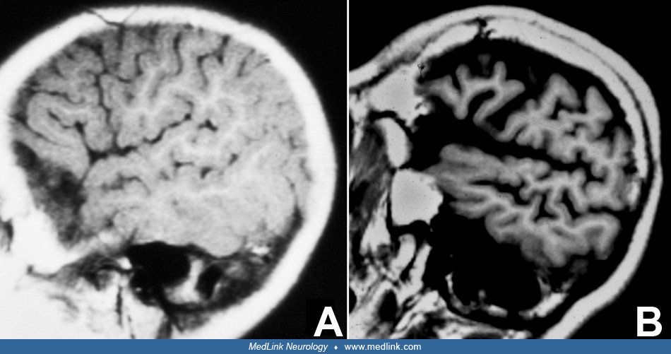
Dystroglycanopathies. This includes Walker-Warburg syndrome (type 2 lissencephaly) and muscle-eye-brain disease. Walker-Warburg syndrome patients have hydrocephalus. Muscle-eye-brain disease patients have typical abnormalities in the cerebellum, such as small cysts, not found in pontocerebellar hypoplasia. Alpha-dystroglycanopathy can be excluded by muscle biopsy and, in a minority of cases, also by genetic analysis.
Merosin deficient congenital muscular dystrophy (106).
mtDNA deletion. Biancheri and colleagues reported cerebellar hypoplasia, together with severe white matter and basal ganglia abnormalities consisting of near absence of putamina, caudate heads, and pallid, and normal metabolic screening, including lactate and deletion of a large part of mtDNA (21).
Serine biosynthesis disorder. Hypoplasia of the vermis and brainstem was documented in a single family with a new serine biosynthesis disorder: phosphoserine aminotransferase deficiency (58), in which amino acid analysis in the cerebrospinal fluid revealed a decrease of serine.
Chromosomal defects with PCH-like MR abnormalities. Chromosomal defects with PCH-like MR abnormalities combined with mental deficiency are occasionally found (12).
Autosomal dominant or recessive TUBB3 mutations. Autosomal dominant or recessive TUBB3 mutations cause a wide and variable spectrum of neuronal migration and axon guidance defects that may include hypoplasia of the brainstem and vermis (109).
Recessive mutation of VLDLR. Recessive mutation of VLDLR (very low density lipoprotein receptor) causes cerebellar hypoplasia with a peculiar flat appearance of the vermis and congenital ataxia, which is described in Turkish families (75).
Cerebellar hypoplasia in extreme prematurity. Cerebellar abnormalities resembling pontocerebellar hypoplasia have been described in preterm infants born at 26 weeks gestation or earlier. This abnormality is becoming increasingly clear as a complication of extreme prematurity (87; 159).
Axon guidance defects that predominantly affect the brainstem. This includes a new disorder: pontine tegmental cap dysplasia. The hallmark of this sporadic disease is ventral flattening of the pons, together with a dorsal bulge overlying the pontine tegmentum (16).
Congenital brainstem disconnection is a fatal disease in newborns presenting on MRI as apparent loss of a segment of the brainstem between the mesencephalon and lower brainstem (110). Accompanying hypoplasia of the cerebellum and flattening of the pons area may wrongly suggest a diagnosis of pontocerebellar hypoplasia.
The pontocerebellar hypoplasias must be distinguished from other cerebellar hypoplasias; Dandy-Walker spectrum disorder; spinocerebellar degenerations; rhombencephalosynapsis; which also can coexist with pontocerebellar hypoplasia (50); and also from acquired cerebellar atrophies, some of which are perinatal or prenatal: hypoxic-ischemic encephalopathy, congenital infections such as cytomegalovirus, which cause ischemia and multiple microinfarcts due to endothelial cell involvement, and certain congenital genetic/metabolic diseases. Fetal akinesia deformation sequence has multiple etiologies but also can occur in pontocerebellar hypoplasia (70). In PCH1, arthrogryposis occurs secondary to motor neuron involvement, but congenital contractures also can occur in PCH4 without motor neuron disease.
In all types of pontocerebellar hypoplasia, signs and symptoms are present in the first months of life, often already in the first week. The single most important investigation to start with in differential diagnosis is MRI of the brain, which will reveal the typical images of cerebellar hypoplasia, usually accompanied by relative sparing of the vermis and a flattening of the ventral pons. In addition to the images of the posterior fossa, the structure of the cerebral cortex and corpus callosum may hold important clues to diagnosis. Variations in MRI findings occur among the various types of pontocerebellar hypoplasia (24). Longstanding cases of PCH2 are likely to show cortical atrophy. This will aid in distinguishing PCH2 from MicPCH (CASK mutation) as the latter shows normal cortex or some coarsening of cortical gyri. The corpus callosum is thinned in the former and normal in the latter. More advanced techniques, such as diffusion tensor imaging aided by fiber tracking, may reveal the absence of transverse pontine fibers that connect the ventral pontine nuclei with the opposite cerebellar hemispheres (55). CASK (calcium/calmodulin-dependent serine protein kinase gene) loss of function often results in autistic spectrum disorder (91) but can also cause microcephaly, pontocerebellar hypoplasia, and a strong family history of early spontaneous embryonic or fetal loss (114).
Also to be considered in differential diagnosis of pontocerebellar hypoplasia is the X-linked Hoyeraal-Hreidarsson syndrome, due to defects in the DKC1 gene, which causes progressive disease with growth defects, bone marrow failure, and progressive neurologic impairment (43).
Biochemical investigations should exclude deficient N-glycosylation by transferrin electrophoresis in plasma. Cerebrospinal fluid should be investigated for lactate and amino acids in the search for RARS2 mutation (PCH6) and serine biosynthesis disorder. Electromyography and motor- and sensory nerve conduction velocities may aid in the diagnosis PCH1.
No specific therapies are available. Grosso and colleagues mention positive results of levodopa treatment in pontocerebellar hypoplasia type 2 (56).
There are no specific contraindications for the use of anesthesia in any of the types described. Special attention during general anesthesia for MRI should be given in metabolic encephalopathies with pontocerebellar hypoplasia to avoid prolonged fasting. Although increased creatine kinase has been found in patients with pontocerebellar hypoplasia type 2, no anesthetic accidents are on record.
All contributors' financial relationships have been reviewed and mitigated to ensure that this and every other article is free from commercial bias.

Harvey B Sarnat MD FRCPC MS
Dr. Sarnat of the University of Calgary has no relevant financial relationships to disclose.
See ProfileNearly 3,000 illustrations, including video clips of neurologic disorders.
Every article is reviewed by our esteemed Editorial Board for accuracy and currency.
Full spectrum of neurology in 1,200 comprehensive articles.
Listen to MedLink on the go with Audio versions of each article.
MedLink®, LLC
3525 Del Mar Heights Rd, Ste 304
San Diego, CA 92130-2122
Toll Free (U.S. + Canada): 800-452-2400
US Number: +1-619-640-4660
Support: service@medlink.com
Editor: editor@medlink.com
ISSN: 2831-9125
Developmental Malformations
Apr. 14, 2024
Developmental Malformations
Apr. 09, 2024
Childhood Degenerative & Metabolic Disorders
Apr. 07, 2024
Childhood Degenerative & Metabolic Disorders
Mar. 22, 2024
Developmental Malformations
Mar. 22, 2024
Developmental Malformations
Mar. 22, 2024
Developmental Malformations
Mar. 22, 2024
Developmental Malformations
Mar. 22, 2024