


Peripheral Neuropathies
Carbon disulfide neuropathy
Apr. 07, 2024
MedLink®, LLC
3525 Del Mar Heights Rd, Ste 304
San Diego, CA 92130-2122
Toll Free (U.S. + Canada): 800-452-2400
US Number: +1-619-640-4660
Support: service@medlink.com
Editor: editor@medlink.com
ISSN: 2831-9125
Toll Free (U.S. + Canada): 800-452-2400
US Number: +1-619-640-4660
Support: service@medlink.com
Editor: editor@medlink.com
ISSN: 2831-9125
Worddefinition
At vero eos et accusamus et iusto odio dignissimos ducimus qui blanditiis praesentium voluptatum deleniti atque corrupti quos dolores et quas.
Peripheral nerve injuries require the coordinated management of neurologists and surgeons to fully appreciate the indications for and timing of surgical intervention. The author thoroughly addresses the presentation, epidemiology, and surgical and nonsurgical outcomes of commonly encountered brachial plexus and peripheral nerve injuries. In this update, the author discusses the role of ultrasound in diagnosing and treating peripheral nerve injury.
|
• Nerve injury can range from minor and transient alteration in sensation to severe and permanent interruption of sensory and motor function. | |
|
• Timing of nerve injury repair depends on the mechanism of injury, the presence of recovery, and the anatomical location of the injury. | |
|
• Nerve grafting techniques and augmentation strategies promise to improve rates of functional recovery in the future. |
Historically, the classification of nerve injuries was developed and used primarily for the assessment of war-related injuries. In 1943, Seddon introduced a classification system based on three types of nerve injury (67). Later, Sunderland expanded on this system by creating a classification based on five degrees of injury severity (72). Both systems base themselves on peripheral nerve anatomy with particular emphasis on the endoneurium, perineurium, and epineurium, which are the key connective tissue elements within a peripheral nerve. Both systems also attempt to correlate the severity of the injury with the patient’s clinical manifestations.
Seddon’s simple classification yielded three terms that have become widely accepted as descriptors of the different types of nerve injury. Neurapraxia, the mildest form, is often incomplete and produces a transient loss of function. Its key feature is spontaneous recovery that occurs within hours to several months of injury and corresponds with Sunderland grade 1 injury. Axonotmesis, or Sunderland grade 2 injury, refers to a complete interruption of the axon and myelin sheath while preserving the endoneurial core connective tissue structure of the nerve. Often described as a “neuroma-in-continuity,” this type of injury results in an immediate loss of motor, sensory, and autonomic function distal to the lesion. Spontaneous recovery can occur but requires more time than a neurapraxic injury and depends on several factors that affect the rate of nerve regeneration. The final type of injury, neurotmesis, implies a complete disruption of both the neural and connective tissue elements of a peripheral nerve. This type of injury is incompatible with spontaneous recovery and will ultimately require surgical intervention if any function is to be regained (67; 27). Sunderland grade 3 injury describes transection of the nerve axon and sheath with preservation of the perineurium and, thus, includes elements of both axonotmesis and neurotmesis. The distinction between Sunderland grade 4 and 5 involves preservation of the epineurium in the former.
The various clinical presentations of patients with peripheral nerve injuries are obviously dependent on the particular nerve or nerves that have been injured as well as the severity and level of the lesion. It is the case with most traumatic peripheral nerve injuries that pain and loss of sensory and motor function are noted either immediately or within days of the inciting traumatic event.
Brachial plexus injuries. The clinical manifestations of brachial plexus injuries are particularly dependent on the specific elements of the plexus that have been injured. The plexus originates from the roots of C5 through T1. For the most part, the C5 root generally gives rise to deltoid function, the C6 root to biceps function, the C7 root to triceps function, the C8 root to the deep flexors of the hand and forearm, and the T1 root to the intrinsic muscle function of the hand (23). Using these motor functions as a guideline, along with the appropriate sensory distributions, the extent of injury can often be determined by clinical examination.
Median nerve injuries. The most significant and debilitating clinical manifestation of a median nerve injury is loss of pinch and grip strength secondary to denervation of the flexor pollicis longus muscle and the intrinsic muscles of the thenar eminence (38). A median nerve injury occurring high in its course through the arm will also decrease pronation strength, result in atrophy of forearm muscle mass, and cause sensation deficit of both the volar and palmar aspects of the lateral 3.5 digits (38).
Radial nerve injuries. The radial nerve originates in the distal axilla and is susceptible at that site to compression injuries, which produce “Saturday night palsy.” The hallmark of such a proximal radial nerve injury is the patient’s inability to extend the forearm secondary to loss of triceps innervation (43). Most radial nerve injuries occur at the midhumeral level secondary to fracture and present with sparing of triceps muscle function in the face of more distal radial distribution loss such as a wrist drop and inability to extend the fingers and thumb (43). Typically, the sensory deficits involved with radial nerve injury are minimal, involving only the dorsum of the hand and the anatomical snuffbox region (37).
Akhavan-Sigari and associates reported a series of 318 patients with radial nerve injuries from military injuries (02). About a third of the injuries were associated with open radial fractures, and about two thirds were closed injuries. All 191 patients with closed injuries recovered well, with a median time to full recovery of 6 months. Nerve injuries associated with open fractures recovered in 113 of 127 cases.
Ulnar nerve injuries. The ulnar nerve receives its entire contribution from the medial cord of the brachial plexus. In an injury producing a total ulnar palsy, a patient will present with a classic hand posture in which there is partial flexion or “clawing” of the little finger and, to a lesser extent, the ring finger (43). In the case of a compression syndrome, sensory symptoms in the form of paresthesias and numbness in the little and ring fingers may be the patient’s first complaint (43). With time, patients will experience atrophy of the intrinsic muscles of the hand, producing a characteristic appearance and significant hand weakness.
Spinal accessory nerve injuries. In 1993, Donner and Kline found the two most common complaints of 83 patients with an extracranial spinal accessory nerve injury to be the inability to raise the arm above the horizontal plane and a “dragging pain” in the shoulder. The inability to raise the arm was secondary to a degree of trapezius weakness (particularly the upper trapezius) seen in all patients, whereas the dragging pain was thought to be due to downward tension on the shoulder joint secondary to chronic sagging of the shoulder. Also, on physical exam, scapular winging is a frequent finding distinguishable from the winging of serratus anterior palsy by its disappearance when the patient’s arm is raised forward, fully extended, and pushed against resistance (18).
Sciatic nerve injuries. Injury to the sciatic nerve at the proximal buttock level or more distally at the midthigh level can produce various symptoms depending on the amount of functional loss in the peroneal and tibial divisions of the nerve (48). The peroneal division lies posteriorly within the sciatic nerve, making it prone to injury from injections or hip fractures. Patients will present with a foot drop secondary to loss of dorsiflexion and eversion of the foot as well as an inability to extend the toes (43; Kim and 43). Injury to the tibial division of the sciatic produces loss of plantar flexion and inversion of the foot as well as loss of toe flexion. Patients with primarily tibial division injury complain mostly of a burning, hyperesthetic sole of the foot (43).
Femoral nerve injuries. The femoral nerve is the largest branch of the lumbar plexus, arising from the anterior divisions of L2, L3, and L4 spinal nerves (39). Injury to this nerve within the pelvis will produce a loss of hip flexion due to denervation of the iliopsoas muscle and a loss of knee extension secondary to denervation of the quadriceps muscles. Patients will find it difficult, if not impossible, to climb stairs with this injury (43). Injury to the femoral nerve at the level of the pelvis will produce a variable loss of hip flexion and variable sensory loss in the anteromedial thigh; however, absence of quadriceps function with loss of the patellar reflex will be uniform (43). Thigh-level injury of the femoral nerve within the femoral triangle region will also produce a complete loss of quadriceps function.
Recovery of useful function distal to a nerve injury depends on a number of factors. The mechanism of injury is probably the most important determinant. Other factors include the age of the patient, the type of surgical intervention required, and the timing of the surgical intervention. In general, close to 90% of patients with recorded intraoperative nerve action potentials experienced good functional recovery. End-to-end epineural repairs produced good results in 70% of patients, compared with 50% of patients who had graft repairs. Of those patients with graft repairs, shorter repairs had better results than grafts greater than 3 inches in length. Finally, patients who are treated earlier do better than those treated later, and children tend to have far better outcomes than adults (69).
He and colleagues examined factors predictive of sensory and motor recovery after upper extremity peripheral nerve injury (29). In a meta-analysis, this group determined that predictors of improved functional recovery for sensory deficits included female sex and a shorter length of the nerve defect. Length of the nerve defect was the strongest predictor of outcome for sensory defects, with a 1 cm increase in the gap leading to an odds ratio of 0.59 favoring poor recovery. Regarding motor recovery, younger age, earlier, direct repair, female gender, and radial nerve involvement were positive predictive factors (29). A prospective analysis by Hundepool and associates in upper extremity injuries found that male gender, older age, lower level of education, arterial injury, more extensive nerve injury, and stress after trauma at 1 and 3 months were predictive of less complete recovery (31). Wang and colleagues looked specifically at timing of repair excluding brachial plexus injuries and found 258 patients at a single center who underwent repair over a 5.5-year period (78). In this study, favorable predictive measures included male sex, worse injury severity scores, lower extremity or proximal involvement, and direct repair with timing of surgery not influencing outcome. The studies show that many of these data are conflicting, and extensive database creation would help better understand predictive factors after nerve injuries.
Brachial plexus injuries. These injuries present a unique situation in that it is critically important to determine the proximity of the lesion to the spinal cord. If proximal, it can result in death of the cell body, causing damage that is irreparable (20). The presence of Horner syndrome, winging of the scapula due to long thoracic nerve palsy, EMG evidence of paraspinal muscle denervation, dorsal scapular or phrenic nerve palsy, or myelographic evidence of pseudomeningoceles are all bad prognostic findings that suggest extremely proximal injury to or avulsion of the spinal nerve (20; 69). In general, infraclavicular repairs do better than supraclavicular repairs; C5, C6, and some C7 repairs fare better than the lower plexus elements of C8 and T1; and lateral cord repairs are more successful than posterior cord repairs. Both lateral and posterior cord repairs fare better generally than those involving the medial cord (47; 69).
Samii and colleagues reported a series of 44 brachial plexus injuries involving the musculocutaneus nerve. Reinnervation of the biceps muscle was achieved in 72% of cases using a spinal accessory branch for neurotization. Shorter segment nerve transfers had a higher success rate than patients treated within 6 months of their injury (65).
Dubuisson and Kline note improvement to a grade 3 (contraction of proximal muscles against some resistance) recovery in 78% of patients with penetrating wounds and 58% of patients with stretch injuries. C5 and C6 stretch injuries carried the most favorable prognosis. A posterior approach with some form of nerve grafting was used in all but a few patients (21).
Reports have focused on long-term functional outcomes of these patients. Ahmed-Labib and colleagues reported on outcomes of 31 individuals with a mean follow-up of over 3 years. They found improved disability scores in patients undergoing surgery within 6 months of injury and poorer scores in patients with root avulsion injuries both in terms of pain and functional outcome (01). Riess and colleagues looked at the same issue in a different manner. They examined 29 individuals with brachial plexus injuries and compared groups with and without associated scapulothoracic dissociation. They found short- and long-term differences between the groups with worsened outcomes with scapulothoracic dissociation in terms of hospital stay, neurologic recovery, and mean muscle strength with a median 60-month follow-up (61). Moiyadi and associates opined that early detection and intervention within 5.5 months of injury led to improved functional outcomes (58).
Sulaiman and colleagues evaluated brachial plexus injuries managed with nerve transfer operations with or without direct brachial plexus element repair. They found excellent (90%) improvement in elbow flexion when medial pectoral to musculocutaneous transfers were performed and 60% improvement in intercostals grafting procedures. Improvement in shoulder adduction was seen more often with spinal accessory to suprascapular nerve transfers (95%) than with axillary nerve restoration procedures. Repair of brachial plexus elements at the time of nerve transfer tended to lead to better outcomes (71).
Not every lesion is amenable to success with microsurgical repair, and chronic pain issues cripple many victims of brachial plexus injury or avulsion. Sindou and colleagues performed microsurgical dorsal root entry zone lesions in 55 patients and found good to excellent pain control in 66% of patients; 71% showed an improvement in activity level (68). Lefaucheur and colleagues investigated transcranial magnetic cortical stimulation in 12 patients with arm pain from brachial plexus injuries and discovered superior results compared to patients with brainstem stroke but inferior results in individuals with facial pain (51).
Kretschmer and colleagues assessed patient satisfaction after brachial plexus surgery. They found that 83% of patients were satisfied with the results of renervation surgery. However, only 48% of patients could return to work at a mean of 9 months after surgery. This study suffered from a low response rate, potentially skewing the data (49).
Median nerve injuries. At all levels (arm, elbow and forearm, anterior interosseous nerve, and wrist) the success of the repair and ultimate functional recovery was closely related to whether the initial lesion was a complete transection or a neuroma-in-continuity (38). Cleanly lacerated median nerves that were primarily repaired in the acute stage achieved better functional recovery than bluntly lacerated nerves that required a delayed secondary repair. With neuromas-in-continuity, those lesions that transmit nerve action potential recordings recover better than those that do not; the latter require resection with graft repair (38).
Radial nerve injuries. Similar to median nerve injuries, functional recovery of radial nerve injuries depends on the mechanism of injury and the type of surgical repair, with clean transections acutely repaired by end-to-end epineurial anastomosis achieving the best results. Although the overall results of radial nerve injury repair are encouraging, one avoidable complication occurs frequently. Surgical repair of laceration injuries to the superficial sensory radial nerve results in a high incidence of painful neuroma formation. To prevent this complication, the treatment of choice is to simply resect the nerve. This has stopped pain symptoms in over 70% of patients (37).
Ulnar nerve injuries. Of all the upper extremity peripheral nerve injuries, repair of the ulnar nerve yields the poorest outcome (20). Combined median and ulnar nerve injuries have an even worse prognosis than an ulnar nerve injury alone. In the functional recovery that does occur, it has been found that good motor recovery occurs more often than good sensory recovery (34).
Spinal accessory nerve injuries. In most cases of extracranial spinal accessory nerve injury, recovery is the rule. In a study of 83 patients with this type of injury, 39 were treated conservatively, and 44 were treated surgically. Of those treated conservatively, the majority exhibited a strong tendency for spontaneous improvement and eventually achieved normalization of shoulder posture and, therefore, relief from the dragging pain that was their initial presenting complaint. Of those treated surgically, most exhibited excellent postoperative recovery of arm abduction, normalization of shoulder posture, and cessation of preoperative pain syndromes (18).
Sciatic nerve injuries. Regardless of the mechanism of injury, a study of 380 patients with sciatic nerve injuries revealed that patients treated conservatively had an 80% chance of recovery of useful function in the tibial division distributions and only a 60% chance of recovery of useful function in the peroneal division distributions. The majority of patients with injection injuries recovered function spontaneously. Similarly, in the surgically treated patients, recovery in the tibial division distributions was greater than recovery in the peroneal division distributions. Also, the prognosis was better for surgical patients when their injuries were at the thigh level rather than at the buttock level (48).
Jones and colleagues looked specifically at sciatic nerve injuries associated with military injuries (36). Thirteen patients were identified with sciatic nerve injuries due primarily to gunshot wounds and blast injuries. All underwent surgery with cadaveric allografts with five early operations and eight on a delayed basis. Functional outcome was poor, with substantial motor function reestablished in only three individuals, although neuropathic pain was reduced by surgery in the majority.
In isolated peroneal nerve injuries after the split from the sciatic nerve, functional recovery after graft repair is largely dependent on the length of the grafts. Seventy-five percent of patients with grafts less than 6 cm recovered enough function to walk without requiring a foot brace. Only 35% of patients with graft lengths between 6 and 12 cm and 14% of patients with graft lengths more than 12 cm were able to make this type of functional recovery within 3.5 years postoperatively (Kim and 43). In a follow-up study, the authors reported excellent function (88% excellent return of function) in patients requiring neurolysis alone with documented nerve action potentials. The ability to perform end-to-end suture repair also led to a high (84%) functional recovery rate. Once again, longer grafts were associated with poorer outcomes (41).
Femoral nerve injuries. In a study of 78 patients with traumatic femoral nerve injuries, the majority required surgical intervention due either to a lack of clinical and electrical spontaneous recovery or partial injuries complicated by pain. Despite a proximal pelvic level for most injuries and, as a result, lengthy graft repairs, the recovery of some useful function was the rule rather than the exception. Most patients had some nerve regeneration and decent recovery of function by 2 years postoperatively (39).
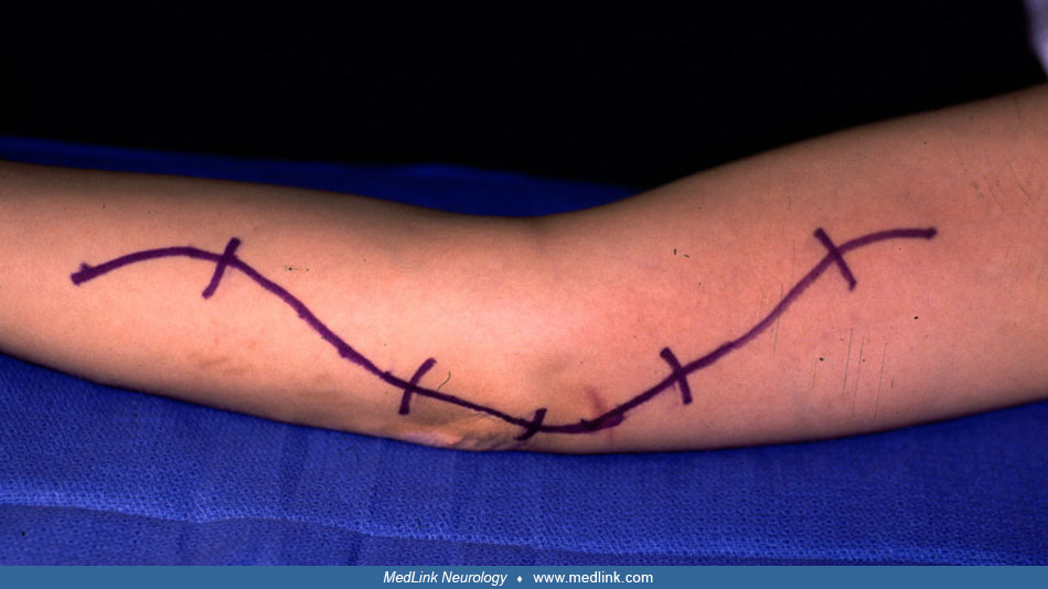
A 29-year-old female was involved in a motor vehicle accident in July 2001, in which she suffered a laceration to the right arm with subsequent ulnar nerve dysfunction. Physical examination found a well-healed transverse incision proximal to the medial malleolus.
There was a Tinel sign over the distal ulnar distribution and sensory loss in a typical ulnar pattern. Motor loss was appreciated in the ulnar innervated intrinsic hand musculature, and a Froment sign was present. Preoperative EMG showed right ulnar motor conduction delay. Sensory ulnar transmission was absent. Operative exploration was planned.
Operative repair was attempted approximately 5 months postinjury. Positioning was performed identically as for an ulnar nerve release with the incision.
Intraoperative nerve action potentials, somatosensory evoked potentials, and EMG were monitored. At surgery, the proximal and distal branches of the right ulnar nerve were found, as well as a large neuroma in continuity.
Nerve action potentials did not cross the neuroma. Microsurgical excision of the neuroma was performed. The proximal and distal branches were then freed, transposed, and reanastomosed using 7-0 proline sutures.
Postoperatively, pain was improved. Motor and sensory function was unchanged but awaits longer follow-up.
There are two major categories of nerve injury, each with a variety of etiologies. In transection, the nerve is completely severed, leaving two separate ends. In the second category, the injury creates a neuroma-in-continuity, in which the gross nerve remains intact despite variable mechanisms of injury and variable loss of function.
Transecting injuries are most often the result of a soft tissue laceration by such sharp, metal objects as surgical instruments, knives, glass, and propeller blades. Fortunately, sharp and clean transections of nerve are favorable for early surgical repair. However, only 30% of serious nerve injuries are caused by this mechanism of injury (44). Transected nerves, by definition, produce a neurotmetic lesion in which there is complete loss of function with no hope for recovery without surgical intervention (27).
Between 60% and 70% of serious nerve injuries seen in a clinical practice will be neuromas-in-continuity. A number of different etiologies may be responsible: stretch, traction, contusion, and ischemia from compression, injection, electrical, and thermal injuries. Stretch, traction, and contusion injuries are by far the most common and can result from a number of mechanisms. Fractures with and without joint dislocation, birth injuries secondary to difficult delivery, gunshot wounds or other high-velocity missiles, and iatrogenic injuries from surgical retraction are all mechanisms that can produce a stretch, traction, or contusion injury to a peripheral nerve. Ischemia and compression injuries are often seen together because peripheral nerves depend on blood flow. Common mechanisms include pressure palsies due to unconsciousness or anesthesia, acute compression by hematoma formation or vascular aneurysms, compartment syndromes, and entrapment neuropathies. Injection injuries are a surprisingly common form of iatrogenic injury to peripheral nerves. The nerve injury itself results from either the needle placement or the injected toxic agent. Electrical and thermal injuries are less common forms of peripheral nerve injury. Electrical injuries can result from accidental contact of an extremity with a high-tension wire, whereas thermal injuries often result from exposure to flame, steam, or other hot elements (44; 27).
Some specific peripheral nerve injuries are more often caused by particular mechanisms, which are discussed below.
Brachial plexus injuries. Brachial plexus injuries are not rare, and the majority affect relatively young people involved in traffic (especially motorcycle) accidents. Stretch injuries constitute the largest category, as many as 70% in one study (20). The remaining categories include gunshot wounds, glass, knife, or metal lacerations, and iatrogenic injuries (47). Obstetric brachial plexus palsies represent a special subgroup of stretch and contusion injuries, the management of which remains a highly controversial area (69). In a series of 100 consecutive cases, Dubuisson and Kline report that 27 were due to motorcycle injuries and 23 were from penetrating trauma; nine wounds were iatrogenic. No clear overriding pattern of injury was seen (21). Another large series of brachial plexus injuries documented that most lesions (52%) involved the trunks, with 36% at the level of the cords and 12% at the trunks. Distribution of injury was more even for gunshot wounds and more slanted toward the trunks in injuries resulting from motor vehicle accidents (09).
Median nerve injuries. In a study of 250 patients with median nerve injuries, the most common mechanism of injury seen at all levels of the nerve was laceration. At the arm level, gunshot wounds and humerus fractures were also associated, whereas at the elbow, forearm, and wrist, stretch and contusion injuries were prominent. The highest percentage of iatrogenic injuries occurred at the elbow level secondary to injection in the cubital fossa (38).
Radial nerve injuries. In a study of 180 patients with radial nerve injuries requiring surgical intervention, the mechanism of injury in over half involved midhumeral fractures or lacerations of the upper extremity. The remaining patients suffered injuries primarily from gunshot wounds, contusions, and compressions (37).
Ulnar nerve injuries. Most ulnar nerve lesions that require surgical intervention are secondary to entrapment and compression at the elbow. Isolated and acute ulnar nerve injuries due to trauma are relatively rare. Often, they are found in combination with median nerve injuries at the wrist level. The most frequent mechanism of injury is accidental laceration by glass, followed by knife wounds and lacerations from attempted suicide (34).
Spinal accessory nerve injuries. Injury to the extracranial portion of the spinal accessory nerve is relatively uncommon. When it does occur, the injury is usually to the portion of the nerve running in the posterior cervical triangle. The most common etiology is iatrogenic injury secondary to minor surgery for benign lymph node biopsy. Injury to the nerve is also caused by other neck-related operations, stretch or contusion injuries, and stab or glass wounds (18).
Sciatic nerve injuries. The sciatic nerve is the most frequently injured lower extremity nerve (43). In one series of 380 patients, 60% of these injuries occurred at the buttock level with injection injuries being the etiology in over half. The remaining 40% occurred at the thigh level and were usually the result of gunshot wounds, femur fractures, lacerations, or contusion (48). At the knee, the peroneal and tibial division of the sciatic nerve split to form distinct nerves. The peroneal nerve in particular takes an oblique course that places it near the fibular neck. This superficial location makes it extremely vulnerable to injury, especially to stretch and contusion injuries with or without fracture of the fibula as well as iatrogenic injury during open or arthroscopic procedures on the knee joint (Kim and 43). In a study of 318 common peroneal nerve injuries, 44% of injuries occurred due to stretch or contusion without bony injury, 12% involved direct laceration, 9% were entrapments, 7% were due to stretch injuries with bony fracture, 13% involved nerve or soft tissue tumors, 7% were caused by external nerve compression from other causes, and 4% were due to gunshot wounds or iatrogenic injuries (41).
Femoral nerve injuries. In a study of 78 patients with traumatic femoral nerve injuries, 60% of patients suffered iatrogenic injury secondary to inguinal herniorrhaphy, total hip replacement, intra-abdominal vascular or gynecologic operations, and other operative procedures within the vicinity of the nerve. Additional injuries resulted from penetrating gunshot and stab wounds, laceration by glass, and stretch or contusion injuries secondary to pelvic fracture (39).
Missile injuries. Wartime experience with painful peripheral nerve injury from missile wounds dates back to Weir Mitchell and the American Civil War. Roganovic and Mandic-Gajic reported their experience with 326 such injuries at the Belgrade Military Medical Academy. Out of a cohort of 3091 overall nerve injuries, 12.3% of patients had painful nerve injuries. Pain was more common following injuries to the median, ulnar, and tibial nerves and less likely with involvement of the peroneal, femoral, and radial nerves. Both the time of onset of the pain and the clinical characteristics were difficult to predict from the nerve involved or the severity of the neurologic deficit (63).
Response to peripheral nerve injury is at first degenerative and then ultimately regenerative. With complete nerve disruption, the resulting gap fills with either hematoma or adjacent soft tissue. Within an hour of injury, the proximal end of the injured nerve swells, and a variable amount of retrograde degeneration occurs depending on the mechanism of injury, less in clean lacerations and more in gunshot wounds (22). At the same time, the open end of the proximal stump heals over and forms a neuroma with the aid of connective tissue proliferation. The distal end of the injured nerve also undergoes early degenerative change; however, it is the severed axons themselves that are degenerating. A distal stump neuroma forms at the nerve end but primarily comprises reactive mesenchymal tissue cells without neural elements (23).
Wallerian degeneration is a term that applies to changes in the distal nerve stump only. Initiated on axonal death, this process involves complete breakdown of the axons within 2 weeks, digestion of the surrounding myelin fragments, and phagocytosis of all cellular debris by metabolically active Schwann cells within 6 weeks (23). Although the fascicular anatomy persists in the distal stump, the nerve itself shrinks, and with time, this shrinkage becomes irreversible, making surgical repair nearly impossible.
In injuries that do not completely disrupt the nerve (neuromas-in-continuity) or in nerves that have been surgically reapproximated, there exists the possibility of axonal regeneration. There is a variable delay before new axonal sprouting occurs at the proximal stump, the length of which is determined by the mechanism of injury. In neurapraxic lesions where the nerve is simply contused, axons may begin to sprout across the injury site in as early as 10 days (23).
The regeneration process requires the restoration of large quantities of axonal lipids and proteins, the synthesis of which occurs in the nerve cell body before being transported down the axon via axoplasmic flow (22). These synthesized materials accumulate at the injury site and form growth cones, from which the axonal sprouting originates. Regeneration of axons down a peripheral nerve trunk averages about 1 mm per day or 1 inch per month (23; 22). Other groups have established the importance of cell adhesion molecules such as NCAM in guiding axonal regrowth. Franz and colleagues found that highly polysialylated NCAM allowed appropriate targeting of muscle pathways for nerve regeneration compared to knockouts for NCAM and wild type treated enzymatically to please the polysialylated moiety that regenerated without specific guidance to muscle after experimental femoral nerve injury (26).
Lad and colleagues performed a comprehensive review of epidemiological trends for upper extremity peripheral nerve injuries to the median, ulnar, and radial nerves as well as the brachial plexus. They found that between 1993 and 2006, the overall incidence of these injuries decreased slightly, whereas the cost associated with these injuries increased significantly. The majority of the injuries were seen in metropolitan areas (95%) and were treated at academic institutions (75%) (50).
War has always been an unfortunate source of peripheral nerve injury research. Birch and associates comprehensively examined injuries and outcomes in peripheral nerve injuries suffered by British troops in the Middle East. They describe 100 patients with 261 injuries. Most of the wounds were caused by explosive injuries, and most also involved open wounds. Most patients (69%) also had an associated vascular or orthopedic injury. The nerve lesions were fairly evenly divided between neuropraxia (45%), axonotmesis (35%), and neurotmesis (20%) injuries. Only nine patients were able to return to full duty. Approximately two thirds of patients had good outcomes, and only 7% had poor results at 28 months (06; 07).
Clinical examination. Few disorders deserve such a thorough history and physical examination as do peripheral nerve injuries. During the workup, it is vital to answer three major questions. First, what was the mechanism of the injury? The answer will provide important information about the severity of the injury and possible management of the lesion. Secondly, was the injury likely to produce a transected nerve or a neuroma-in-continuity? Again, the answer will provide important information about the management of the lesion. Finally, where is the level of the lesion, and how complete is the patient’s loss of function? Answering this requires intimate knowledge of the motor, sensory, and autonomic distribution of the nerve in question. The information gathered may hint at the patient’s prognosis for recovery of function (46).
On physical examination, in addition to close inspection for muscle atrophy, it is important to grade both the loss of sensory and motor function. A grading system not only allows for an initial evaluation of functional loss, but also provides a way to monitor the recovery of function over time. A useful system developed at the Louisiana State University Medical College includes motor grades for contraction with various amounts of resistance and sensory grades that place greater and more practical emphasis on the patient’s ability to localize stimuli (46; 69).
A common error made during motor examination is the failure to recognize substitution of other muscles to produce the action being tested. For example, a patient with a complete palsy of the biceps and brachialis muscles secondary to a musculocutaneous nerve injury can still flex their forearm using the brachioradialis muscle, which is innervated by the radial nerve (45). This type of mistake highlights the need for a thorough examination of all major muscles in the limb in which nerve injury is suspected.
During testing of sensory function, it is essential to test for sensation in the zones of a nerve that are not clouded by overlap from adjacent nerves. This ensures that the findings are attributable to a single nerve only. Similarly, a nerve can be tested for autonomic function by looking at the presence or absence of beads of sweat in a nerve’s autonomous zones (46).
One key physical finding is the presence or absence of Tinel sign. Eliciting a Tinel sign involves sharp percussion over the course of the injured nerve distal to the site of injury (45). If doing so produces paresthesias, Tinel sign is said to be positive. Acutely, Tinel sign can be useful in locating the level of nerve injury because any area of focal nerve injury will respond with local paresthesias on percussion (46). With time, an advancing positive Tinel sign indicates at least fine fiber growth through the lesion and into the distal stump; however, this provides no predictive value with regard to the potential for significant distal functional recovery. A negative Tinel sign several months postinjury may be a telling finding, as it provides strong evidence against significant neural regeneration (45).
Electrophysiological evaluation. Electrophysiological evaluation of a peripheral nerve injury is almost as important as careful clinical examination. EMG is the most often used method of electrophysiological evaluation. In a normal EMG, three phases occur. The first phase is a brief burst of electrical (insertional) activity due to the placement of the needle within the muscle. In the second phase, the muscle is at rest. When it is normally innervated, the tracing at rest should be flat. In the third phase, an electrical response in the form of a motor unit action potential is produced when the patient attempts voluntary muscle contraction or when the technician stimulates the nerve that supplies it (46).
For the first few days postinjury, the distal nerve can still be stimulated to produce distal muscle function because Wallerian degeneration requires time to proceed down the distal stump of a nerve. However, after 48 to 72 hours this response to stimulation no longer occurs. Denervational changes on EMG do not appear until at least 2 to 3 weeks postinjury. Typical denervational changes include: (1) loss or extreme reduction of insertional activity during the first phase of EMG testing, (2) spontaneous firing of rapid, biphasic, low-amplitude waves, or (3) fibrillations instead of a flat tracing when the muscle is at rest. A poorly formed or absent motor unit action potential is obtained on attempted muscle contraction (46; 45). Reversal of these changes can occur and provides evidence of some reinnervation (particularly in muscle most proximal to the lesion). However, clinical muscle contraction may exist despite the presence of continued denervational changes on EMG, making it essential to test muscles clinically rather than rely on EMG alone (69).
In addition to EMG evaluation, the clinician can also perform nerve stimulation, sensory conduction studies, or somatosensory studies to further delineate the level of a particular lesion and follow its progress with respect to regeneration and ultimate functional recovery.
Radiologic evaluation. Due to the common association between fractures and peripheral nerve injuries, plain film radiographs of the affected limb are indicated. It is important to note, however, that the level of nerve lesion does not always correlate to the level of the fracture (46).
Myelography remains an important study when evaluating supraclavicular brachial plexus stretch injuries. The finding of a meningocele suggests that the force of the injury was enough to produce an arachnoidal tear, suggesting that the spinal root itself may be avulsed. Bertelli and Ghizoni examined the ability of CT myelography combined with clinical examination to differentiate between brachial plexus root avulsion and root rupture. This guided surgical planning when considering root grafting for rupture as opposed to nerve transfers for brachial plexus injuries when there was root rupture. They assessed a supraclavicular Tinel sign and retention of shoulder retraction along with an absence of root rupture on CT myelogram to predict a graftable C5 or C6 cervical root 96% of the time. They had poorer results with MRI in a limited number of patients (04).
Doi and colleagues have described a novel MRI imaging technique known as overlapping coronal-oblique slice imaging. In a series of 35 patients with brachial plexus injuries, MRI was equivalent to CT myelography in detecting root avulsions. Theoretically, this could provide an advantage in terms of ease of accessibility and patient comfort (17).
Another MR-based imaging modality that has been employed to evaluate peripheral nerve injuries is MR neurography. Du and associates reported use of this technique in 191 patients with peripheral nerve injuries (19). MR neurography showed abnormal signal and could localize the site of nerve injuries, correlated well to EMG, and provided additional/supplemental localization in 45% of patients. MR neurography was useful both pre- and postoperatively and was especially useful when EMG was inconclusive. Marquez Neto and colleagues used the same technology to correlate with postoperative recovery and restoration of function after traumatic ulnar nerve repair (54). Upadhyaya and associates demonstrated the utility of MR neurography in distinguishing individual root and trunk injury in patients with brachial plexus lesions (75).
High-resolution ultrasonography is another emerging technology in the evaluation of peripheral nerve injuries. Toros and colleagues used this technology in 26 cases (74). In every case that was explored surgically, there was a correlation between the ultrasonographic images and the clinical findings. Nerve bundle transaction, axonal swelling, neuroma formation, and partial lacerations could all be identified with ultrasound. Toia and associates studied the use of ultrasound in 119 nerve injuries in 108 patients with tumors, traumatic injuries, and compression neuropathies. Preoperative ultrasound was helpful in 72% of cases and was concordant with operative findings (73). Elshewi and colleagues examined 69 individuals with 96 peripheral nerve injuries in the upper extremities, including median, ulnar, and radial neuropathies. They compared the ability of ultrasound to diagnose the extent of injury to electrophysiological studies and found comparable diagnostic accuracy. Ultrasound could reliably show discontinuity and degree of nerve injury defined by Sunderland grade as well as the degree of nerve retraction and thickening (24).
Two options exist for managing peripheral nerve injuries: nonoperative and operative. Surgical management, in particular, has long been a controversial area. It is critical to understand both the indications for surgery based on the mechanism of injury and especially the timing for surgical intervention, as it is timing that appears to have the most impact on successful outcomes (20).
Timing. Injuries suspected of producing complete peripheral nerve transection are one indication for relatively early operation. In clean transections, typically following knife, glass, scalpel, or razor blade injuries, surgery within 72 hours allows an excellent opportunity for end-to-end suture repair (69). This type of primary repair by epineurial anastomosis yields the best results with regard to functional recovery (20). In some injuries where the nerve is bluntly transected by auto metal, machinery, propeller blades, etc., it is best to delay surgical repair for several weeks. This delay allows the initial degenerative changes to occur in both nerve ends, making the proximal and distal neuromas easy to identify by the time of operation. Resection back to healthy tissue can then be carried out during the repair (69). If an injury is explored acutely and found to be a ragged or blunt transection, the two nerve ends can be “tacked” to adjacent soft tissue to prevent their retraction. Secondary surgical repair can then be done after 2 to 4 weeks have passed (42; 20; 69).
Wang and colleagues analyzed 311 patients with acute penetrating and nonpenetrating peripheral nerve injuries (78). Of the 258 patients who underwent operative repair, there was no statistical difference in functional recovery between patients with repair within or after 24 hours. The authors concluded that peripheral nerve repair could be staged and was unnecessary during abdominal, chest, or cranial damage control procedures.
The timing of repair for traumatic brachial plexus injuries remains controversial. A database analysis by Martin and associates reviewed 43 individual studies and concluded that there was a clear delineation with better results with surgery within 6 months of the traumatic event (55). Motor outcomes, pain scores, and quality of life were all improved with earlier intervention. Because of the potential for spontaneous recovery, a 3-month delay was considered warranted, but optimal recovery was advised with intervention between 3 and 6 months post-injury.
The vast majority of peripheral nerve injuries will not be transections. Instead, they will produce a neuroma-in-continuity, and it is within this category of injuries that allowing time for spontaneous regeneration to occur before attempting surgical repair is so important. Several months of serial clinical and electrophysiologic examinations are needed to recognize subtle evidence of recovery (69). During this period, physical therapy focusing on range-of-motion exercises is key to preventing joint fixation and contractures and optimizing functional state once surgical repair is finally attempted.
In patients who demonstrate clinical or electrical evidence of return of proximal muscle function, nonoperative management can be continued because the injury was likely neurapraxic or axonotmetic in nature (20; 69). Injuries that do not show evidence of functional recovery are likely neurotmetic in nature and should be surgically explored. In suspected focal injuries from gunshot wounds, contusions, or fractures, surgical exploration should occur around 2 to 3 months, whereas suspected lengthier lesions due to stretch injury should be explored at 3 to 5 months (69).
Surgical options. Surgical exploration of the neuroma-in-continuity has been revolutionized by the introduction of intraoperative nerve action potential recording. Nerve action potentials can be recorded from a whole nerve or from individual fascicles. They provide a clearer picture of the extent of injury and regeneration, which is important because the gross intraoperative appearance of an injured nerve can be misleading (20). Bipolar stimulating and recording electrodes are used to obtain nerve action potential recordings. Initially, both electrodes are placed proximal to the injury site, and a nerve action potential is elicited. The nerve action potential is then observed as the electrodes are moved into the neuroma-in-continuity and ultimately beyond (42). The presence of a nerve action potential distal to the site of injury provides evidence of either preserved axonal function or significant axonal regeneration, both of which bode well for future functional recovery (42; 69). Similarly, the absence of a nerve action potential distal to the site of injury suggests a purely neurotmetic lesion that will have little or no capacity for spontaneous regeneration.
The presence or absence of intraoperative nerve action potentials is important for determining the extent of surgical repair required. When a nerve action potential is present, external neurolysis of the injured nerve (ie, circumferential dissection to remove external scar tissue) will be sufficient to maximize functional recovery (69). When a nerve action potential is absent, the entire neuroma-in-continuity should be resected both proximally and distally to healthy neural tissue. The length of the gap between the new proximal and distal stumps determines the repair at this stage.
When resection of the neuroma-in-continuity is required, all efforts should be made to repair the gap with an end-to-end epineurial anastomosis. However, a repair that places the nerve under undue tension must be avoided, as distraction is the major cause of repair failure postoperatively. Methods to decrease the nerve gap and, therefore, the tension on the repair site include generous mobilization of the nerve ends, transposition of the nerve, and flexion of the limb. When the length of the nerve gap makes an end-to-end repair impossible, it becomes necessary to use nerve grafts to fill the gap. Ten percent is added to the measured nerve gap, and then either the patient’s sural nerve or antebrachial cutaneous nerve is harvested. The proximal and distal stumps are separated into fascicular groups, and the grafts are sutured between them (69).
Occasionally, surgical exploration reveals a portion of the nerve to be intact and transmitting a nerve action potential, whereas the portion immediately adjacent does not transmit a nerve action potential. In this case, a split repair becomes necessary. The portion transmitting a nerve action potential is left alone to continue its spontaneous recovery, whereas the adjacent, more severely injured portion is resected back to healthy tissue and a nerve graft is placed (69).
Special circumstances. In the case of an extremely proximal injury to the brachial plexus causing a spinal root avulsion, the technique of nerve transfers (neurotization) has become useful. In this technique, another nerve or plexus element that is functional serves as a proximal donor for one that has an irreparable injury (20). Commonly used nerve transfers include the thoracic intercostals, medial or lateral pectorals, distal spinal accessory, and descending cervical plexus. In the case of obstetric brachial plexus palsy, the length of required regeneration is considerably shorter than in adults with brachial plexus injuries; therefore, initial conservative management is strongly encouraged (20). Babies who show signs of recovery within the first few months often achieve excellent spontaneous results; those who do not should undergo surgical intervention (69).
In the case of fracture-related radial nerve injuries, a slightly longer period of observation has been recommended because spontaneous recovery rates have been found to be as high as 76% (37). Similarly, a longer period of observation is recommended for injection injuries most commonly involving the proximal radial and sciatic nerves. Patients typically will have acute onset of symptoms after the injection; however, these injuries have a good chance of achieving excellent functional recovery. Surgical intervention after 3 to 5 months is indicated only in those cases with persistent and severe neurologic deficit or severe neuritic pain that is unrelieved by sympathetic blocks (69).
Preganglionic injuries have generally been considered untreatable. Wu and colleagues described a novel strategy employing cervical laminectomy followed by reconnection of the injured roots to the spinal cord using a combination of sural nerve grafts with a mixture of fibrin glue and acidic fibroblast growth factor (79). They found improvement in muscle strength postoperatively in 16 of 18 patients.
Missile injuries. Deficits and pain following penetrating injuries are felt to be relatively refractory to available treatment modalities. In the wartime experience of Roganovic and Mandic-Gajic, 79% of patients with painful penetrating peripheral nerve injuries were treated with surgery – 82 as an initial treatment modality and 176 after failed pharmacological therapy. Multimodality therapy was least effective for deafferentation pain but successful with neuralgia either due to neuroma formation, foreign body compression, or compression syndromes. For complex regional pain syndrome, direct surgery was ineffective, and a combination of drug therapy and sympatholysis was most effective. The authors speculate that dorsal root entry zone (DREZ) ablation might help the patients with deafferentation pain (64).
New horizons. Peripheral nerve transplantation has long been investigated as an alternative or adjunct to autologous grafting techniques in the peripheral nervous system (66). Dam-Hieu and colleagues describe using peripheral nerve grafts to bridge between the lumbar dorsal roots and the dorsal columns of rats after thoracic cordotomy. Three months after surgery, half the animals had regained some nociceptive potential, and retrograde labeling into the dorsal root ganglia via the nerve grafts by 9 months was seen in two thirds of animals (16). Cui and colleagues have described the use of autogenic and allogenic peripheral nerve sheaths populated with purified neonatal Schwann cells or adult olfactory ensheathing glia. They witnessed evidence of both myelinated and unmyelinated axons along the nerve sheath in their rat model (15).
Brooks and colleagues expanded this work into a clinical trial. Processed allograft was employed in a multicenter trial involving 132 nerve injuries. Only 76 patients had adequate follow-up. The mean graft length was 22 mm. Meaningful recovery was seen in 87% of patients. Only a 5% revision rate was seen (10). Squintani and associates used processed allograft in 10 patients with brachial plexus injuries and saw regeneration of motor function in every instance (70). Cho and colleagues reported on upper extremity injuries treated with allograft as part of a prospective registry and found meaningful recovery in 75% of median and 67% of ulnar repairs (13).
Another modality employed experimentally is the use of therapeutic ultrasound to enhance nerve regeneration. Raso and colleagues found a significant improvement in nerve fiber density and functional sciatic regeneration in animals treated with pulsed ultrasound compared to controls at 21 days (60).
Rochkind describes the use of low-power laser radiation as an adjunct in the repair of peripheral nerves (62). He describes preclinical research showing nerve cell growth in response to phototherapy both in culture and a rat sciatic nerve injury model, as well as promising results in a pilot clinical trial. This strategy has been employed successfully in experimental models of nerve injury. Medalha and colleagues found improvement in recovery after sciatic nerve injury in rats with low-level laser therapy (56). In a critical review, Al-Shengiti and Oldham document the success in preclinical studies but caution that current clinical research is inadequate to address the clinical efficacy (03).
Liu and Liu performed a meta-analysis of 41 studies, including 3304 patients using nerve growth factor. They concluded that nerve growth factor resulted in superior outcomes with an increase in generally mild adverse events (52).
Collagen-coated tubes have become commercially available to aid in nerve regeneration. Two clean ends of a damaged nerve are placed in either end of a tube and gently sutured to the tube. The tube itself promotes direct regrowth and reinnervation. Ichihara and associates describe improved regeneration in a canine peroneal nerve model with a novel tube that adds poly (L-lactic) acid to the commercially available polyglycolic acid product (32). Hood and colleagues have coupled axon guidance tubes with autologous Schwann cells to try to enhance axonal regeneration (30).
Some reports have described encouraging progress in the use of stem cells to guide peripheral nerve regeneration after injury. Walsh and colleagues showed improved regeneration of nerve gaps with skin-derived stem cell augmentation (77). Wakao and associates showed survival of bone marrow-derived stem cells that were induced into Schwann cell differentiation up to a year after treatment (76). Gu and associates were able to demonstrate the formation of new neuromuscular junctions and the arrest of muscle atrophy after stem cell implantation in a tibial nerve injury model (28). Cheng and colleagues were able to track and quantitate nerve recovery using MRI after experimental nerve injury (12). However, the mechanism of the treatment effect is not yet clear. Erba and colleagues used adipose-derived stem cells to enhance axonal growth in an experimental model of sciatic nerve injury (25). They found few viable cells after transplantation and opined that growth factor release by the stem cells was more important than differentiation into neural tissue.
Zhang and colleagues have reported preclinical trials successfully using bone marrow-derived mesenchymal stem cells to improve sciatic nerve recovery in experimental models (81; 82) These and other reports have engendered enthusiasm about combining this technology with Schwann cell conduits for improved nerve regeneration (80; 05).
Other suggestions for conduits for nerve repair and regeneration include human umbilical cord membrane. Bourgeois and colleagues examined preclinical and limited clinical data and postulated that wrapping nerves in amniotic tubes may reduce adhesions and fibrosis, speed nerve healing, and optimize function (08). Cox and associates reviewed 20 patients with surgical reconstruction of nontransected nerves (14). The majority of the patients had upper extremity injuries. Favorable outcomes were seen in terms of pain measures and functional recovery in this anecdotal report.
Exosomes are nano-vesicles secreted by Schwann cells in response to injury, which help increase and enhance axonal regeneration (53). Bucan and associates showed increased regeneration using exosomes derived from adipose-derived mesenchymal stem cells in experimental sciatic nerve injury (11). Pan and colleagues showed that stem cell-derived exosomes promote motor recovery equivalent to nerve autografts in experimental models (59).
Symptomatic management. Not all peripheral nerve injuries are ultimately salvageable, and neither motor nor sensory function is restored in many individuals. Treatment strategies for painful peripheral neuropathy for some individuals can be just as beneficial as restorative strategies. Multiple putative mechanisms for pain relief have been postulated (33). Meyer-Frieβem and colleagues looked at perineural injection of botulinum-A toxin in painful peripheral nerve injuries (57). Sixty patients were treated with a 57% success rate of achieving at least 30% pain relief, with only one case of transient weakness. Nonpharmacological treatments, such as peripheral nerve stimulation, have also been offered in ongoing trials to address these injuries (35).
All contributors' financial relationships have been reviewed and mitigated to ensure that this and every other article is free from commercial bias.

Richard S Polin MD
Dr. Polin of Polin Neurosurgery has no relevant financial relationships to disclose.
See Profile
Randolph W Evans MD
Dr. Evans of Baylor College of Medicine received honorariums from Abbvie, Amgen, Biohaven, Impel, Lilly, and Teva for speaking engagements.
See ProfileNearly 3,000 illustrations, including video clips of neurologic disorders.
Every article is reviewed by our esteemed Editorial Board for accuracy and currency.
Full spectrum of neurology in 1,200 comprehensive articles.
Listen to MedLink on the go with Audio versions of each article.
MedLink®, LLC
3525 Del Mar Heights Rd, Ste 304
San Diego, CA 92130-2122
Toll Free (U.S. + Canada): 800-452-2400
US Number: +1-619-640-4660
Support: service@medlink.com
Editor: editor@medlink.com
ISSN: 2831-9125
Peripheral Neuropathies
Apr. 07, 2024
General Child Neurology
Apr. 05, 2024
Peripheral Neuropathies
Apr. 02, 2024
Peripheral Neuropathies
Apr. 01, 2024
Peripheral Neuropathies
Apr. 01, 2024
Peripheral Neuropathies
Apr. 01, 2024
Neuroimmunology
Mar. 24, 2024
Infectious Disorders
Mar. 24, 2024