
Peripheral Neuropathies
Carbon disulfide neuropathy
Apr. 07, 2024
MedLink®, LLC
3525 Del Mar Heights Rd, Ste 304
San Diego, CA 92130-2122
Toll Free (U.S. + Canada): 800-452-2400
US Number: +1-619-640-4660
Support: service@medlink.com
Editor: editor@medlink.com
ISSN: 2831-9125
Toll Free (U.S. + Canada): 800-452-2400
US Number: +1-619-640-4660
Support: service@medlink.com
Editor: editor@medlink.com
ISSN: 2831-9125
Worddefinition
At vero eos et accusamus et iusto odio dignissimos ducimus qui blanditiis praesentium voluptatum deleniti atque corrupti quos dolores et quas.
In this article, the author discusses the anatomy of the spinal accessory nerve and injury of the nerve. The spinal accessory nerve is most commonly injured as the result of surgical procedures in the neck. This review discusses physical examination, electrophysiologic testing, and management of injury to the spinal accessory nerve.
|
• Spinal accessory nerve palsy is usually iatrogenic, related to surgery in the posterior triangle of the neck. | |
|
• Shoulder shrug is a poor method for testing the function of the trapezius muscle; arm abduction over the head is much better. | |
|
• Spinal accessory nerve lesions cause marked disability and pain. |
The spinal accessory nerve, cranial nerve XI, is a purely motor nerve that has both cranial and spinal components. The motor fibers originate from two separate motor nuclei in the brainstem and spinal cord. The cranial portion arises from cells adjacent to the nucleus ambiguous and involves fine fibers that join the vagus nerve to supply the muscles of the pharynx via pharyngeal and recurrent laryngeal branches (67; 18); however, a cadaveric study failed to find evidence for a cranial portion of the spinal accessory nerve in 15 specimens (28). The spinal nucleus typically originates in the first to fifth cervical segments. Motor fibers from several cervical levels join to form the spinal portion of the accessory nerve. The nerve then courses upward through the foramen magnum and joins the cranial portion briefly before exiting the skull. Cranial nerve XI exits the skull via the jugular foramen along with the glossopharyngeal (IX) and vagus (X) nerves. The accessory nerve then courses downward and posteriorly in one of the three compartments of the parapharyngeal space (56). It lies anterior to the internal jugular vein in 55% to 70% of cases and posterior to the internal jugular vein in 30% to 45% of cases (18; 23). However, the relationship of the spinal accessory nerve to the internal jugular vein has been disputed depending on whether the determination is made in cadavers or in surgical series and depending on what level in the neck one is evaluating. In a surgical series, the nerve was found lateral to the internal jugular vein in 95% of cases (60). There are rare cases where the nerve appears to pierce the jugular vein (45). It then either pierces or passes posterior to, and gives off, branches to the sternocleidomastoid muscle. The spinal accessory nerve then runs obliquely across the posterior triangle of the neck to the undersurface of the trapezius muscle that it also supplies. There is considerable anatomic variability (62).
Iatrogenic injury of the nerve is most common in the posterior triangle of the neck, where it assumes a coiled appearance in most people (63). Symes and Ellis dissected the nerve bilaterally in 25 cadavers in order to determine whether there was a consistent anatomical arrangement of nerve fibers that might allow surgeons to avoid injuring the nerve in this region (58). They found that there was considerable variability in the anatomic relationships of the nerve, making the general surgical approach to the posterior triangle valueless. Branches from the second, third, and fourth cervical segments unite with the spinal accessory nerve either in the posterior triangle or on the undersurface of the trapezius muscle and contribute to the nerve supply of that muscle.
The clinical manifestation of spinal accessory neuropathy depends on the location of the injury. Injury is most common in the posterior triangle of the neck. Injury in this area produces weakness and atrophy of the trapezius muscle. Resultant weakness of shoulder elevation (shrug) occurs in some cases, although large series of confirmed cases have shown that most have normal shoulder shrug (35). As a result of inadequate shoulder stabilization, weakness of shoulder abduction is often present. This is most evident in the last 90 degrees of arm abduction, in which the actions of the trapezius and serratus anterior muscles are particularly important. Trapezius weakness also results in winging of the superior and medial border of the scapula. This is present at rest and may be accentuated by shoulder abduction. A provocative maneuver involving external rotation of the shoulder against resistance has been reported to reliably accentuate the winging (10). Other clinical signs known as the active elevation lag and the triangle sign are reported to reliably differentiate between spinal accessory and long thoracic nerve palsies (30). Eventually, shoulder height discrepancy becomes evident. A prominent accompanying symptom is shoulder pain, the primary complaint of the patient, which may be severe.
If the injury of the spinal accessory nerve occurs more proximally, there will also be weakness of the sternocleidomastoid muscle (06). This muscle is involved in rotating the chin to the opposite side and, to a lesser degree, in flexion of the neck. This muscle is not solely responsible for these movements; thus, the patient almost never notices this weakness. The spinal accessory nerve is intimately associated with cervical nerves from the second to the fourth levels. Therefore, accompanying sensory loss is often present in the anterolateral neck with injury of the accessory nerve.
The prognosis for spontaneous recovery in the more common cases of nerve transection is rather poor. A review of the natural history of spinal accessory neuropathy determined that the outcome was generally good; however, based on the review’s criteria, only 45% of patients had a good outcome (16). In this study, electrodiagnostic findings did not seem to correlate well with outcome, except for an improvement in spinal accessory compound motor action potential recorded from the trapezius. Those patients who were believed to have spinal accessory nerve injury related to neck dissection for well-differentiated thyroid carcinoma were all said to have only transient weakness, although both the diagnostic criteria for the injury and recovery were poorly defined (27). Patients are frequently left with debilitating residual shoulder pain and weakness. Inability to properly elevate the scapula limits the strength and range of motion of abduction of the shoulder. Studies in patients undergoing radical neck dissections for cancer have shown that quality of life is markedly impacted by "sacrifice" of the spinal accessory nerve and resultant shoulder pain (61). One study has clearly shown that ability to abduct the arm and reach above are markedly better in cases where the spinal accessory nerve is preserved (43). That study also suggested that rehabilitation may improve arm abduction after radical neck dissection, particularly in those patients who have the spinal accessory nerve resected.
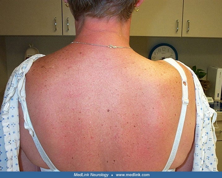
A 59-year-old man presented with right shoulder pain. He had undergone a right carotid endarterectomy 10 months prior. Immediately after surgery, he noticed a "knot" sensation in the upper-shoulder and neck area. After discharge from the hospital, he noticed trouble lifting objects with his right arm. He then developed a deep, aching pain in his right shoulder. He noticed that his right shoulder was lower than his left and that it was smaller in the upper shoulder area. On physical exam, there was a clear shoulder height discrepancy; the left was higher than the right by at least 1 inch. There was no scoliosis, and there was marked atrophy of the upper portion of the trapezius muscle. There was mild protrusion of the superomedial border of the scapula that was accentuated with shoulder abduction. The patient was unable to completely abduct his right arm above his head actively, but passive range of motion was normal. In spite of the atrophy, shoulder shrug appeared to have normal strength. There was mild asymmetry in bulk of the sternocleidomastoid muscle, but no weakness noted on head turn or neck flexion. Pinprick and light touch appreciation were reduced only in the area immediately adjacent to his surgical scar. Palatal and tongue function were normal. Nerve conduction studies were performed with stimulation of the accessory nerve at the sternocleidomastoid muscle and recording over the upper trapezius. There was a 10-fold reduction in the motor-evoked amplitude on the right as compared to the left. Needle EMG revealed chronic motor unit reinnervation in the form of long duration, polyphasic motor unit potentials with reduced voluntary motor unit recruitment in both the sternocleidomastoid and trapezius muscles. The deltoid muscle was electromyographically normal. The patient was referred for physical therapy for the right shoulder to improve strength and mobility.
This case was most likely due to stretch or compression injury of the spinal accessory nerve during retraction. This complication is more common in patients who have a high-riding carotid bifurcation (69), necessitating more retraction of the sternocleidomastoid for exposure. There was relatively robust recruitment of motor unit potentials, and the patient had only mild limitation of abduction of his arm; thus, in this case, the prognosis with conservative management is better. The more common and severe injuries resulting from "sacrifice" of the nerve in radical neck dissection for cancer or accidental transection in lymph node biopsy have a worse prognosis; surgical intervention is more often required to regain good function or achieve pain relief in these cases.
The most common cause of spinal accessory nerve palsy is iatrogenic injury (08). Surgical procedures in the posterior triangle are responsible for the majority of cases. The two most common surgical procedures that result in iatrogenic injury of the spinal accessory nerve are lymph node biopsy and radical neck dissection for head and neck cancer (04; 14; 26). The cervical lymph nodes are intimately associated with the spinal accessory nerve, and great care needs to be taken to prevent injury (67). In many cases of squamous cell carcinoma, the spinal accessory nerve is intentionally "sacrificed" to assure complete removal of malignant nodes (61). Shoulder pain after neck dissection surgery is a common complaint, and more than half of the patients have injury of the spinal accessory nerve (65). In a series where “super selective dissection” was used, two of six patients had spinal accessory nerve injury compared to six of 11 patients who had selective dissection (17). A randomized study of 30 patients compared different levels of dissection (15 per group) and found that by omitting level 2b dissection, patients had a significantly lower neck dissection impairment index and better range of motion 6 months after surgery (15). Neck dissection performed in cases of differentiated thyroid carcinoma also commonly results in spinal accessory nerve injury (27). The injury of the spinal accessory nerve is more often accidental in cases of lymph node biopsy. With these posterior triangle spinal accessory nerve lesions, the degree of injury is often severe, and the mechanism is direct transection of the nerve. A large series looking at trapezius muscle palsy found a surprisingly low number of cases caused by surgery (55). In this series, neuralgic amyotrophy was determined to be the etiology in 22 of 54 cases, whereas surgery was felt to be the cause in only 16 of 54.
Other surgical procedures that have been reported to result in iatrogenic injury include carotid endarterectomy (69). The mechanism of injury is more often compression or stretch. This type of injury is more often reported in those with high-riding carotid bifurcations. In this instance, more vigorous retraction is required to achieve and maintain adequate exposure. Case reports are also available of spinal accessory nerve injury associated with repair of carotid aneurysm at the base of the skull (34), thoracotomy (41), face lift surgery (14; 53; 39), internal jugular vein cannulation (20; 07), posterior fossa decompression surgery for Chiari malformation (49), deep-brain stimulation surgery for Parkinson disease, impulse generator placement (50), and tunneling of extension leads (32).
Other potential etiologies for spinal accessory nerve palsy include tuberculosis (scrofula) or leprosy involving the cervical lymph nodes (14). The nerve may also be injured by direct trauma from a gunshot or stabbing or as a late complication from irradiation. Idiopathic inflammatory damage of the spinal accessory nerve has been described (probably related to brachial neuritis or Parsonage-Turner syndrome). This syndrome may be seen after any surgical procedure and often produces severe shoulder pain. Thus, differentiating direct surgical injury from idiopathic inflammatory damage may be difficult. Compression of the nerve may occur as a result of wearing a sling (68) or a failed hanging (57). A case of spinal accessory nerve injury from a bite to the neck during lovemaking has been reported (46). Stretch injury that resulted from sudden head turn when holding heavy objects has been described (33), and injury of the nerve due to power lifting has also been reported (64). Injury of the spinal accessory nerve as a result of whiplash has been described as the cause of persistent pain (05). Spinal accessory nerve injury has also been reported in association with deep tissue massage (01).
Finally, the spinal accessory nerve may be injured alone or in combination with cranial nerves IX and X at the jugular foramen. This is known as Vernet syndrome (57). Possible etiologies of injury at this level include skull base cancers or metastases (52), primary nerve tumors (ie, schwannomas) (21), inflammatory processes (ie, sarcoidosis and glomus jugulare tumors), or infection (including herpes zoster) (19). Schwannoma has been reported in the spinal portion of the accessory nerve, causing a foramen magnum syndrome (59).
Most cases of iatrogenic injury involve transection of the nerve during surgery. Excessive traction or bite may cause crush injury of the nerve. Stretch injuries have also been described and are thought to result from ischemia to the nerve. Inflammation of the nerve may be the result of a more generalized process, such as sarcoidosis, or it may be idiopathic and localized. Scarring from prior surgery or radiation may also lead to injury.
In a series of 84 cases (14), the most common cause of spinal accessory nerve palsy was lymph node biopsy followed by tumor excision. Trauma-induced stretch and laceration were also relatively common in this series.
The spinal accessory nerve is the most common nerve damaged accidentally during surgical procedures. In a large series of peripheral nerve iatrogenic injuries, the spinal accessory nerve accounted for 11% of the cases. The next most likely to suffer iatrogenic injury were the common peroneal, superficial radial, and genitofemoral nerves, accounting for 9%, 8%, and 7%, respectively (26).
Injury of the spinal accessory nerve during surgical procedures can be prevented by careful dissection. Previously, it was felt that guidelines for anatomical relationships between the nerve and other structures are not practical due to considerable variability (58). However, a constant anatomic relationship between the greater auricular nerve and the spinal accessory nerve was identified in 100% of 100 cases of radical neck dissection (48). This relationship forms an “X” behind the sternocleidomastoid muscle and the authors coined the term “X-pointer” and feel this is an extremely valuable method of reliably identifying the nerve to prevent iatrogenic injury. Use of a hand-held stimulator (25) may allow the surgeon to identify the nerve by activating the trapezius muscle. As the muscle is superficial in the posterior triangle, a very low intensity of stimulation is needed. Use of a peripheral nerve monitor, similar to those used to monitor the facial nerve, has been employed successfully to prevent spinal accessory nerve injury (38). Ultrasound has been proposed as a means for mapping the course of the spinal accessory nerve and was successful in 50 healthy controls with a mean age of 37 years (40). A review of the use of intraoperative monitoring of the spinal accessory nerve and trapezius muscle during surgery of the neck revealed no conclusive data to recommend or refute the utility (36).
The differential diagnosis of shoulder pain includes injury of other individual peripheral nerves in the region. These include the axillary, suprascapular, dorsal scapular, and long thoracic nerves. Cervical radiculopathy involving the fourth, fifth, or sixth cervical roots may also produce shoulder pain and weakness of abduction. Intrinsic shoulder disease, such as rotator cuff tears or impingement, needs to be considered. Shoulder height discrepancy may also be seen in scoliosis or shoulder dislocation.
The differential diagnosis of scapular winging includes long thoracic nerve injury with weakness of the serratus anterior or weakness of the rhomboid muscles with injury of the nerve to rhomboids. In a large series of 128 cases of scapular winging, the authors found that long thoracic nerve lesions were more frequently the etiology (55%) compared to spinal accessory lesions (30%) (54). The most helpful differentiating features are the portion of the scapula that is winging and the movements that accentuate the winging. Winging due to spinal accessory nerve palsy with resultant trapezius weakness is most prominent at the upper, medial border of the scapula and is increased by arm abduction. Winging caused by serratus anterior weakness from long thoracic nerve injury is most prominent at the inferomedial border and is accentuated by having patients press their palms firmly against a wall directly in front of them. The winging produced by rhomboid weakness involves the entire medial border of the scapula and is accentuated by having patients place their hands on their hips and try to touch their elbows behind their backs. Several forms of muscular dystrophy may present with asymmetric scapular winging such as that seen with spinal accessory nerve injury, particularly facioscapulohumeral muscular dystrophy (55).
A prospective study regarding cranial neuropathies in PCR-positive patients hospitalized with COVID-19 reported that 135 of 302 patients developed new cranial nerve abnormalities, of which 11 affected the spinal accessory nerve (13). However, 86% of the reported cranial nerve deficits were based on patient reports, and only 14% were based on physician evaluation; for the spinal accessory nerve, the screening question was “Is there temporary or permanent impairment in rotating the head or lifting the shoulder.”
The clinical history of shoulder and neck pain following surgical procedures in the neck is highly suggestive. The physical examination findings of atrophy of the trapezius muscle, shoulder height discrepancy, and winging of the superomedial border of the scapula help to confirm the diagnosis.
Electrodiagnostic testing may be helpful, especially in cases of mild nerve injury. Comparison of the motor-evoked responses in the upper trapezius with stimulation of the accessory nerve at the sternocleidomastoid muscle frequently demonstrates a marked asymmetry. As with most other motor nerve conduction studies, a difference of more than 50% in the motor amplitude is considered significant. Delay of motor latency on the affected side has also been used as evidence for spinal accessory nerve dysfunction (33). Although a report of 60 normal, healthy individuals has been performed (02), the variability of equipment, technique, patient population, and other factors makes the generalization of these “normal” values problematic. A study evaluating 69 healthy subjects (48 women and 21 men) aged 20 to 42 concluded there is significant variation among subjects and lower amplitude in those with higher BMI, thus, side-to-side differences should be used (12). Needle EMG is useful to determine whether recruitment of motor unit potentials remains robust enough to consider conservative management or whether more aggressive intervention is required. Nerve conduction studies of the upper extremity nerves and EMG of other shoulder muscles help eliminate the possibility of associated brachial plexopathy or nerve root injury. A review of 16 cases referred for suspected spinal accessory nerve transaction by Laughlin and colleagues showed that all had low amplitude CMAPs that required higher stimulation to elicit, and all cases showed spontaneous activity in the upper trapezius (29). MRI imaging in cases of spinal accessory nerve injury includes atrophy and T2/STIR signal hyperintensity of the trapezius (31).
The primary decision involves whether to manage the patient conservatively or to recommend a surgical procedure. The degree of nerve injury is the primary determinant. This is often extremely apparent clinically. Electrodiagnostic testing may be helpful in making this decision. If relatively robust recruitment of motor unit potentials is present on EMG, physical therapy may be the treatment of choice. In cases where absent or markedly reduced motor unit recruitment is present, a surgical procedure is probably appropriate. As with many peripheral nerve lesions, ultrasound has been demonstrated to be helpful in demonstrating whether there is continuity of the nerve, which may influence the decision to recommend surgical repair (09). Potential procedures that may help regain function of the trapezius muscle include end-to-end repair (44), a cable graft of the sural nerve (66), or reconstruction using thoracodorsal or long thoracic nerve (51). The outcome has been rated as good or excellent in the majority of cases subject to surgical intervention. The outcome does not seem to be severely compromised by delaying repair (22; 42; 08), although this point is controversial (11). A large retrospective analysis of surgical management of spinal accessory nerve injuries showed a positive outcome (improvement to at least antigravity function) in 75% of patients (24). Similarly, whether just neurolysis was performed in those with evidence for recordable nerve action potentials, or end-to-end repair or grafting was performed, approximately 90% of patients regained the ability to at least elevate their arm to 90 degrees of abduction in a series of 156 iatrogenic spinal accessory nerve injury cases caused by lymph node biopsy (47). A small series of patients showed benefit of neuromuscular electrical stimulation for treatment after spinal accessory nerve injury; however, those patients had relatively mild dysfunction before treatment, and there was no comparator group (03). A randomized trial of intensive physical therapy was shown to offer some short-term benefit at 3 months in terms of active abduction of the shoulder but not in other measures, and the benefit compared to standard therapy did not persist (37).
During insertion of central venous catheters in the jugular vein, care needs to be taken to avoid injuring the spinal accessory nerve, as this has been a described complication (20; 07).
All contributors' financial relationships have been reviewed and mitigated to ensure that this and every other article is free from commercial bias.
Michael T Pulley MD PhD
Dr. Pulley of the University of Florida, Jacksonville received consulting fees from Argenx, Alexion, and UCB/Ra.
See Profile
Louis H Weimer MD
Dr. Weimer of Columbia University has received consulting fees from Roche.
See ProfileNearly 3,000 illustrations, including video clips of neurologic disorders.
Every article is reviewed by our esteemed Editorial Board for accuracy and currency.
Full spectrum of neurology in 1,200 comprehensive articles.
Listen to MedLink on the go with Audio versions of each article.
MedLink®, LLC
3525 Del Mar Heights Rd, Ste 304
San Diego, CA 92130-2122
Toll Free (U.S. + Canada): 800-452-2400
US Number: +1-619-640-4660
Support: service@medlink.com
Editor: editor@medlink.com
ISSN: 2831-9125
Peripheral Neuropathies
Apr. 07, 2024
General Child Neurology
Apr. 05, 2024
Peripheral Neuropathies
Apr. 02, 2024
Peripheral Neuropathies
Apr. 01, 2024
Peripheral Neuropathies
Apr. 01, 2024
Peripheral Neuropathies
Apr. 01, 2024
Neuroimmunology
Mar. 24, 2024
Infectious Disorders
Mar. 24, 2024