Stroke & Vascular Disorders
Cardiovascular intervention: neurologic complications
Aug. 23, 2023
MedLink®, LLC
3525 Del Mar Heights Rd, Ste 304
San Diego, CA 92130-2122
Toll Free (U.S. + Canada): 800-452-2400
US Number: +1-619-640-4660
Support: service@medlink.com
Editor: editor@medlink.com
ISSN: 2831-9125
Toll Free (U.S. + Canada): 800-452-2400
US Number: +1-619-640-4660
Support: service@medlink.com
Editor: editor@medlink.com
ISSN: 2831-9125
Nearly 3,000 illustrations, including video clips of neurologic disorders.
Every article is reviewed by our esteemed Editorial Board for accuracy and currency.
Full spectrum of neurology in 1,200 comprehensive articles.
Listen to MedLink on the go with Audio versions of each article.
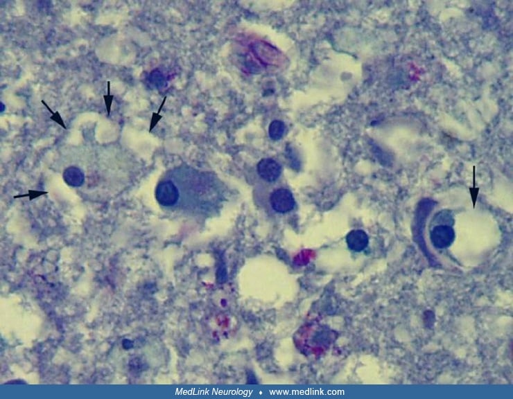
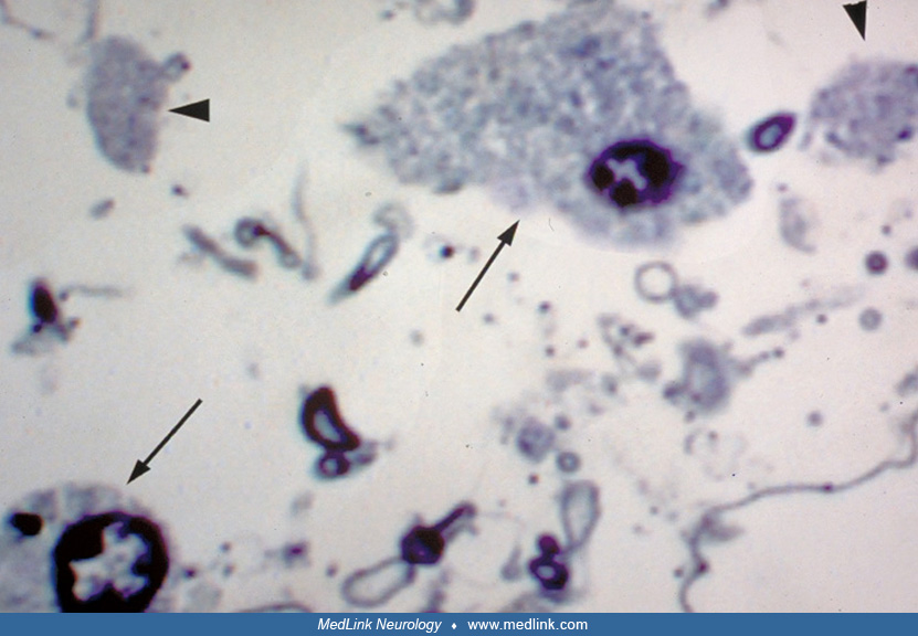
Fluorescent light microscopy (200 magnification) double labeling with 2,3 cyclic nucleotide 3' phosphodiesterase (CNP, red, oligodendroglial membrane marker) and CD68 (blue-white, macrophage marker) antibodies. (Arrow) Red membrane marking by CNP of a foamy oligodendroglial cell. Phase contrast (not shown) of same histologic section demonstrated a small, round oligodendroglia-type nucleus within the CNP-stained membrane. (Contributed by Dr. Regina Armstrong.)