Behavioral & Cognitive Disorders
Hyposmia in neurodegenerative disorders
May. 19, 2023
MedLink®, LLC
3525 Del Mar Heights Rd, Ste 304
San Diego, CA 92130-2122
Toll Free (U.S. + Canada): 800-452-2400
US Number: +1-619-640-4660
Support: service@medlink.com
Editor: editor@medlink.com
ISSN: 2831-9125
Toll Free (U.S. + Canada): 800-452-2400
US Number: +1-619-640-4660
Support: service@medlink.com
Editor: editor@medlink.com
ISSN: 2831-9125
Nearly 3,000 illustrations, including video clips of neurologic disorders.
Every article is reviewed by our esteemed Editorial Board for accuracy and currency.
Full spectrum of neurology in 1,200 comprehensive articles.
Listen to MedLink on the go with Audio versions of each article.
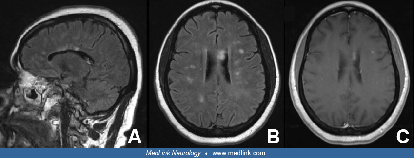
Parenchymal lesions of neurosarcoidosis seen on MRI mimicking multiple sclerosis plaques. Label: A. Sagittal fluid-attenuated inversion recovery (FLAIR) image showing T2 hyperintense lesions involving the subcortical white matter and callososeptal interface. Some of these lesions mimic Dawson fingers, characteristic demyelinating plaques seen in multiple sclerosis. B. An axial FLAIR sequence showing ovoid T2 hyperintense lesions in juxtacortical, periventricular and deep white matter. C. T1 weighted image with gadolinium contrast illustrating some lesions with associated enhancement. (Courtesy of Dr. Surabhi Ranjan, Neuro-Oncology Branch, National Institutes of Health.)