Infectious Disorders
Post-polio syndrome
Jan. 09, 2024
MedLink®, LLC
3525 Del Mar Heights Rd, Ste 304
San Diego, CA 92130-2122
Toll Free (U.S. + Canada): 800-452-2400
US Number: +1-619-640-4660
Support: service@medlink.com
Editor: editor@medlink.com
ISSN: 2831-9125
Toll Free (U.S. + Canada): 800-452-2400
US Number: +1-619-640-4660
Support: service@medlink.com
Editor: editor@medlink.com
ISSN: 2831-9125
Nearly 3,000 illustrations, including video clips of neurologic disorders.
Every article is reviewed by our esteemed Editorial Board for accuracy and currency.
Full spectrum of neurology in 1,200 comprehensive articles.
Listen to MedLink on the go with Audio versions of each article.
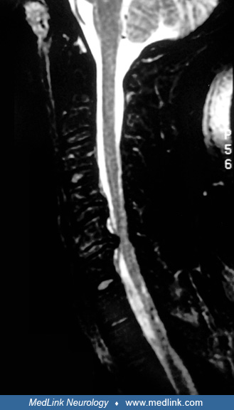
This MRI of the cervical spine was obtained from a 55-year-old female who presented with left neck and shoulder pain. Sagittal T2- (A) and T1- (B) weighted images show degenerative disc disease at both the C4-C4 and C5-C6 (white arrow) levels. This resulted in spinal stenosis and abnormal signal in the cord (A). The axial gradient image (C) shows a focal left paracentral disc herniation at the C5-C6 level (arrowhead) deforming the cord and resulting in spinal stenosis and marked left neural foraminal narrowing. (Contributed by Dr. Diana Gomez-Hassan.)