Epilepsy & Seizures
Epilepsia partialis continua of Kozhevnikov
Feb. 12, 2023
MedLink®, LLC
3525 Del Mar Heights Rd, Ste 304
San Diego, CA 92130-2122
Toll Free (U.S. + Canada): 800-452-2400
US Number: +1-619-640-4660
Support: service@medlink.com
Editor: editor@medlink.com
ISSN: 2831-9125
Toll Free (U.S. + Canada): 800-452-2400
US Number: +1-619-640-4660
Support: service@medlink.com
Editor: editor@medlink.com
ISSN: 2831-9125
Nearly 3,000 illustrations, including video clips of neurologic disorders.
Every article is reviewed by our esteemed Editorial Board for accuracy and currency.
Full spectrum of neurology in 1,200 comprehensive articles.
Listen to MedLink on the go with Audio versions of each article.
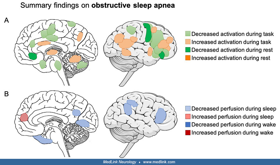
(A) Summary of findings from fMRI studies in obstructive sleep apnea. During tasks, activation is reduced in the dorsolateral prefrontal cortex, anterior cingulate cortex, middle frontal gyrus, inferior frontal gyrus, cingulate and parietal regions, bilateral parahippocampal regions, right insula, bilateral claustrum, left precentral gyrus, and right precuneus, depending on the cognitive domain involved. Activation is increased in the right anterior prefrontal gyrus, bilateral inferior and middle frontal gyri, cingulate gyrus, junction of the inferior parietal and superior temporal lobes, thalamus, cerebellum, parietal cortex, medial precuneus, hippocampus, and left temporo-occipital area. (B) Summary of findings from PET and SPECT studies in obstructive sleep apnea. During sleep, reduced regional perfusion is present in the left parietal region, bilateral parahippocampal gyri, left lingual gyrus, precuneus, middle and posterior cingulate gyrus, parieto-occipital cortex, and prefrontal cortex. Hyperperfusion is present in the frontal cortex during sleep. (Contributed by Dr. Nathan Cross and Dr. Florence Pomares.)