Movement Disorders
Fragile X-associated tremor/ataxia syndrome
Mar. 23, 2023
MedLink®, LLC
3525 Del Mar Heights Rd, Ste 304
San Diego, CA 92130-2122
Toll Free (U.S. + Canada): 800-452-2400
US Number: +1-619-640-4660
Support: service@medlink.com
Editor: editor@medlink.com
ISSN: 2831-9125
Toll Free (U.S. + Canada): 800-452-2400
US Number: +1-619-640-4660
Support: service@medlink.com
Editor: editor@medlink.com
ISSN: 2831-9125
Nearly 3,000 illustrations, including video clips of neurologic disorders.
Every article is reviewed by our esteemed Editorial Board for accuracy and currency.
Full spectrum of neurology in 1,200 comprehensive articles.
Listen to MedLink on the go with Audio versions of each article.
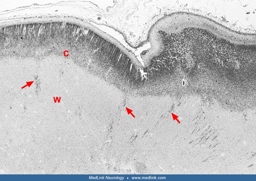
The cortex (C) shows abnormal lamination with many neuron-deficient columns in its upper part. In areas where the plane of section is near transverse, the tangentially cut neuronal columns look like islets of cells surrounded by a cell-deficient neuropil, giving a glomerular appearance (as seen on the right). The underlying white matter (W) shows many arrested heterotopic neurons (arrows), trapped along their migration corridors. (Magnification x20, cresyl violet staining) (Contributed by Dr. Hazim Kadhim.)