General Neurology
Neurologic disorders associated with behavioral symptoms
May. 10, 2023
MedLink®, LLC
3525 Del Mar Heights Rd, Ste 304
San Diego, CA 92130-2122
Toll Free (U.S. + Canada): 800-452-2400
US Number: +1-619-640-4660
Support: service@medlink.com
Editor: editor@medlink.com
ISSN: 2831-9125
Toll Free (U.S. + Canada): 800-452-2400
US Number: +1-619-640-4660
Support: service@medlink.com
Editor: editor@medlink.com
ISSN: 2831-9125
Nearly 3,000 illustrations, including video clips of neurologic disorders.
Every article is reviewed by our esteemed Editorial Board for accuracy and currency.
Full spectrum of neurology in 1,200 comprehensive articles.
Listen to MedLink on the go with Audio versions of each article.
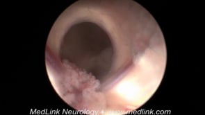
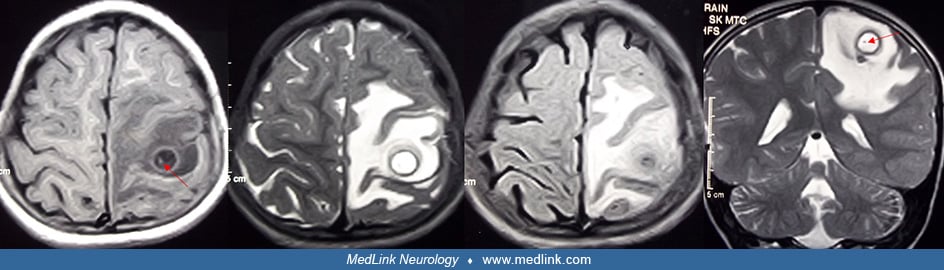
Computed tomography of brain revealed a dot-like hyperdense lesion located adjacent to skull bone. There is perifocal edema as well. The patient presented with recurrent seizures and required antiepileptics. Seizures in these patients are usually easily controllable. (Contributed by Dr. Ravindra Kumar Garg.)