General Neurology
Brachial plexus injuries
Oct. 19, 2023
MedLink®, LLC
3525 Del Mar Heights Rd, Ste 304
San Diego, CA 92130-2122
Toll Free (U.S. + Canada): 800-452-2400
US Number: +1-619-640-4660
Support: service@medlink.com
Editor: editor@medlink.com
ISSN: 2831-9125
Toll Free (U.S. + Canada): 800-452-2400
US Number: +1-619-640-4660
Support: service@medlink.com
Editor: editor@medlink.com
ISSN: 2831-9125
Nearly 3,000 illustrations, including video clips of neurologic disorders.
Every article is reviewed by our esteemed Editorial Board for accuracy and currency.
Full spectrum of neurology in 1,200 comprehensive articles.
Listen to MedLink on the go with Audio versions of each article.
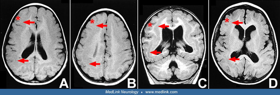
(A) and (B) Axial T1-weighted images from a single patient with subcortical band heterotopia. (A) is more caudal than (B) and demonstrates an approximately 1 cm-thick band in the subcortical white matter. In (B), due to an averaging artifact, the band is somewhat more distinct at this more rostral level. (C) Coronal image from a member of a subcortical band heterotopia pedigree, demonstrating a distinct band and an essentially normal-appearing cerebellum. This patient appears to have two layers of heterotopic band, as has been reported by Barkovich and colleagues (Barkovich AJ, Guerrini R, Battaglia G, et al. Band heterotopia: correlation of outcome with magnetic resonance imaging parameters. Ann Neurol 1994;36:609-17.) (D) Axial image from a sporadic patient with subcortical band heterotopia, demonstrating that the band may be indistinct from the overlying cortex. The band is more distinct posteriorly. Also note mild ventricular dilatation in all patients. Asterisk=outer cortex, arrow=band. (Contributed by Dr. Joseph Gleeson.)


