Epilepsy & Seizures
Familial focal epilepsy with variable foci
Jul. 06, 2022
MedLink®, LLC
3525 Del Mar Heights Rd, Ste 304
San Diego, CA 92130-2122
Toll Free (U.S. + Canada): 800-452-2400
US Number: +1-619-640-4660
Support: service@medlink.com
Editor: editor@medlink.com
ISSN: 2831-9125
Toll Free (U.S. + Canada): 800-452-2400
US Number: +1-619-640-4660
Support: service@medlink.com
Editor: editor@medlink.com
ISSN: 2831-9125
Nearly 3,000 illustrations, including video clips of neurologic disorders.
Every article is reviewed by our esteemed Editorial Board for accuracy and currency.
Full spectrum of neurology in 1,200 comprehensive articles.
Listen to MedLink on the go with Audio versions of each article.
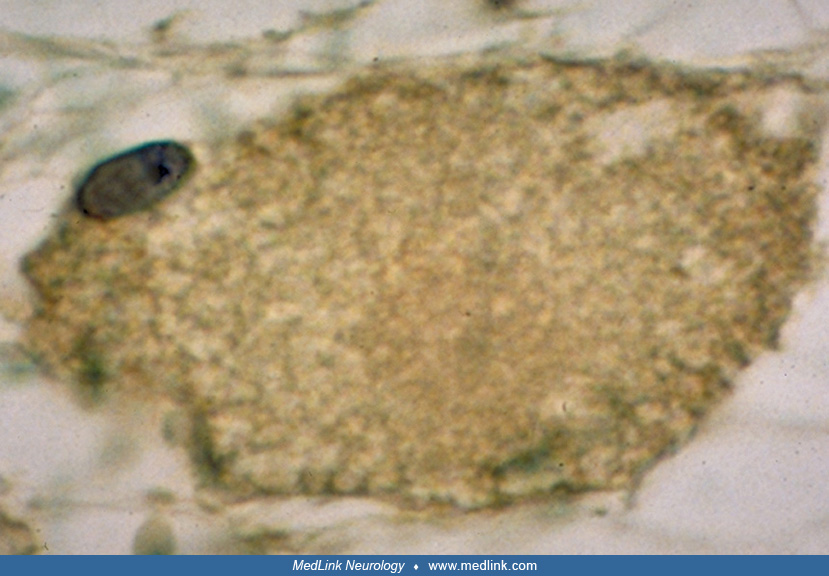
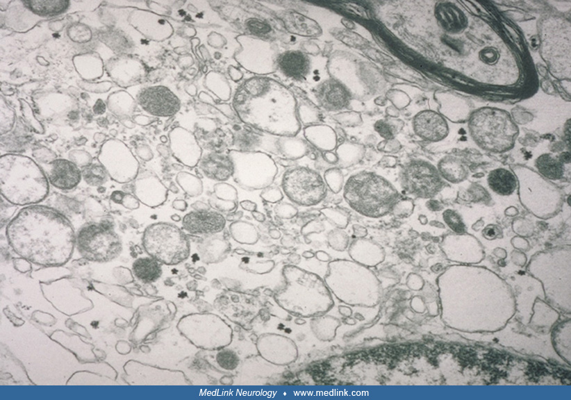
Cytoplasmic membranous structures and abnormal mitochondria. A narrow rim of the nucleus is visible at the right lower border. Because the volume of the foamy oligodendroglial cells is much greater than the size of the nucleus, there were numerous cell cytoplasmic collections in the white matter. These sections containing the nucleus most likely represent an eccentric, tangential, small section of the entire foamy oligodendroglial cell. (Contributed by Dr. Raphael Schiffmann.)