Neuropharmacology & Neurotherapeutics
Gene therapy of neurogenetic disorders
Aug. 24, 2021
MedLink®, LLC
3525 Del Mar Heights Rd, Ste 304
San Diego, CA 92130-2122
Toll Free (U.S. + Canada): 800-452-2400
US Number: +1-619-640-4660
Support: service@medlink.com
Editor: editor@medlink.com
ISSN: 2831-9125
Toll Free (U.S. + Canada): 800-452-2400
US Number: +1-619-640-4660
Support: service@medlink.com
Editor: editor@medlink.com
ISSN: 2831-9125
Nearly 3,000 illustrations, including video clips of neurologic disorders.
Every article is reviewed by our esteemed Editorial Board for accuracy and currency.
Full spectrum of neurology in 1,200 comprehensive articles.
Listen to MedLink on the go with Audio versions of each article.
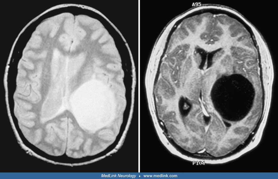
Unenhanced MRI depicting the more rostral cystic regions of the tumor. Left: T2-weighted image (TR 2900/TE 31) demonstrating the well-circumscribed cyst, filled with fluid that is slightly hyperintense compared to cerebrospinal fluid. Note the lack of surrounding edema. Right: T1-weighted image (TR 14.3/TE 3.1) demonstrating the cyst. The cyst fluid has similar intensity to cerebrospinal fluid. (Contributed by Dr. Herbert Newton.)