

Sleep Disorders
Sleep-related urologic dysfunction
Jul. 06, 2026
MedLink, LLC
3525 Del Mar Heights Rd, Ste 304
San Diego, CA 92130-2122
Toll Free (U.S. + Canada): 800-452-2400
US Number: +1-619-640-4660
Support: service@medlink.com
Editor: editor@medlink.com
ISSN: 2831-9125
Toll Free (U.S. + Canada): 800-452-2400
US Number: +1-619-640-4660
Support: service@medlink.com
Editor: editor@medlink.com
ISSN: 2831-9125
Worddefinition
At vero eos et accusamus et iusto odio dignissimos ducimus qui blanditiis praesentium voluptatum deleniti atque corrupti quos dolores et quas.
Acute hydrocephalus is a life-threatening condition that is usually treatable with prompt surgical intervention. Various alterations in the normal cerebrospinal fluid dynamics, which result in elevated intracranial pressure, are together termed hydrocephalus. Hydrocephalus is a clinical and neuroradiographic diagnosis marked by the abnormal accumulation of cerebrospinal fluid (15). This condition may present with or without alterations in intracranial pressure. In cases where intracranial pressure remains normal or low, it is hypothesized that compensatory mechanisms are at play. These mechanisms may involve the displacement of cortical tissue, the expansion of the skull, or, in rare instances, both phenomena concurrently (18). There are many causes of hydrocephalus, including aqueductal stenosis, congenital anomalies, tumors, infection, hemorrhage, and inflammatory diseases. Hydrocephalus may present acutely or in a more chronic fashion, depending on the severity of the CSF alterations. Treatment is surgical and may include resection of causative lesions, shunt placement, or third ventriculostomy. Patients who require shunt placement are at a life-long risk of shunt failure and acute neurologic decline. The author reviews the basic characteristics of this condition and its management, including updates about endoscopic third ventriculostomy as an alternative surgical procedure to shunt placement.
|
• Hydrocephalus is an abnormal elevation of intracranial pressure due to altered cerebrospinal fluid dynamics. | |
|
• Various etiologies are associated with hydrocephalus and are classically grouped into communicating and non-communicating categories. | |
|
• Treatment may be limited to removing an obstructive lesion or placing a temporary CSF diversion system but often requires either a shunt or a third ventriculostomy for long-term management. | |
|
• Shunt failure is a complication that often requires prompt recognition due to the high morbidity associated with delayed treatment. |
In the early 1700s, Vesalius was the first to accurately recognize hydrocephalus as an accumulation of fluid within the cerebral ventricles. During the remainder of the 18th century, Morgagni and others described the neuroanatomic and pathologic causes of hydrocephalus. In the next century, the physiology of CSF circulation began to be elucidated. Magendie is credited with developing the concept of an active bulk flow of CSF.
The first half of the 20th century was a time of rapid increases in our understanding of the clinical and radiographic aspects of hydrocephalus. Of particular importance is the contribution of Dandy with the introduction of pneumoencephalography in 1918 and pioneering work in neuroendoscopy (17). The work of D.S. Russell is important with regard to the addition of systematic pathological studies of the causes of hydrocephalus.
The last 3 decades have provided us with better, less invasive techniques for diagnosing hydrocephalus with the developments of computed tomography, cranial sonography, and magnetic resonance imaging. MRI has been particularly important because of its ability to accurately image hindbrain structures and obstructive lesions, as well as its ability to identify areas of CSF flow.
The history of hydrocephalus treatment can be divided into premodern and modern eras. The premodern era has been reviewed by Pudenz (34). Harvey Cushing introduced CSF diversionary procedures in the early 1900s (40). The modern era began with the development of the valve-regulated shunt system by Nulsen and Spitz in the 1950s. Since that time, valve-regulated shunts have become the standard of care. A few studies have reported effective medical treatments of certain forms of infantile hydrocephalus using isosorbide or furosemide and acetazolamide. However, study results have been inconsistent, and these treatments have not received widespread acceptance. There has been a resurgence in endoscopic third ventriculostomy as a treatment method.
Etiology plays the most important role in determining the presentation of hydrocephalus. Depending on the time course of the underlying disease process causing the hydrocephalus and the degree of the brain’s compensatory mechanisms, the clinical manifestations of hydrocephalus can present in an acute (often life-threatening), subacute, or chronic manner. Headache, particularly worse in the morning due to recumbency and CO₂ retention, is one of the most common symptoms of increased intracranial pressure (35).
In infants and newborns, acute manifestations include vomiting, lethargy, irritability, and seizures. This is usually accompanied by a bulging fontanelle, a large or enlarging head, and splaying of the sutures. Other eye movement abnormalities, including strabismus and gaze limitations, may also occur. Developmental delay and irritability may become apparent in more chronic cases (19).
In toddlers and young children, manifestations can include developmental delay, irritability, vomiting, imbalance, and poor feeding. Subtle behavioral changes and cognitive decline may be the earliest clues in slowly progressive hydrocephalus (41). The presentation of hydrocephalus in older children differs from that of newborns and infants. After 3 years of age, abnormal changes in head measurements are less likely. Hydrocephalus of acute onset in older children usually presents with headaches (often worse in the morning), vomiting (often protracted), deficiencies of eye movements (particularly sixth nerve and gaze palsies), and altered levels of consciousness. If onset is particularly rapid or if the condition is unrecognized, coma and herniation syndromes can ensue. Papilledema is seen frequently, although it takes time for it to develop; thus, the absence of papilledema does not rule out increased intracranial pressure. Chronic or occult presentations may involve gradual cognitive decline, poor school performance, behavioral change, gait disturbance, and occasionally, endocrine abnormalities, such as precocious puberty (36). Sometimes hydrocephalus can be more occult in presentation, with ventriculomegaly as the only clue to its presence, as described in a group of children with cerebral palsy (01).
The advent of the valve-regulated shunt system has radically changed the prognosis for individuals with most forms of hydrocephalus. In the United States, virtually all children with hydrocephalus, with the possible exception of some situations with massive in utero acquired hydrocephalus, receive intervention. Before the introduction of shunts, untreated hydrocephalus carried a dismal prognosis, with less than 20% of infants surviving to adulthood and most survivors having severe intellectual impairment (22).
Also of concern was the finding that even clinically “compensated” cases may deteriorate suddenly, with episodes of acute decompensation or even sudden death. There is little question that for most individuals, untreated hydrocephalus is bad for health and intellect. Poor neurodevelopmental outcomes have been associated with the development of post-hemorrhagic hydrocephalus versus those without hydrocephalus in premature babies (42). How well do patients do with treatment? Studies show that ventricular decompression is important, although absolute ventricular measurements are not reliable predictors of outcome. The most important prognostic factors are the cause of hydrocephalus, the duration of the condition prior to shunting, and the associated brain abnormalities. In a study of neuropsychological outcomes in children with spina bifida, there was no major difference between shunted hydrocephalus versus arrested hydrocephalus patients (14). In spina bifida infants in Uganda, there was no significant difference in neurocognitive outcome between shunt-treated and endoscopically treated children, and ventricular volume did not correlate with performance (46).
A key question remains: is there a critical point during the development of hydrocephalus where irreversible damage occurs? There seems to be a correlation between early CSF diversion and functional outcomes in children with congenital hydrocephalus (43). The evidence is clear that shunt infections are a risk factor for below-normal IQ scores (32). When children with infection and related complications are excluded from the sample, studies yielded normal IQ scores within groups of uncomplicated, shunted hydrocephalic children. Wills points out in her review of the neuropsychological function of hydrocephalic children that the effects of infection, as well as serious birth trauma, are so robust that they are likely to “wash out” more subtle or complex influences of other medical factors on neuropsychological function (49).
A study looking at very long-term outcomes showed that 29% of patients had severe mental handicaps, whereas 48% had normal verbal intelligence. Shunt infections, epilepsy, and the number of shunt operations were associated with reduced verbal intelligence (11). Another study showed that long-term follow-up is necessary for pediatric shunted patients because patients survive into adulthood and continue to be at risk for shunt malfunction as a cause of death (33).
Additional long-term complications include over-drainage (slit ventricle syndrome, subdural hematoma, acquired craniosynostosis), catheter obstruction or migration, and seizures. Even with treatment, patients require lifelong neurosurgical follow-up, as shunt malfunction remains a major cause of morbidity and mortality in adulthood. Quality of life outcomes are variable: although many survivors achieve normal schooling and social independence, others struggle with learning difficulties, psychosocial challenges, or psychiatric comorbidities (19).
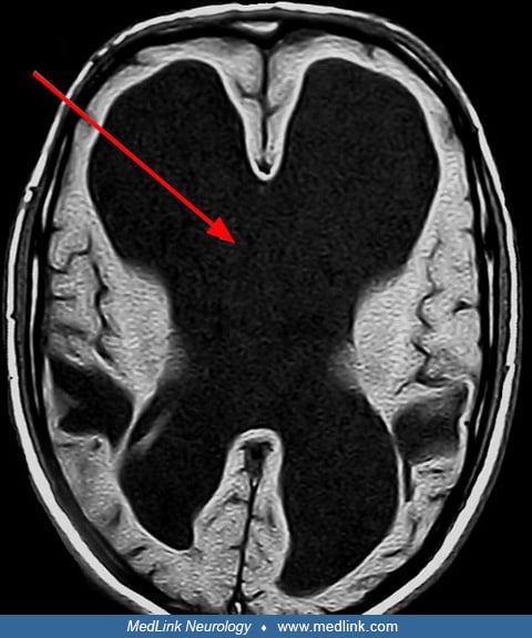
An 8-year-old child presented with morning headaches and papilledema several months after a traumatic brain injury with subarachnoid hemorrhage. CT scan showed a dilated ventricular system and transependymal edema. Ventriculoperitoneal shunting was done, and the ventricular distension and periventricular edema resolved. The headaches also resolved. The patient will require regular monitoring of shunt function.
There are multiple causes of hydrocephalus. Any condition that alters normal CSF dynamics to cause increased intracranial pressure is a cause of hydrocephalus. Traditionally, they are divided into obstructive and communicating hydrocephalus. Obstructive causes include any lesion that can prevent the normal progress of CSF flow, causing increased pressure proximal to the lesion. The various causes of obstruction include tumors, cysts, congenital aqueductal stenosis, intraventricular hemorrhage, and adhesions resulting from hemorrhage or infection.
Communicating hydrocephalus is most commonly attributed to inadequate function of the arachnoid granulations for CSF absorption. There is no discrete, identifiable lesion causing the hydrocephalus that can be targeted for treatment. Often, this form occurs in a delayed fashion after some other insult to the brain. It may be seen after any type of intracranial hemorrhage or intracranial surgery and is even occasionally reported after spinal procedures. Infections or other inflammatory conditions may also cause this type of hydrocephalus.
The pathogenesis and pathophysiology of hydrocephalus encompasses the anatomic, biochemical, and physiologic abnormalities that can result in an increased accumulation of CSF. Equally important are the anatomic, biochemical, and physiologic processes that attempt to provide accommodation (compensation) to the resulting CSF accumulation. Molecular genetic findings have implicated mutations of the L1CAM (neural adhesion molecule) gene in some inherited forms of hydrocephalus. Also, experiments in animal models are advancing our understanding of cellular damage in this condition and its similarities to ischemic white matter injury (06).
Basic physiology and anatomy of CSF circulation. The normal circulation of CSF requires both a balance between the CSF formation and absorption and the unimpeded flow of CSF through its normal travel routes. Most CSF formation (at least 80%) occurs in the choroid plexus (28). This complex tissue is found in the lateral, third, and fourth ventricles, with some of the tissue extending via the foramina of Luschka into the cerebellopontine angles. The bulk of this tissue resides within the lateral ventricles. It appears that brain extracellular fluid is the source of nonchoroidal CSF. Brain extracellular space accounts for about 15% of brain volume under normal conditions (28). CSF is formed by the ultrafiltration of plasma through choroidal capillary epithelium as well as by the uptake of the ultrafiltrate and secretion of CSF (by an active metabolic process). It should be noted that several enzymes are believed to be important in the process of CSF formation, with sodium-potassium-adenosine triphosphate pumps, carbonic anhydrase, and aquaporin (20) being particularly prominent.
Pathophysiology of abnormal CSF circulation. Etiology plays a critical role in determining such factors as the age of onset, presence, and degree of blockade to CSF circulation, the degree and course of ventricular dilatation, brain and cranial compliance, and the type of associated intracranial pathology. It should be emphasized at this juncture that the term congenital hydrocephalus can be deceiving because the presentation of hydrocephalus caused by a congenital malformation has been reported at almost any age. Therefore, age of onset does not always lead immediately to easy determination of the etiology.
Many classification systems for the etiology of hydrocephalus have as their main branch point a separation between congenital cases and other causes, such as inflammatory or neoplastic. However, it would be better to abandon the term congenital hydrocephalus when discussing etiology. The issue is really whether the cause of the hydrocephalus is a malformation (such as Chiari malformation), a genetic process (such as X-linked or autosomal recessive types), inflammation of either the leptomeninges or ventricular lining, chemical irritation (such as subarachnoid or ventricular blood), or neoplasm.
It is first important to identify the site of the blockade to CSF circulation or absorption. The terminology that best describes CSF blockade is communicating versus obstructive. Obstructive (noncommunicating) hydrocephalus denotes a blockade of CSF pathways at, or proximal to, the outlet foramina of the fourth ventricle (foramina of Luschka and Magendie). Communicating hydrocephalus denotes a blockade distal to this point, in the basal subarachnoid cisterns, in the subarachnoid spaces over the brain surface, or within the arachnoid granulations (specialized units where CSF is absorbed back into the circulation) (29).
The next step in proper diagnosis is to determine the etiology of the blockade. The site of the blockade is often helpful in narrowing the search for causes of the hydrocephalus. However, it is often the case that a given pathologic process will demonstrate elements of both communicating and noncommunicating hydrocephalus, although one type usually predominates. Tables 1 and 2 summarize potential etiologies for hydrocephalus.
|
• Aqueductal stenosis | ||
|
• Chiari malformation with or without myelodysplasia | ||
|
- Myelomeningocele | ||
|
• Dandy-Walker malformation | ||
|
- Neoplasia | ||
|
• Inflammatory ventriculitis from | ||
|
- Infection | ||
|
Congenital anomalies that sometimes cause hydrocephalus without obvious blockade of proximal CSF pathways such as: | ||
|
• Chiari malformation with or without myelodysplasia | ||
|
Leptomeningeal inflammation from: | ||
|
• Viral infection | ||
|
- Vascular malformation | ||
|
• Chemical arachnoiditis | ||
|
Neoplasia causing carcinomatous meningitis | ||
Studies of the prevalence of infantile hydrocephalus show a rate of between 0.5 and 0.8 per 1000 live births globally, with higher rates reported in low- and middle-income countries (19; 07; 37). About one-half of these cases are the result of premature births, with almost 90% of these cases caused by intraventricular hemorrhage. Benign external hydrocephalus is estimated to affect 0.3 to 0.5 per 1000 live births in population studies, most often presenting as macrocephaly in infancy but usually self-limiting (48). In developed nations, post-hemorrhagic hydrocephalus constitutes the predominant etiology of secondary hydrocephalus in pediatric populations. The primary cause of post-hemorrhagic hydrocephalus is intraventricular hemorrhage associated with prematurity, occurring mainly in very preterm (< 32 weeks) and very low birth weight infants, with an incidence ranging from 15% to 25% depending on neonatal care resources (45; 37).
By contrast, in sub-Saharan Africa and other low- and middle-income countries, post-infectious hydrocephalus (particularly following neonatal meningitis) remains a leading cause, accounting for more than 60% of pediatric hydrocephalus cases in some regions (47; 07). These global differences highlight the importance of both neonatal intensive care and infection prevention in shaping hydrocephalus epidemiology.
One of the common causes of hydrocephalus is posthemorrhagic hydrocephalus due to intraventricular hemorrhages in premature infants. Therefore, efforts to reduce the incidence of premature births or the incidence of intraventricular hemorrhages in these infants will reduce the incidence of posthemorrhagic hydrocephalus.
Additional preventive strategies include antenatal folic acid supplementation and improved prenatal care, which have reduced the incidence of neural tube defects and associated congenital hydrocephalus (12). In neonatal intensive care, measures such as administration of antenatal corticosteroids, prophylactic indomethacin, delayed cord clamping, and gentle ventilation techniques significantly lower the risk of intraventricular hemorrhage in preterm infants (45; 03).
Infectious causes of hydrocephalus remain important globally, particularly in low- and middle-income countries. Prevention of neonatal meningitis through vaccination programs (Haemophilus influenzae type b, pneumococcus) and prompt treatment of neonatal sepsis are critical steps in reducing post-infectious hydrocephalus (47; 07).
Systematic reviews and meta-analyses conclude that preoperative antibiotic agents reduce the risk of shunt infection in children with hydrocephalus, with observed infection rates dropping from 10.7% without prophylaxis to 5.9% with antibiotics. The relative risk reduction is approximately 44.9% (21). Also, a meta-analysis of 19 clinical trials found that antibiotic-impregnated shunt catheters significantly reduce shunt infection rates compared to standard catheters, particularly within the first 3 to 6 months post-implantation (50).
Macrocephaly is one of the clinical features of hydrocephalus, particularly in infants. Benign macrocephaly in infancy, also referred to as external hydrocephalus or benign enlargement of the subarachnoid spaces in infancy, is a common cause of macrocephaly without the increased intracranial pressure associated with altered CSF dynamics. The differential diagnosis of nonhydrocephalic macrocephaly also includes thick calvarium, leukodystrophies such as Canavan disease, and benign familial megalencephaly. In older children and adults, the presentation of hydrocephalus is similar to that of other situations that result in increased intracranial pressure, such as brain tumors and idiopathic intracranial hypertension. As headache is a frequent symptom of adult hydrocephalus, there is overlap with the differential diagnosis of the various headache syndromes.
Ventriculomegaly on imaging may result from non-hydrocephalic etiologies. There may be ex-vacuo dilation of the ventricles from generalized brain volume loss. In these cases, the intracranial pressure is not elevated from the excess fluid. In addition, there may be congenital morphological characteristics of the brain, often in the setting of chromosomal disorders, which may include ventriculomegaly without altered CSF dynamics.
Neuroimaging with CT, MRI, or head ultrasound is the primary mode of diagnosis. The diagnosis of hydrocephalus is based on one of the following situations:
(1) Ventriculomegaly with signs and symptoms of increased intracranial pressure, such as vomiting, headaches, irritability, lethargy, poor feeding, and changes in muscle tone.
(2) Progressive increase in head or ventricular size without signs and symptoms of increased intracranial pressure.
(3) Static ventriculomegaly in an individual with a large head. Some authors refer to this situation as arrested or compensated hydrocephalus (31).
Along with helping to identify new-onset hydrocephalus or episodes of shunt failure in a timely manner, the neurologist can best benefit the patient with hydrocephalus and his or her family by helping them to understand the proper evaluation, interpretation, treatment, and management of the cognitive and academic problems associated with hydrocephalus (49). Newer work demonstrates specific neuropsychological impairments, such as memory deficits, discourse and pragmatics abnormalities (25), executive dysfunction (26; 24), and cognitive visual problems associated with childhood acquired hydrocephalus (04; 38; 16). Often, these issues are unresolved by school personnel and others not familiar with the neuropsychological deficits common in this condition. Individuals with hydrocephalus often do not meet the strict diagnostic criteria that school systems set for learning disabilities and, therefore, qualify for little or no supportive educational services. However, these children often show patterns of learning deficiencies and inefficiencies that severely affect their ability to reach their educational and, ultimately, their vocational potential. In addition, these individuals can have evidence of deficits in the so-called executive functions. Difficulties in such areas of function as attention, organizational skills, and problem-solving can profoundly affect a child’s day-to-day school functioning and stamina for academic work. The clinician must help families and schools recognize these impairments as being neurologically based and steer them from labeling a child with hydrocephalus as lazy, unmotivated, or attention-seeking. These labels imply a conscious decision on the part of the child to fail, and this is usually not the case. On the other hand, it is important to recognize that children with hydrocephalus are at risk for concomitant social and emotional problems. These must also be diligently screened for, identified, and addressed if we are to help these individuals achieve their maximal potential.
In some patients with clearly identifiable obstructive lesions, such as tumors or cysts, surgical removal of the offending lesion may be adequate to eliminate the hydrocephalus. However, even in these cases, hydrocephalus may persist or recur in a delayed fashion. For cases where such a simple solution is not available or effective, the mainstay of surgical management is CSF diversion. This can be done in two basic ways.
First is the placement of a shunt. A shunt with an integrated valve and anti-siphon system can be placed from the ventricular system, typically the right lateral ventricle (although this can vary based on the individual case), to a distal site for CSF drainage and absorption. The most common distal site is the peritoneal space, although the pleural space or even the right atrium of the heart may be used.
Second is a fenestration within the brain to allow CSF to bypass an identified obstructive lesion, such as aqueductal stenosis. The most common procedure in this category is the endoscopic third ventriculostomy (ETV), in which a fenestration is made in the floor of the third ventricle to allow egress of CSF from the lateral and third ventricles into the basal cisterns.
This procedure has the advantage of requiring no long-term hardware and no long-term dependence on hardware function. Successful treatment with this option results in low complication rates, especially a low infection rate as compared to shunt placement (05). In one study, success rates for this procedure in managing hydrocephalus vary by age, with 58% of infants under 6 months, 65% of infants between 6 months and 1 year old, and 86% of children over 1 year of age having successful long-term control of hydrocephalus, likely due to an increased rate of ostomy closure from growth in younger children (10). Another review of 100 consecutive patients with mean follow-up over 5 years had a 75% long-term efficacy rate (44). The best results were seen for patients with Chiari I malformation, aqueductal stenosis, and tumors. Failure of the ETV is extremely rare beyond 6 months after the procedure, but it has been reported even at 16 years after surgery (44). Failure is more common in patients with a history of cerebral hemorrhage or infection (10). In rare cases, sudden death from delayed closure of the ostomy has been reported, especially in the 6-month postoperative period. This potential complication may be underrecognized by emergency room physicians due to the lack of shunt hardware in these patients, and it may be more preventable with proper recognition. Importantly, sudden death is also a possible complication of shunting and is not unique to ETV as a treatment modality for hydrocephalus.
Due to the lower success rates of ETV in children under 6 months of age, many neurosurgeons proceed to shunt placement without first attempting an ETV. However, in a study of 91 patients with hydrocephalus of multiple etiologies, success rates at 1 year were comparable to those of shunts (39). Should the ETV fail, a shunt can then be placed. A task force concluded that ETV and ventriculoperitoneal shunts demonstrated equivalent outcomes (23).
There are five key components to the management of hydrocephalus:
(1) Diagnosis and evaluation of suspected cases of hydrocephalus. The standard test to evaluate a patient for the presence of hydrocephalus is either a CT scan or an MRI. Head ultrasounds can be employed in infants with open anterior fontanelles. Once the type of hydrocephalus is identified, the decision of whether or not to surgically treat it needs to be made.
(2) Recognition of acute shunt failure (or ETV failure) and prompt referral to a neurosurgeon for definitive treatment of the increased intracranial pressure. Causes for shunt malfunction include obstruction of the tubing by proteinaceous materials or choroid plexus, breakage or disconnection of the line, severe kinking, pullout of the shunt from its distal site, and valve malfunction. Evaluation in this setting includes CT or MRI to assess ventricular caliber for comparison with prior studies as well as a plain film shunt series to look for areas of breakage or kinking that may have caused the malfunction. It is also valuable to recognize situations where individuals are at increased risk for repeated shunt failures, such as the slit ventricle syndrome (30).
(3) Recognition of chronically “uncompensated” hydrocephalus requiring evaluation of the integrity of any in-place shunt system. These situations can be difficult to manage. Some neurosurgeons occasionally monitor intracranial intraventricular or intrathecal pressure (for cases of communicating hydrocephalus) in an intensive care unit in an attempt to predict whether shunting or shunt replacement is likely to alleviate any of the presenting problems.
Sometimes ventricular size remains stable in these cases. Ventricles can be statically enlarged or slit. Although there is no clear set of neuropsychological tests that can predict with sensitivity and specificity which children are indeed suffering from increased intracranial pressure, at least some authors suggest that serial ordering behavior may be affected by hydrocephalus status. Such tests as verbal fluency and word retrieval as well as such complex timed motor tasks as the WISC-R Picture Arrangement may be particularly useful (49).
(4) Awareness of the seriousness of shunt infections and how to evaluate and treat them. This requires the standard workup for shunt malfunction but may also include tapping the shunt or surgical revision. Shunt infection rates are high, typically in the 5% to 10% range, but they become extremely rare by 6 months or more after the most recent shunt procedure or shunt access.
(5) Reinforcement to individuals with shunts and their families of the life-threatening nature of shunt problems and the need to take all symptoms and signs of potential shunt failure or infection seriously. A review of the 50-year history of CSF shunt usage discusses their successes and complications (09).
Pregnancy in women with ventriculoperitoneal shunts is generally well tolerated, and most women can have normal pregnancies and deliveries without major complications (02). A small number may experience shunt malfunction or infection during pregnancy or postpartum, but these are uncommon. Vaginal delivery is usually safe, and cesarean section should be reserved for obstetric indications rather than shunt-related concerns. Multidisciplinary follow-up with neurosurgery and obstetrics is recommended (08).
Beyond pregnancy, long-term considerations include the transition of pediatric hydrocephalus patients into adult care, where specialized neurosurgical follow-up is essential but often underdeveloped (19). Adults with shunted hydrocephalus may also face challenges related to education, employment, mental health, and quality of life, requiring psychosocial and neurocognitive support (27). In low-resource settings, endoscopic third ventriculostomy with or without choroid plexus cauterization has emerged as an important alternative to shunting, reducing lifelong dependence on implanted hardware (47).
Anesthetic considerations in selecting agents with hydrocephalic patients include the possibility of increased intracranial pressure, aspiration risk with rapid induction, and the benefit of modest hyperventilation until the intracranial pressure is normalized (13).
All contributors' financial relationships have been reviewed and mitigated to ensure that this and every other article is free from commercial bias.

Fardin Nabizadeh MD
Mr. Nabizadeh of Iran University of Medical Sciences has no relevant financial relationships to disclose.
See Profile
Alcy R Torres MD FAAP
Dr. Torres of Boston Medical Center and Boston University Chobanian and Avedisian School of Medicine has no relevant financial relationships to disclose.
See ProfileNearly 3,000 illustrations, including video clips of neurologic disorders.
Every article is reviewed by our esteemed Editorial Board for accuracy and currency.
Full spectrum of neurology in 1,200 comprehensive articles.
Listen to MedLink on the go with Audio versions of each article.
MedLink, LLC
3525 Del Mar Heights Rd, Ste 304
San Diego, CA 92130-2122
Toll Free (U.S. + Canada): 800-452-2400
US Number: +1-619-640-4660
Support: service@medlink.com
Editor: editor@medlink.com
ISSN: 2831-9125
Sleep Disorders
Jul. 06, 2026
Sleep Disorders
Jul. 05, 2026
General Child Neurology
Jun. 24, 2026
General Child Neurology
Jun. 10, 2026
Epilepsy & Seizures
Jun. 02, 2026
General Neurology
May. 13, 2026
General Child Neurology
May. 12, 2026
Developmental Malformations
May. 08, 2026