Neuroimmunology
Balo concentric sclerosis
Jul. 02, 2026
MedLink, LLC
3525 Del Mar Heights Rd, Ste 304
San Diego, CA 92130-2122
Toll Free (U.S. + Canada): 800-452-2400
US Number: +1-619-640-4660
Support: service@medlink.com
Editor: editor@medlink.com
ISSN: 2831-9125
Toll Free (U.S. + Canada): 800-452-2400
US Number: +1-619-640-4660
Support: service@medlink.com
Editor: editor@medlink.com
ISSN: 2831-9125
Worddefinition
At vero eos et accusamus et iusto odio dignissimos ducimus qui blanditiis praesentium voluptatum deleniti atque corrupti quos dolores et quas.
Myasthenia gravis is a potentially fatal neuromuscular disorder; however, myasthenic patients can typically lead normal lives when properly diagnosed and adequately managed. In this article, the author reviews immunopathogenesis, clinical features, diagnostic evaluation, and treatment of myasthenia gravis. Developments include the expansion of novel targeted intravenous and subcutaneous immune therapies, including neonatal Fc receptor (FcRn) blockers (efgartigimod, rozanolixizumab, nipocalimab), complement inhibitors (eculizumab, ravulizumab, zilucoplan), and directed B-cell therapies (inebilizumab), which have been FDA approved for use in AChR generalized myasthenia gravis patients. The use of novel agents is being expanded to younger populations, with eculizumab being FDA-approved for adolescents 12 years and older, and nipocalimab being approved for children 6 years and older.
|
• Myasthenia gravis is fatal in up to one third of patients if untreated. | |
|
• The most dangerous manifestation of myasthenia is bulbar and respiratory crisis due to rapidly progressive muscle weakness. | |
|
• Hospitalization and observation with respiratory function monitoring and support are essential in myasthenic crisis. | |
|
• Acute therapy for myasthenic crisis is best achieved with IVIG or plasma exchange. | |
|
• Chronic immunomodulatory therapy can effectively control symptoms in the vast majority of patients and lead to remission. | |
|
• Ten percent of patients with myasthenia gravis will have a thymoma. | |
|
• Myasthenia gravis is the most common neuromuscular junction disorder, with a rapidly growing selection of effective treatment options and strategies. |
Initial descriptions of myasthenia gravis date back to the 17th century, with Thomas Willis (London) describing fatigable muscle weakness. Later clinical descriptions were made by Samuel Wilks (London 1877), followed by more complete descriptions by Wilhelm Erb (Heidelberg 1878) and Samuel Goldflam (Warsaw 1893). It was German physician Friedrich Jolly who coined the term “myasthenia gravis pseudo-paralytica” in 1895. Although often misinterpreted as meaning severe, the “gravis” was intended to convey a heavy, painful weakness (09). As early as 1904, Elliot proposed that neurotransmitter release at the neuromuscular junction could mediate muscle contraction (01). In 1934, the specific release of acetylcholine at the neuromuscular junction was demonstrated (111). During this same period, a number of reports of pathologic thymic abnormalities in myasthenic patients and of symptomatic improvement following thymectomy appeared, prompting Blalock to further investigate and ultimately recommend removal of the thymus as a primary therapy (12; 15). In 1960, Simpson proposed an autoimmune pathogenesis for myasthenia gravis based on the high prevalence of immunologic disorders in myasthenic patients, the transient neonatal form of the disease, and the well-described thymic abnormalities.
Later studies demonstrated antibodies in the sera of affected patients that reacted with the cross striations of skeletal muscle, as well as muscle membrane damage following the application of myasthenic sera to nerve-muscle preparations. In 1962, alpha-bungarotoxin (a snake alpha-toxin) was found to specifically bind and irreversibly inactivate the acetylcholine receptor (AChR) in skeletal muscle. The density of AChRs is particularly high in the electric organs of the Torpedo marmorata electric fish (143), providing a rich source of AChRs for basic scientific investigation. In 1973, a group of rabbits was immunized with solubilized membranes from torpedo electric organs in an attempt to create anti-AChR antibodies for labeling studies. These animals developed a syndrome that closely paralleled human myasthenia gravis (134). The detection of antibodies in these animals that cross-reacted with rabbit AChRs confirmed the first animal model of experimental allergic myasthenia gravis. In 1974, Almon, Andrew, and Appel identified anti-AChR antibodies in human sera (04), further opening a promising new immunologic frontier in the pathogenesis of human disease. Subsequent animal models have been created in rats, mice, goats, monkeys, frogs, and hens (90). Passive transfer has also been accomplished by injecting human myasthenia gravis IgG into mice and of experimental allergenic myasthenia gravis sera and purified monoclonal anti-AChR antibodies into normal mice or rats (175). The data from these early experiments confirmed an autoimmune pathogenesis for myasthenia gravis, satisfying the criteria proposed by Milgrom and Witebsky for an autoimmune etiology (104).
|
• Myasthenia gravis most commonly presents with subacute- to acute-onset muscular weakness. | |
|
• Ptosis and binocular diplopia are the most common early symptoms. | |
|
• Respiratory muscles may be affected, leading to fatal respiratory failure. | |
|
• Respiratory muscle weakness rarely, if ever, occurs in isolation. | |
|
• Other bulbar symptoms (dysphagia, dysarthria, jaw, and facial weakness), proximal more often than distal limb muscle weakness, and axial weakness may also appear. |
The principal manifestations of myasthenia gravis are muscle weakness and fatigability, variably affecting the ocular, bulbar, and limb muscles. Although weakness of the extraocular and limb muscles can be disabling, dysfunction of the swallowing and respiratory muscles is particularly dangerous because of the risk of aspiration or respiratory failure. Consequently, the appearance of dysphagia and dysarthria requires a high degree of vigilance as well as aggressive early intervention.
Diplopia and ptosis are early primary features in 50% to 60% of patients, and an additional 30% will develop these symptoms later. Intraocular causes of diplopia are ruled out if it resolves when covering one eye. Isolated extraocular and palpebral muscle weakness may be the only initial manifestations in some patients (ocular myasthenia gravis). However, 85% to 90% of patients presenting with ocular symptoms will eventually develop more generalized weakness (50; 124; 127; 10). The likelihood that pure ocular myasthenia will progress to generalized disease decreases the longer those symptoms remain isolated, such that those patients with pure ocular disease for at least 2 years have only a 10% chance of further progression (50). A higher prevalence of this form of the disease has been reported in male patients older than 40 years (50; 93; 126).
Combined weakness of the extraocular muscles most commonly appears, but weakness of a single extraocular muscle may also sometimes be seen, and central disorders such as internuclear ophthalmoplegia, supranuclear ophthalmoplegia, and one-and-a-half syndrome may be suspected in some patients. However, prominent fatigability with shifting diplopia and ptosis, worsening with sustained gaze and improving with rest, usually enables distinction from the more fixed deficits of CNS dysfunction. Such fatigability frequently prevents localization of weakness to specific extraocular muscles during neurologic examination, particularly on more sensitive studies, such as the red lens test. Upgaze at a fixed target for 30 to 60 seconds is a somewhat specific provocative test for myasthenic ocular weakness. Worsening diplopia, ptosis, and dysconjugate gaze during this maneuver are positive signs. Many patients also report a mild degree of photophobia on careful questioning, and quantitative pupillary reflex studies have documented fatigability of the constrictor pupillae following repetitive exposure to light.
Myasthenics often have additional weakness of muscles innervated by other cranial nerves. Facial muscle weakness may diminish facial expression, interfere with eyelid closure (or with burial of the eyelashes with forceful eye closure), and cause difficulty whistling or inflating a balloon. Attempts to smile may result in the "myasthenic snarl" (decreased horizontal excursion of the lips and preserved vertical excursion), and masseter weakness may interfere with chewing or closing of the mouth. Patients with more severe masseter weakness may hold their hands to their chins in a seemingly pensive gesture in order to keep the jaw closed. Weakness of the neck flexors and extensors may manifest as head drop or, more commonly, posterior neck stiffness and cramping, especially toward the end of the day. Progressive dysarthria with sustained conversation may result in unintelligible attempts at articulation due to weakness of the tongue, lip, and palate. Hypernasality due to posterior pharyngeal and palatal weakness and (less commonly) hoarseness may also appear. Significant swallowing problems, especially with large boluses of coarse foods (eg, steak), are also common symptoms, along with nasal regurgitation of liquids. Substantial weight loss or aspiration pneumonia can be the presenting feature in patients with early dysphagia. With generalized disease, extremity weakness, usually involving the proximal upper and lower extremities and the extensor muscles, is common and typically worsens with exertion. Because of its proximal distribution, such weakness may lead to a misdiagnosis of a limb girdle myopathy if extremity symptoms are the presenting feature.
The most serious complication of myasthenia gravis, however, is respiratory muscle weakness, which may progress to hypoventilation and respiratory failure. This is termed “myasthenic crisis” and is a true neuromuscular emergency. Dyspnea on exertion may be the initial manifestation, followed by dyspnea at rest, progressive hypoventilation, carbon dioxide retention, and death in some untreated patients. Fatal respiratory dysfunction may develop rapidly, within hours. Consequently, pulmonary function must be carefully assessed and closely followed in newly diagnosed patients or in confirmed myasthenics experiencing a symptomatic exacerbation. Hospitalization for observation and serial vital capacity measurements is often required, and intubation and mechanical ventilation may ultimately be needed in severe cases. With rapid deterioration in respiratory function, controlled intubation should be considered before respiratory failure develops.
Older studies done prior to the advent of more efficacious contemporary therapies and advances in critical care reported a 20% to 30% mortality in untreated myasthenics in the first 3 years after disease onset due to primary respiratory failure (125). Another 20% to 25% of patients experienced clinical remission at some later point, with improvement in 20% to 25% and unchanged symptoms in the remaining 20% (125). True ocular myasthenia has no mortality. However, 85% to 90% of those presenting with ocular symptoms will eventually develop generalized disease (50; 124; 10; 127). The patient's chances of having symptoms permanently isolated to the extraocular and eyelid muscles increase the longer those symptoms remain isolated, such that those patients with pure ocular disease for at least 2 years have only a 10% chance of further progression (50). Overall, many patients have good clinical outcomes, achieving pharmacologic remission within 3 years. They may then be able to be weaned off immune medications.
Myasthenic exacerbations frequently occur without any apparent precipitating factor but are more likely when a patient becomes infected or has another intercurrent severe medical illness, surgery, or injury.
A 64-year-old male with a medical history notable for hypertension and diabetes mellitus developed horizontal binocular double vision over a period of 1 week. Initially, it was intermittent; however, by the end of the week, the diplopia was present each time he looked to the right. He was sent to the emergency room by his primary care doctor, and his neurologic exam was consistent with a right cranial nerve six palsy. Brain imaging was unremarkable, without evidence of acute stroke, mass, or cranial nerve enhancement. ESR and CRP were normal. His hemoglobin A1C was 7.9. A lumbar puncture was obtained, which showed a slightly elevated protein and otherwise unremarkable cerebrospinal fluid studies. He was given an alternating eye patch and was referred to outpatient ophthalmology. In the next month, he experienced ptosis of the right eye as well as dysarthria. He began to have difficulty chewing solid foods, walking up stairs, and maintaining a full smile.
He was assessed by his ophthalmologist, who noted fatigable right-eye ptosis that improved after the ice pack test. An AChR binding antibody assay was positive. MuSK antibody testing was negative. CT chest did not reveal evidence of thymoma. The patient was started on prednisone 60 mg per day. Double vision, proximal limb muscle, and facial weakness improved after 1 week of steroids. The dysarthria and ptosis persisted. Pyridostigmine bromide 30 mg three times daily therapy was added; however, it resulted in only a mild improvement of his symptoms. The patient could not tolerate a higher dose of pyridostigmine, 60 mg three times daily, due to cramping. After the first month of corticosteroids, prednisone dosage was adjusted from 60 mg per day to 100 mg every other day. The dosage was then reduced to 80 mg every other day in the second month and 60 mg every other day in the third month; however, the end-of-the-day dysarthria and ptosis continued.
At the 3-month neurology follow-up, the gentleman reported developing worsening glucose levels, irritability, and insomnia. He was referred to a diabetologist and was prescribed melatonin and a proton pump inhibitor. Vitamin D and calcium supplementation were added. Mycophenolate mofetil was added to the medication regimen as a steroid-sparing agent. The patient developed leukopenia and anemia on complete blood count monitoring. Mycophenolate mofetil was discontinued, and his blood counts were corrected. Target immunotherapy options, including FcRn inhibitors and C5 cleavage inhibitors, were discussed, given the side effects of steroids and the patient’s continued symptoms. Given that the patient had a history of difficult venous access, he opted for a subcutaneous medication option: subcutaneous efgartigimod. He received a weekly efgartigimod injection for 4 weeks without significant side effects. By the end of the treatment cycle, there was noted improvement of his dysarthria and ptosis. He was able to remain minimally symptomatic while continuing his steroid taper to a low dose and subsequently reducing the adverse effects of the steroids.
|
|
• Autoimmune myasthenia gravis is due to antibody-mediated attacks at the neuromuscular junction, most commonly targeting the acetylcholine receptor alpha subunit. |
|
|
• Other pathogenic antibody targets include muscle-specific kinase (MuSK). |
|
|
• The thymus plays a key role in the immunopathogenesis of myasthenia gravis in some patients. |
|
|
• B and T cells and the complement cascade are all engaged in autoimmune myasthenia gravis and provide therapeutic targets. |
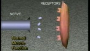
Myasthenia gravis is the prototypic autoimmune antireceptor antibody disorder. It is caused by autoantibodies directed against epitopes on or around the AChR in the muscle membrane. These anti-acetylcholine antibodies may block the binding of acetylcholine molecules to their receptors (following release from the terminal motor axon) and may also initiate immune-mediated degradation of the AChRs themselves, thus, reducing their numbers. Reduced AChR activation results in reduced total muscle membrane depolarization in an individual muscle fiber, sometimes to the extent that the depolarization threshold for contraction is not reached. Such blocking of neuromuscular transmission, when it occurs in a sufficient number of muscle fibers within a muscle, produces the clinical weakness that is the hallmark of the disease.
Understanding the pathogenesis and pathophysiology of myasthenia gravis requires knowledge of the molecular biology and cellular physiology of the neuromuscular junction and the immunologic mechanisms for autosensitization of the immune system against the AChR.
Neuromuscular junction structure and physiology. In healthy subjects, motor nerves continue to branch until they form single nerve fibers, each of which ultimately divides into several smaller branches (the terminal spray) that end in swollen tips (the terminal boutons) just before reaching the muscle end plate (71). Individual nerve terminals contain numerous vesicles aligned near a docking region in the presynaptic membrane known as the active zone, each of which holds from 5000 to 12,000 acetylcholine molecules (187). Following neuronal depolarization, calcium channels open, and calcium influx activates a network of proteins that prompt vesicle fusion with the terminal membrane and subsequent exocytosis of a number of vesicular packets (or quanta) of acetylcholine.
After extrusion, acetylcholine molecules must diffuse across the synaptic cleft to reach the postsynaptic end plate, a specialized region of the muscle membrane having involuted crests and valleys with an abundance of AChRs concentrated on the shoulders of each crest. Acetylcholine molecules then bind with these receptors, resulting in numerous local depolarizations known as miniature end-plate potentials. Summation of these miniature end plate potentials results in the total end plate potential, which must reach the muscle fiber's depolarization threshold to trigger the wave of electrical propagation known as the action potential, which ultimately results in muscle fiber contraction. Acetylcholine is released from its receptor shortly after depolarization but is hydrolyzed by synaptic acetylcholinesterase in a fraction of a millisecond, preventing further receptor activation until the next presynaptic release.
In acquired myasthenia gravis, the structure and function of the neuromuscular junction are significantly altered. Anti-acetylcholine antibodies both block the receptor site and initiate an immune cascade, which damages the postsynaptic membrane, flattening its folds, widening the synaptic cleft (191), and decreasing AChR number and density (194; 42; 39; 68; 34). In the normal subject, sustained activation of the motor nerve results in declines in acetylcholine release as the population of vesicles positioned for immediate use is exhausted. However, the healthy neuromuscular junction has such an abundance of AChRs (the "safety margin" for neuromuscular transmission) that depolarization of the muscle fiber to threshold still occurs despite declines in acetylcholine output. However, in the myasthenic patient, these declines eventually prevent many muscle fibers from reaching depolarization threshold because of the reduced numbers of total and open AChRs (a lowered "safety margin" for neuromuscular transmission).
Immunology. The nicotinic AChR is a transmembrane protein with five subunits designated alpha, beta, gamma, delta, and epsilon arranged radially around a central ion channel. In myasthenia gravis and its animal models, antibodies (largely of the IgG class) to multiple sites on the extracellular portion of the AChR are generated, but antibodies directed against the alpha subunit predominate. The reasons for this unexpected finding (all subunits share a high degree of amino acid sequence homology) remain unclear but may be related to differences in secondary and tertiary structures among different subunits, which can be greatly influenced by small changes in amino acid sequences (170). Numerous pathogenic epitopes on and around the AChR may be involved in the pathogenesis of myasthenia gravis, the best studied of which is the extracellular heptapeptide found on both alpha subunits to which alpha-bungarotoxin binds. Antibodies against this region block cholinergic binding and prevent proper ion channel function, producing an acute myasthenic response in experimental animals. Antibodies that prevent alpha-bungarotoxin binding, presumably by binding directly to the receptors themselves ("binding antibodies"), have been identified in the sera of patients with severe disease (160). Autoantibodies may induce myasthenia gravis through a number of mechanisms. Antibodies reacting at or near the acetylcholine binding site may prevent neurotransmitter docking by directly covering the site or via steric hindrance. Alternatively, antibodies against other portions of the receptor may interfere with ion flux through other, less clear mechanisms. Autoantibodies can also increase receptor degradation or complement-mediated focal lysis (160). Clustered acetylcholine receptor antibodies (clustered AChR-Abs) have been detected in approximately 50% of patients in whom standard assays for acetylcholine are negative. These clustered antibodies are associated with neuromuscular junction dysfunction by electrophysiological assays and can be passively transferred in animals (69).
Patients lacking acetylcholine receptor antibodies may have antibodies to muscle-specific kinase (MuSK) or to low-density lipoprotein 4 (LRP4). MuSK is a tyrosine kinase essential in the steps required for maintaining AChRs and their functional clusters at the neuromuscular junction, as well as for initiation of both pre- and postsynaptic differentiation. This sequence of events begins when nerve-derived agrin binds to LRP4, which then activates MuSK; phosphorylation of another compound (cortactin) then occurs, and together these compounds mediate functional AChR clustering and also control presynaptic differentiation via direct retrograde signaling. All of these compounds have important roles, and their absence significantly disrupts the structure and function of the postsynaptic apparatus (22; 48).
The anti-MuSK antibody is a high-affinity antibody that binds to the extracellular Ig-like domains of native MuSK and is predominantly of the IgG4 subclass. Passive transfer experiments have demonstrated reduced miniature end plate potential amplitude in murine models of myasthenia gravis in a fashion similar to that induced by anti-AChR IgG (46). Antibodies have also been identified against LRP4 in patients seronegative for both MuSK and AChR Abs, and LRP4 antibodies also appear to have a pathogenic role in myasthenia gravis (48). Similarly, antibodies directed against cortactin have also been detected in patients seronegative for both MuSK and AChR antibodies, and these antibodies also appear to be pathogenic for myasthenia gravis (28).
Both B- and T-cells are involved in the immunopathogenesis of myasthenia. Thymic cell cultures consistently produce the highest level of AChR antibodies, despite low B-cell frequency in this tissue; significant experimental evidence supports a critical role for the T-lymphocyte in myasthenia gravis. Thymectomy and thymic irradiation prevent the development of experimental allergic myasthenia gravis following appropriate inoculation, and in vitro T-cell dependence of AChR antibody production has been demonstrated in numerous experiments. The thymus has shown significant involvement in myasthenia gravis both clinically and histologically. Further attempts to characterize T-cell epitopes demonstrated that T-cells primed with AChR alpha subunits were most efficient in stimulating helper activity for antibody production in experimental allergic myasthenia gravis, as compared to T-cells primed with other AChR subunits. This suggests a dominant role for the alpha subunit T-cell epitope in this process. T-cell epitopes in human myasthenic subjects, however, are not so dramatically dominant because the patients, unlike carefully bred animal subjects, have differing human leukocyte antigen (HLA) types and probably have a variety of differing immunodominant epitopes (141). Such heterogeneity might also explain the successful stimulation of human T-cells by a wide variety of antigenic peptide sequences (14). As discussed previously, AChR epitopes stimulating B-cell antibody production are also heterogenous, producing a variety of potentially pathogenic antibodies. The multitude of apparent epitopes stimulating both B- and T-cell activity in human myasthenia gravis adds yet another dimension of complexity to the immunopathogenesis of this disease and may make selective approaches to immunotherapy more difficult to achieve than previously thought. However, autoantibodies produced by these processes (especially AChR autoantibodies) trigger complement activation, causing further injury to the neuromuscular junction.
The normal thymus. The thymus serves as the primary organ for T-cell development and differentiation. It is located in the anterior mediastinum in a substernal fat pad and is covered by a connective tissue capsule. This capsule penetrates the thymus, dividing it into lobules approximately 0.5 to 2.0 mm in diameter. The epithelial cell network within the thymus is invaded by lymphocyte-forming cells, which intensely proliferate throughout early life and childhood, pushing the epithelial cells apart to form a reticular pattern. Each fully developed lobule consists of a peripheral cortex of densely packed lymphocytes and a central medulla. The medulla contains a large number of epithelial reticular cells, which sometimes form aggregates known as Hassall corpuscles. Other medullary cells include dendritic cells (interdigitating cells in the corticomedullary junction) and myoid cells, similar in many ways to skeletal muscle and sharing epitopes with the nicotinic AChR and the major immunogenic region (91; 30).
Prototypic stem cells first enter the cortex, the most active site of T-cell differentiation, and begin maturation, later migrating centrally. Here, genetic rearrangement of the T-cell receptor locus produces antigen specificity (30). Dendritic cells contribute to T-lymphocyte development through antigen presentation and epithelial reticular cells via thymic hormone and lymphokine elaboration. Functional T-cells initially develop both CD4 and CD8 surface antigens, losing one or the other with further maturation to become CD4+CD8- or CD4-CD8+ (60). This process, known as "positive selection," restricts recognition by these T-cells to specific antigens only in the presence of certain types of major histocompatibility complex molecules ("restriction"). The next step is induction of self-tolerance, during which T-cells with high affinity for self-antigens are eliminated ("negative selection"), although some survive to migrate to the peripheral lymph system, where further selection occurs. Mature T-cells move toward the medulla (containing only 5% of the total thymic lymphocyte population) and exit the thymus via venules and the lymph system, migrating to other lymphoid organs. With aging, the thymus involutes, shrinking from 30 to 40 g at puberty to 10 to 15 g in the elderly, but it retains significant proliferative capability.
The myasthenic thymus. The myasthenic thymus demonstrates frequent abnormalities. Of myasthenics, 10% to 15% have a lymphoepithelial thymoma (122), whereas 70% of those without tumor demonstrate lymphoid follicular hyperplasia, especially younger patients and HLA-DR3 positive females (25; 02; 60). With thymic hyperplasia, active germinal centers form between the cortex and the medulla, and perivascular spaces distend with proliferating lymphoid tissue (17; 60). Myasthenics demonstrate an increased percentage of mature T-lymphocytes and thymic B-cells, with active and robust AChR antibody production following culture and stimulation, in excess of such antibody production by B-cells from control patients and peripheral B-cells from myasthenics (141). AChR-specific T-cells can be isolated from the thymus of myasthenic patients but not from peripheral blood, suggesting a higher proportion of sensitized T-cells within the thymus itself.
There is a greatly increased incidence of thymic tumors in patients with myasthenia gravis. Thymomas are classified as lymphomas; according to their cells of origin, they are epithelial, carcinoid, or mesenchymal tumors. The epithelial thymic tumor is the classic "thymoma." It is often referred to as a lymphoepithelial tumor and graded according to lymphocytic infiltration, though the epithelial cell is the neoplastic element or classified by cortical, medullary, or mixed cell type (02). Collections of lymphocytes (lymphorrhages) between muscle fibers, both in the perivascular and parenchymal regions (with associated muscle fiber necrosis in patients with thymoma), were initially thought to represent metastases. Further studies demonstrated no neoplasia, and similar collections were identified in up to two thirds of all myasthenic patients, with a somewhat higher prevalence in those with thymic tumors (02). Analysis of thymoma epithelial cells and cell cultures demonstrates the expression of numerous AChR epitopes (95; 96). Patients with thymoma and myasthenia also demonstrate heterogenous antibodies cross-reacting to striated skeletal muscle, cardiac muscle, and thymic myoid cells that do not react to AChR epitopes (178). The HLA associations delineated for myasthenia gravis without tumor have not been found in thymoma patients.
A number of possible immunologic mechanisms might initiate myasthenia gravis within the thymus. Myoid cells, which share epitopes with the AChR, are located in the thymic medulla close to mature lymphocytes and dendritic cells (75; 30). Persistence of the embryonic gamma subunit (not normally expressed in adult skeletal muscle) within the thymus has also been demonstrated (114). Dendritic cells expressing HLA-DR are also found within this organ and may play a role in AChR antigen presentation. Under the right conditions, the myoid and dendritic cells could provide both an antigen and a mechanism for autosensitization to the AChR. Loss of self-tolerance might be facilitated by the persistence of the embryonic gamma AChR subunit, with subsequent generation of wider autoimmunity against the receptor. In the altered microenvironment accompanying tumor growth, neoplastic epithelial thymoma cells having numerous AChR epitopes may result in both loss of self-tolerance and autoimmunity. The frequency of antibodies with cross-reactivity to myoid cells and other striated muscle components in patients with thymic tumors further supports the plausibility of such a mechanism.
Genetic modulation. Different families of major histocompatibility antigen types are produced by a given animal's genome and expressed in various tissues. These major histocompatibility antigen types or haplotypes play a role in antigen presentation and are critical for the immune system's ability to distinguish between foreign and self-antigen (174). Individual strains of mice demonstrate susceptibility to experimental allergic myasthenia gravis following inoculation with specific AChR peptides (ie, immunodominant epitopes) and resistance to experimental allergic myasthenia gravis following inoculation with other AChR peptides. This susceptibility to immunization with specific peptide sequences consistently correlates with different mouse H-2 haplotypes. Human haplotypes are more difficult to study, and a number of AChR peptides will stimulate lymphocyte proliferation in human tissue. Though not as dramatic as in experimental allergic myasthenia gravis subjects, there is some degree of varying immunodominance that appears to be linked to the patient's HLA type and T-cell receptor V-gene activity. A number of HLA associations have been noted among different ethnic groups with myasthenia gravis. In Caucasians, HLA-A1, B8, DR3, and DR5 predominate, especially in those with thymic hyperplasia as well as in females with early-onset disease (11). HLA-DR3 is also found with increased incidence in other autoimmune disorders, including diabetes mellitus (186). In the older age groups, HLA-A2, A3, D7, and Dw2 have been found with increased incidence. Other associations include HLA-DR9 and DRW13 in pediatric Japanese patients and HLA-BW46 in pediatric Chinese patients. Other genetic and epigenetic influences may also play a role in the immunopathogenesis of myasthenia gravis. One study identified a point mutation in the ecto-NADH oxidase 1 gene (ENOX1), which decreased expression of ENOX1 in lymphoblastoid cells in a cohort with familial autoimmune myasthenia gravis, suggesting a possible pathogenic role on lymph cell dysfunction in this kindred and raising the question of a candidate gene for myasthenia gravis (81).
A retrospective study of myasthenic patients in North America revealed that among 1032 patients, 5.6% had a family history of myasthenia and 28.4% had a family history of autoimmune disease (49).
The prevalence of acquired myasthenia gravis is estimated to range from 100 to 200 per million (136; 24; 162). Epidemiological studies have shown prevalence and incidence rates as progressively increasing over the last 70 years (176; 138; 101). This phenomenon is most likely secondary to improved diagnostics and survival as more effective therapies have been applied. The aging global population may also play a contributing factor. It is important to note that the prevalence and incidence of myasthenia widely vary between population studies.
Disease onset has a bimodal distribution, often peaking around the age of 30 years old in women and between 60 and 70 years old in men (24). Prior to more effective therapies and advances in critical care, 20% to 30% mortality was reported in untreated patients within 3 years of onset due to respiratory failure (125). Of the total patients, 20% to 25% experienced clinical remissions at some later point, whereas 20% to 25% eventually improved but continued to have symptoms. The remaining 20% had stable and relatively unchanged symptoms over time (125). Contemporary disease-specific mortality is less than 5%, with higher rates among the elderly. Hospitalization rates amongst patients with myasthenia have been increasing, indicating the growing burden of myasthenia on health care systems (51).
|
• Thus far, there are no known measures to prevent the onset of myasthenia gravis. | |
|
• Once myasthenia gravis has started, avoiding medications that worsen it is very important. | |
|
• Patients need to work closely with their neurologists to maximize therapy and to reduce the risk of a potentially life-threatening exacerbation. | |
|
• Patients should be educated on the risk of respiratory failure so they can seek emergency care when needed. |
There are no known means of preventing myasthenia gravis. However, a number of medications may worsen neuromuscular junction function and should be avoided, if possible, in patients with myasthenia. These medications include certain antibiotics (the aminoglycosides, including neomycin, streptomycin, kanamycin, gentamicin and tobramycin, polymyxin B and colistin, oxytetracycline and rolitetracycline, lincomycin, clindamycin, erythromycin, ampicillin), antiarrhythmics (quinine, quinidine, procainamide, trimetaphan, lidocaine, and beta-adrenergic blockers), and other drugs with ion channel effects (chloroquine and phenytoin). Timoptic eyedrops may also worsen symptoms. Neuromuscular blocking agents, magnesium salts, and anticholinesterases must also be closely monitored when administered to myasthenic patients. A CT scan contrast agent, meglumine diatrizoate, may cause acute exacerbations, and an anesthetic, methoxyflurane, may unmask subclinical myasthenia in some patients. Oxytocin, aprotinin, propanidid, diazepam, and ketamine have all been reported to prolong postoperative recovery.
The aminoglycoside and the peptide antibiotics, as well as oxprenolol, practolol, trimetaphan, phenytoin, trimethadione, carnitine, interferon, and, most notably, D-penicillamine, have all been reported to worsen preexisting myasthenia and induce myasthenia in previously asymptomatic patients. Antineoplastic immune checkpoint inhibitors may also induce myasthenia gravis de novo in some patients (03). Statins are a common lipid-lowering medication that, although safe in most myasthenics, can potentially worsen symptoms (171).
|
• Multiple sclerosis |
Myasthenia gravis has a variety of clinical presentations and may be confused with a number of other neurologic or neuromuscular disorders. It is often called a “snowflake disease,” given that every patient presentation can be unique in its own way. Pure ocular or oculobulbar disease may mimic CNS dysfunction. Diplopia, dysarthria, and dysphagia may also be early manifestations of multiple sclerosis, small vessel ischemia, and even early mass lesions of the brainstem. Other causes of diplopia, such as third, fourth, and sixth nerve dysfunction, must also be considered in cases of isolated diplopia or ptosis. Myasthenia can sometimes present with dysarthria, dysphagia, respiratory muscle weakness, and minimal diplopia with or without extremity weakness, causing confusion with motor neuron disease. More commonly, predominantly proximal arm and leg weakness suggests a possible myopathy. Bilateral ptosis can sometimes lead to the diagnosis of myopathies affecting the ocular muscles (OPMD, CPEO, myotonic dystrophy). Because of these myriad possibilities, an appropriate evaluation is mandatory, including a careful history and physical examination, serologic testing, electrophysiologic studies of neuromuscular junction function, brain imaging, and even genetic testing in some cases. It is wise to acknowledge that myasthenia can concomitantly occur with other neurologic conditions, including the ones listed above.
|
• Autoimmune diseases |
Myasthenia has been associated with a number of disorders. The best-known association is with the thymic epithelial cell tumor or thymoma. Approximately 10% to 15% of myasthenics have a thymoma, and 40% of patients with a thymoma will have myasthenia as well (02). The mean age of patients with a thymoma is 50 years, with a 1:1 male-to-female ratio. Of thymomas, 90% are benign and easily treatable with resection, whereas 10% are malignant and will spread beyond the thymic capsule to local tissue, the lymphatic system, or the blood. Only 1% to 5% metastasize distantly (83; 179). Recurrence of benign tumors is rare, but malignant thymomas that have spread to the pleura or elsewhere have a 5- to 10-year average survival, despite surgery and radiotherapy (83). Most patients with myasthenia and thymoma present with severe generalized extremity and bulbar weakness, whereas pure ocular disease is rare in this patient group (02). Some of these patients may have cardiac involvement with arrhythmia, bundle branch block, or cardiac failure with focal myocarditis (148). When thymoma is diagnosed late, myasthenics typically have more severe symptoms, more frequent exacerbations, and a higher mortality rate. In contrast, myasthenia associated with early diagnosed thymoma has a slightly better prognosis than myasthenia gravis with thymic hyperplasia alone (122).
The frequency of autoimmune diseases is also increased in myasthenics, ranging from 2.3% to 24.2% in different series (128; 189; 130; 123; 125; 107; 172). Hyperthyroidism with associated thyroiditis is the most prevalent, with a frequency of 2.2% to 16.9%. As thyroid disease may exacerbate myasthenia or induce additional neuromuscular disease, these patients may elude diagnosis for longer periods. Rheumatoid arthritis is the second most prevalent associated disorder, with a frequency ranging from 0% to 10.3% (excluding those cases in which myasthenia was induced by d-Penicillamine). Other associations, although less common, include systemic lupus erythematosus (128; 189; 123; 125), Sjogren syndrome, sarcoidosis (189; 125), scleroderma (130; 123), polymyositis (130; 123), and Lambert-Eaton myasthenic syndrome, among others. Although the evidence for associated CNS involvement is weak, a possible increased incidence of multiple sclerosis has been reported in a few series.
In addition, the prevalence of sleep disorders, particularly sleep apnea, is increased in myasthenics, perhaps due to increased weakness of the posterior pharyngeal muscles. A careful sleep history should be obtained in all patients, as sleep deprivation considerably worsens myasthenic weakness and fatigue, and effective treatments (eg, continuous positive airway pressure for sleep apnea) can dramatically improve their symptoms and daily functioning (94).
|
• AChR antibodies are positive in 60% to 80% of generalized myasthenia gravis patients. | |
|
• Anti-MuSK antibodies are found in half of those seronegative for AChR antibodies. | |
|
• Repetitive nerve stimulation is positive in 60% to 70% of generalized myasthenia gravis patients. | |
|
• Single-fiber electromyography remains the single most sensitive electrodiagnostic test for myasthenia gravis; however, it is not entirely specific. | |
|
• Bedside signs and tests, such as the icepack test, curtain sign, Cogan lid twitch, and Bienfang test, have variable sensitivity and specificity. |
Acetylcholine receptor antibodies. Three assays are available for diagnostic evaluation: the AChR binding, modulating, and blocking antibodies (87; 88; 86; 85; 61; 84). In the binding antibody assay, immunoglobulin is coprecipitated from the patient's serum with solubilized human AChR labeled with I125-radiolabeled alpha-bungarotoxin (84). Although it has a high specificity, this assay is sometimes also positive in patients with thymoma alone without myasthenic symptoms. In the AChR-blocking antibody assay, a modified immunoprecipitation technique measures those antibodies blocking the binding of I125-labeled alpha-bungarotoxin to the AChR. Because blocking antibodies are found in only 1% of myasthenic patients without binding antibodies, it is not useful as a first-line screening test. It may have utility in the serial evaluation of patients on immunosuppressive therapy, but false positive results have been reported following curare-like muscle relaxants (85; 61). In the AChR-modulating antibody assay, incubation of the patient's serum with monolayer skeletal muscle cell cultures at physiologic temperatures for 16 hours is followed by the addition of I125 radiolabeled alpha-bungarotoxin. Modulating antibodies (which cross-link AChRs, accelerating endocytosis and degradation) can be estimated through this technique, although blocking antibodies will also hide intact receptors and contribute to the observed value. A more specific assay for isolated modulating antibody activity requires paired incubation, both with inhibition of muscle metabolism (and, thus, endocytosis) and without such inhibition, enabling subtraction of blocking antibody effect on intact receptors. Most useful when the AChR-binding antibody assay is negative, the modulating antibody assay may also be more sensitive in patients with early, mild, or pure ocular disease. However, it is more likely to be falsely positive due to disruption of the cell culture and other extraneous sources (86). The sensitivity and specificity of each of these three assays vary and also change with increasing disease severity. A positive result is up to 99% specific for myasthenia with all three techniques, with sensitivities ranging from 59% (blocking) to 90% (binding and modulating) in generalized myasthenia and from 30% (blocking) to 70% (binding and modulating) in ocular disease (61). Although there is no good correlation between absolute antibody titers and disease severity in the individual patient, mean antibody titers rise with increasing disease severity in populations of myasthenics (88). High titers may be found in early onset disease as well as in patients with thymoma; decreased titers following therapy correlate with symptomatic improvement in some patients (87). In addition to these methods, a cell-based assay (CBA) testing for antibodies to clustered AChRs has also been investigated, demonstrating clustered AChR antibodies in 38% of those patients negative by radioimmunoprecipitation assay, with 100% specificity. Sixty-two percent of patients with these antibodies had childhood onset disease, and they were more likely to have ocular myasthenia gravis, a good response to therapy, and a higher chance of remission compared to patients negative to radioimmunoprecipitation assay (146).
Myasthenia gravis remains a clinical diagnosis, and a percentage of patients with symptomatic disease do not have detectable acetylcholine antibodies by the available assays. Such apparent seronegativity may sometimes be artifactual. As a variety of technical errors can yield false negative results, the use of a reputable, well-recognized laboratory with extensive experience in these methods is critical. In some patients, only one of the available assays is performed (typically the binding antibody assay). However, with a negative binding antibody assay, the other assays may be positive (61) and should be considered. In other patients, high-affinity antibodies may aggressively adhere to their respective antigens in vivo, rendering standard assays negative because of extremely low serum levels of free antibody. Electrophysiologic assays (repetitive nerve stimulation, single fiber EMG) or motor point biopsy with immunochemical analysis of the end plate may be more sensitive in such cases. Finally, immunosuppressive therapy, especially when administered for more than 1 year, may reduce antibody production to undetectable levels (84; 181).
Anti-MuSK antibody. A second population of antibodies identified in myasthenia gravis is those against muscle-specific kinase (MuSK). MuSK is a tyrosine kinase associated with the agrin receptor. Both MuSK and agrin have important roles in regulating and maintaining AChRs and their functional clusters at the neuromuscular junction; their absence significantly disrupts the structure and function of the postsynaptic apparatus. The anti-MuSK antibody is a high-affinity antibody that binds to the extracellular Ig-like domains of native MuSK and is predominantly of the IgG4 subclass. Passive transfer experiments have demonstrated reduced miniature end plate potential amplitude in murine models of myasthenia gravis in a fashion similar to that induced by anti-AChR IgG (46).
Between 40% and 70% of patients who are seronegative for anti-AChR antibodies are anti-MuSK positive, and it is much more common in females than in males. Patients having anti-AChR antibodies almost never harbor anti-MuSK antibodies (and vice versa). Pure ocular myasthenics are much less likely to have anti-MuSK antibodies, regardless of whether they are positive for the anti-AChR antibody or not (though there are reports of pure ocular myasthenia gravis with anti-MuSK antibodies) (23; 26). It is especially important to send anti-MuSK antibodies during the diagnostic workup in patients seronegative for AChR antibodies because anti-MuSK patients are more likely to have normal electrophysiologic studies (including single fiber EMG), especially if the tested muscle(s) are not weak. Anti-MuSK antibodies do not appear in normal controls.
In general, anti-MuSK-associated myasthenia gravis appears at an earlier age than other varieties and disproportionately affects the neck, shoulder, pharyngeal, and respiratory muscles, with less limb weakness and rare ocular symptoms. Reports from different centers have suggested three primary modes of presentation for the syndrome associated with the anti-MuSK antibody: (1) predominant and severe facial and pharyngeal muscle weakness; (2) predominant neck, shoulder, and respiratory weakness; and (3) a more classic presentation closely resembling AChR antibody-associated myasthenia gravis (58; 41; 153; 100; 197; 31).
Almost all anti-MuSK myasthenia gravis patients appear to respond to immunomodulatory therapy, particularly plasmapheresis. Case reports have suggested that anti-MuSK patients who are refractory to conventional therapies may respond well to rituximab infusions as adjunctive therapy (52). Some reports suggest symptomatic resistance to acetylcholinesterase inhibitors (53).
Anti-MuSK myasthenia gravis patients are less likely to have thymic hyperplasia and rarely have thymoma (82). However, the efficacy of thymectomy in these patients is difficult to gauge because of the small number of patients reported thus far.
Future studies will likely reveal even greater antibody heterogeneity in seronegative myasthenics, further increasing our understanding of the disease process and the complexities of normal neuromuscular junction function.
Anti-LRP 4 antibody. LRP4 antibodies have been identified in 5% of patients with generalized myasthenia gravis overall and in 18.7% of patients seronegative for both anti-AChR and anti-MuSK antibodies. Anti-LRP4 antibodies were also identified in patients’ sera concurrent with anti-AChR antibodies (8%) and also with anti-MuSK antibodies (13%). Anti-LRP4 antibodies were not seen in normal controls but were seen in 4% of patients with other neuromuscular diseases. They were associated with milder disease in general (particularly when anti-LRP4 antibodies were found in isolation) and were more common in women and more likely in younger patients. In patients with LRP4 antibodies, response to therapy resembled that of patients with AChR more than those with MuSK antibodies (198).
Other antibody assays. Antistriated muscle antibodies were the first reported autoantibodies in myasthenia gravis. These antibodies are reactive against thymic myoid cells as well as the contractile elements of skeletal muscle, and they are present in 27% of all patients with the disease, with high levels noted in up to 90% of patients with myasthenia gravis and concurrent thymoma (88; 47). Progressive rises in antistriational antibody titers can be the first indication of thymic tumor recurrence following resection. They may also be present in isolation when AChR antibody assays are negative, making them a useful adjunctive test. Other antibodies found in a significant percentage of patients with myasthenia gravis are directed against nucleus (20% to 40%), thyroid (15% to 40%), rheumatoid factor (10% to 40%), gastric parietal cell (10% to 20%), lymphocyte (40% to 90%), and platelet (5% to 50%) antibodies as well as anti-smooth muscle, mitochondrial, red blood cell, and squamous epithelial cells, often without additional disease.
Electrodiagnosis. Two major electrodiagnostic tests are available to assess neuromuscular junction function. Repetitive nerve stimulation involves repeated supramaximal stimulation of a selected peripheral nerve (typically at a frequency of 1, 2, 3, or 5 Hz) while recording the electrical waveforms (compound motor action potentials) produced by the resulting recurrent muscle contractions of a selected muscle innervated by that nerve. Trains of 5 to 10 waveforms are then recorded at baseline, after 30 to 60 seconds of maximal voluntary contraction, and at variable intervals (30 to 60 seconds apart) for several minutes after exercise. The size of the first waveform in a train is compared to one of the later waveforms to assess for a decrease (decrement) or increase (increment) in size within each train. Myasthenic patients typically demonstrate decrements in excess of 10% to 15% in the baseline train. Post-exercise, waveform size may transiently increase by 10% to 50% (65), with improved decrement. This phenomenon, known as post-exercise facilitation, may be due to a temporarily enhanced release of acetylcholine following a brief maximal contraction. A decay of these improvements occurs over the next 3 to 4 minutes, with the disappearance of increment and worsening of decrement often below baseline levels (post-activation exhaustion) as junctional and reserve acetylcholine levels decline (151). Within 5 minutes post-exercise, all parameters typically return to baseline.
A second, more sophisticated test of neuromuscular junction function is single-fiber electromyography. This technique, developed by Eric Stalberg (169), uses a needle engineered to record single muscle fiber discharges. The variability in the time required for signal transmission across an individual neuromuscular junction can be quantified by assessing the firing interval of single muscle fibers. This value, known as jitter, can be recorded in the healthy patient. In the myasthenic patient, jitter is considerably increased and may be associated with intermittent blocking of neuromuscular transmission, in which complete failure of neuromuscular junction transmission results in intermittent failure of contraction in one of the muscle fibers. The sensitivity and specificity of repetitive nerve stimulation and single-fiber electromyography differ considerably. Repetitive nerve stimulation in a hand or shoulder muscle has a sensitivity of approximately 75% in generalized myasthenia gravis and less than 50% in ocular disease, whereas single fiber electromyography has a sensitivity of 95% or greater in generalized and 90% or greater in ocular myasthenia gravis when appropriate muscles are tested (155; 65). Because of its high sensitivity, single-fiber electromyography has its greatest diagnostic utility in cases of mild generalized, ocular, or seronegative myasthenia gravis. As jitter may be increased in a variety of neuromuscular diseases, the specificity of this technique is limited, and other conditions must be appropriately excluded prior to single-fiber electromyography studies.
Other diagnostic tests. Intravenous administration of edrophonium, an acetylcholinesterase inhibitor, to a suspected myasthenia gravis patient may transiently improve certain symptoms, providing supporting evidence for the diagnosis. This test is also commonly known as the tensilon test. For an appropriate examination, a markedly affected, easily testable, and observable muscle should be selected. Frequently, the deltoid or an upper extremity extensor is chosen, or alternatively, in the case of clinically obvious ptosis, the patient may be observed for improvement in ptosis symptoms. Two 1 mL tuberculin syringes should be prepared: one as a placebo injection (1 mL normal saline) and one injection of 10 mg of edrophonium in 1 mL of solution. The placebo is usually administered first, with observation for improvement over a few minutes. The edrophonium is administered next with an initial dose of 2 mg (0.2 mL), with observation and strength testing over 1 minute. If no improvement is noted, the remaining 8 mg (0.8 mL) are given, and the process is repeated. If no effect is noted following the second dose, the test is negative. Any symptomatic improvement in responsive patients is short-lived and lasts no longer than 30 minutes. Ideally, the patient should be off any acetylcholinesterase inhibitor for at least 24 hours prior to the test. Several cholinergic side effects may appear following intravenous edrophonium, including asystole, bradycardia, syncope, nausea, and excessive lacrimation and salivation. The examiner must be aware of these risks, which in certain patients will be relative contraindications for this test. Reported sensitivities and specificities for this test are 90% to 95% and 80% to 95% respectively in generalized, and 80% to 95% and 80% to 90% respectively in ocular myasthenia gravis (180; 129; 117; 40; 137), but these figures are subject to significant subjective error; they depend greatly on patient selection and a carefully performed and judiciously interpreted examination. The ice pack test is a simple bedside examination in which an ice pack is applied to the eye of a patient with ptosis for 2 minutes. Improvement in ptosis is considered supportive of the diagnosis. Although of historic importance, the use of the edrophonium test has become increasingly rare due to the availability of serological and electrodiagnostic testing, as well as the cardiac risks of edrophonium.
There are many useful bedside signs and tests that are helpful in the diagnosis of myasthenia gravis. Many of them are ocular, such as the icepack test (improvement in ptosis after application of ice), Cogan lid twitch (overshoot twitch of eyelid retraction after downgaze), curtain sign (elevation of the ptotic eye leading to ptosis of the other eye), and Bienfang test (improvement in ptosis after eyelid closure) (37). The sensitivity and specificity of individual bedside tests vary; however, they are improved when combined with other clinical signs versus when found only in isolation (193).
|
• Acute myasthenic crisis can be rapidly fatal due to neuromuscular respiratory failure and requires emergent and often critical care intervention. | |
|
• Intervention for acute myasthenia gravis crisis includes hospitalization, respiratory and cardiac monitoring, and intubation and ventilation when indicated. | |
|
• IVIG and plasma exchange are key tools for the management of acute myasthenia gravis crisis. | |
|
• Chronic management of myasthenia gravis may include oral or intravenous steroids, steroid-sparing agents, and a variety of other immunosuppressant drugs. | |
|
• The role and availability of targeted immune therapies, such as FcRn receptor antagonists, complement inhibitors, and targeted B-cell therapy, is rapidly increasing. | |
|
• Ten percent of myasthenics will have a thymoma. | |
|
• Thymectomy has a role in the management of myasthenia gravis in selected patients, even in the absence of thymoma. | |
|
• Myasthenia gravis is often accompanied by other autoimmune disorders, especially thyroid disease and rheumatoid arthritis. |
Thymectomy. For the last half-century, removal of the thymus gland has been considered a standard therapy for myasthenia gravis. However, due to the number of inherent complexities in performing a large prospective, randomized trial of this treatment in myasthenia gravis, such a definitive study was not completed until 2016 (190). This study assessed the response to thymectomy performed via the classic median sternotomy in patients having myasthenia gravis for less than 5 years and who were also on standardized doses of prednisone, compared against those treated with prednisone alone. In patients treated with thymectomy, the following benefits were observed: (1) Moderately greater clinical improvement overall, (2) greater chance of minimal disease manifestations by 3 years, (3) lower chance of needing steroid-sparing agent treatment with azathioprine, (4) fewer myasthenia gravis exacerbations requiring hospitalization, and (5) a lower prednisone dose needed for disease control versus those treated with prednisone alone. However, no remissions were observed in the surgical group during the course of the study, and thymectomized patients as a group still required moderate doses of prednisone.
Historical reports done prior to the 2016 study varied considerably in the surgical procedures used, the adjunctive therapies administered, and the symptomatic scoring systems employed. These studies suggested thymectomy was safe and effective for the treatment of myasthenia, and they focused on which techniques of removal were most beneficial, how responses were influenced by adjunctive immunotherapy, and which patients were most likely to respond. Thymectomy was first reported to yield unexpected benefits following resection of a thymic tumor in patients with myasthenia gravis in the early part of the last century (16). Subsequent studies, particularly those of Blalock, reported an increased rate of clinical remission in thymectomized myasthenics, both with and without thymoma (15). Most older studies used some variation of the Osserman clinical scoring system (128), but meta-analyses were hindered by significant differences in both the variety of scoring used and in the definition of what constituted a clinical response. Stricter criteria of complete remission rates (defined as the absence of any symptoms or signs in a patient on no other therapy) were primarily employed for study-to-study comparisons. Natural history studies prior to the era of effective medical immunotherapy or thymectomy reported spontaneous remission rates of 6% to 20%. Several studies done prior to 1960 in myasthenics without thymoma reported modest remission rates of 11% to 20%, with one study demonstrating a more robust 52%. With advances in critical care during the 1960s and 1970s, improved remission rates appeared in numerous studies, ranging from 27% to 38% (161; 135). Results during the 1980s (in studies using adjunctive immunosuppressive therapy and extended and maximal resections) documented higher remission rates, ranging from 54% to 56% (120).
Since the late 1960s, a variety of surgical methods have been devised for thymic resection, including Blalock's classic transsternal approach (splitting of the sternum with removal of the well-defined mediastinal lobes and partial extraction of the cervical extensions of the thymus from below), the extended transsternal thymectomy (splitting of the sternum with wider exploration of the extrapleural and cervical regions), maximal transcervical and transsternal thymectomy (extensive exploration of the neck and mediastinum with direct visual inspection and removal of all suspected thymic tissue; care must be taken to avoid injury to the phrenic and vagus nerves), and in recent years, the focused transcervical approach (a more limited resection through a cervical incision instead of a sternotomy, with or without mediastinoscopy). Comparative analyses of these techniques, however, suggest steady improvements in remission rates with greater removal of thymic tissue, though these studies, unlike the 2016 MGTX trial, did not include a non-surgical control group. Reported mean remission rates are 20% to 30% for transcervical (77; 97; 98; 131; 33), 30% to 40% for classic transsternal (38; 98; 122; 108), and 50% to 60% for both extended and maximal surgeries (149; 121; 70). Statistically significant differences between remission rates for cervical and maximal thymectomy have been reported, making the cervical approach less attractive (70); most centers routinely use extended or maximal procedures, as used in the 2016 MGTX study. Data showing that less invasive techniques may be equally effective. In fact, a randomized, controlled trial study being proposed to evaluate thymectomy in ocular myasthenia gravis will exclusively use less invasive video-assisted thymectomy. Studies performed in the 1980s, after the adoption of more extensive thymic resections, reported remission rates of 10% to 37% (122; 106). Numerous studies have also documented a decreased medication requirement and improved overall symptoms in patients undergoing the surgery (108; 45). Lasting clinical improvement is typically delayed at least 6 to 12 months, and in some patients may not appear for several years (74; 122; 106; 89). Traditional transsternal and extended thymectomies are safe when performed in experienced centers with appropriate preoperative and perioperative management, and the complication rate of these procedures now approaches that of general anesthesia alone (34). Myasthenics now undergoing thymectomy, with careful anesthetic and critical care and appropriate preoperative stabilization, remain intubated only 1.4 days on average (103). Newer methods of surgery utilizing robotic and minimally invasive approaches began appearing after 2000 and continue to proliferate. A 5-year follow-up study of extended robotic thymectomy in nonthymomatous myasthenia demonstrated a high level of safety, with shorter postoperative recovery than traditional trans-sternal methods (44). Overall improvement was noted in more than 80% of patients, though concurrent medical therapy was not carefully controlled in the analysis (44).
The judicious selection of patients for thymectomy remains a critical part of the initial evaluation. Imaging studies of the chest with CT or MRI are mandatory; all patients with localized thymic tumors should undergo a complete resection. Patients with widely metastatic disease, however, are an exception, as they may not have significant symptomatic benefit. Removal in this group should be considered primarily as a debulking maneuver in conjunction with other therapies and may not be indicated in many of these patients. Thymectomy should be performed in most patients with new-onset myasthenia. However, patients with pure ocular disease have not traditionally been treated with thymectomy, and its benefits in this group remain questionable. Age is also an important consideration; there is a general consensus that patients from early adulthood to 60 years of age are most likely to benefit from the surgery. As the thymus plays an integral role in the developing pediatric immune system, thymectomy is not usually recommended in children, although significant improvement without apparent adverse effects has been reported in a few series in juvenile myasthenia (165; 145). Because the thymus progressively shrinks with advancing age, and patients older than 60 to 65 years carry a greater surgical risk, thymectomy is rarely performed in the elderly. However, when resection has been studied in this age group, results comparable to those in younger patients have been reported (120; 108).
Appropriate preparation for any surgery, including thymectomy, is highly important in the myasthenic patient. Preoperative pulmonary function testing (including vital capacity measurements) should be done in addition to other routine preoperative tests. Ideally, the patient's disease should be stable enough to enable them to forego acetylcholinesterase inhibitor therapy for at least 24 hours prior to surgery. Acetylcholinesterase inhibitors should, in any case, be stopped no later than the morning of surgery. Muscle relaxants should be avoided, if possible, as they may induce prolonged paralysis. Postoperatively, a transient but dramatic increase in strength may be observed that may persist for several days. Patients on acetylcholinesterase inhibitor therapy may report up to a 25% reduction in dosage requirement during this period (15).
Thymoma will be found in 10% to 15% of all myasthenic patients, whereas 40% of patients with thymoma will develop myasthenia (02). The mean age of thymoma patients is somewhat higher than that of patients with myasthenia alone, at 50 years. Approximately 90% of these tumors can be easily treated with resection. The remaining 10% are malignant and spread throughout the mediastinum, although distant metastasis occurs in only 1% to 5% (83; Verley and Hollman 1985). Patients with malignant thymoma should undergo thorough surgical resection followed postoperatively by radiotherapy with or without chemotherapy. Despite these measures, patients having malignant thymomas that have spread to the pleura or elsewhere have a 5- to 10-year average survival (83). Antistriated antibodies are strongly associated with concurrent myasthenia and thymoma and are elevated in 90% of these patients. As such, antibodies are seen in only in 27% of all patients with myasthenia, their presence raises suspicion for a possible tumor (88; 47). With successful tumor therapy, antistriated antibody levels fall. Consequently, progressively increasing titers may serve as the first evidence of tumor recurrence.
The mechanisms through which thymectomy works remain controversial. Immunologically active tissue is so abundant elsewhere in the body that a gross reduction in immune system mass seems an unlikely explanation for the observed effects. Resection of the thymus does decrease the levels of the immunoactive thymic hormones, perhaps accounting for a more widely distributed immunosuppression (177). However, the presence of the quintessential elements for B- and T-lymphocyte autosensitization against the AChR within the thymus makes it a key location for the continued production of myasthenic antibodies, making its removal particularly efficacious in quelling that specific immune response (182).
Corticosteroids. Successful oral prednisone therapy for myasthenia was initiated in the early 1970s (76). Following confirmation of the autoimmune etiology in myasthenia, steroid treatment became increasingly popular. Unfortunately, its dramatic efficacy resulted in widespread adoption before placebo-controlled studies could be initiated. Subsequently, there are no randomized clinical trials to support corticosteroid use in myasthenia even though they remain a mainstay of therapy.
A mild, early exacerbation of myasthenia gravis symptoms in 50% and a more severe exacerbation in 10% of patients occur within 1 to 17 days after starting glucocorticoids (most commonly starting on day 5) but last only 4 days on average (72).
Such therapy should begin with an intensive course, but different methods of induction have been proposed. One approach involves initiation with 60 to 80 mg oral prednisone per day with in-hospital observation for possible exacerbation until three consecutive days of improved symptoms are documented (156; 163; 166). Others have proposed induction with gradual increases in an attempt to minimize the chances of an exacerbation; one such regimen begins with a starting dosage of 15 to 20 mg of oral prednisone daily with gradual upward adjustments (increases of 5 mg every 3 to 5 days) to a maximum of 50 to 60 mg (34), whereas a second recommends initiation with 25 mg every other day with upward adjustments (12.5 mg increments every third dose) to 100 mg every other day (164; 163; 166). Despite extensive experience with each of these methods in different centers, limited comparative data are available regarding the risks or efficacy of immediate, high-dose induction versus progressive dosage increases; the time of onset of exacerbations, though, may be less predictable during incremental dosage adjustments (21). After high-dose therapy is initiated, sustained improvement appears in most patients within 2 weeks, with improvement in 90% of patients within 3 weeks. Even more substantial improvement usually appears within 4 to 12 weeks, after which patients on daily therapy may be switched to alternate-day therapy. This may be accomplished by giving slightly less than twice the daily dose on alternate days with no treatment on "off" days (eg, 60 mg of prednisone every day to 100 mg every other day).
Alternatively, a slower, more gradual transition to therapy every other day over 3 to 6 months with maintenance of a small dose on the "off" day may be attempted (34). Maximal improvement appears within 6 months in most patients (72), after which the alternate dose is further reduced. Further reduction may be accomplished by decreases of approximately 20 mg every 2 months (given a sustained clinical response), until a level of 40 to 50 mg every other day is reached. Dosage reduction thereafter may be accomplished in 10 mg increments every 2 months to a total dose of 20 mg every other day, with further reductions by 5 mg increments every 2 months. As many patients experience symptomatic recurrence at these dosage levels, this phase of therapy must be approached cautiously. Clinical recurrence may be delayed weeks to months after a dosage change and may require reinstitution of higher-dose therapy before control is again achieved. Some patients may require indefinite low doses of prednisone for maintenance purposes. Once a dose of prednisone 5 mg daily or less is attained, the side effects of steroids are often minimal while still providing valuable symptomatic effect (67). However, if steroids cannot be reduced to an acceptable level, alternative immunosuppressive therapies should be considered. Steroid treatment is usually highly effective, inducing remission in up to 80% of patients (132). However, as adjustment of steroid dosages to lower levels may take 1 to 2 years, the side effects of long-term therapy must be considered in individual cases.
Azathioprine. Azathioprine is typically used when steroid therapy fails or when excessive steroid maintenance doses are required. Often, under such conditions, a combination of steroids and azathioprine is needed; some have also used this combination for induction therapy (59; 92). Unfortunately, a response to azathioprine may not begin for 3 to 12 months (99; 188; 43), and a maximal response may not appear for 1 to 2 years. After it is taken orally, the drug is converted to 6-mercaptopurine, which interferes with purine biosynthesis, reducing the number of proliferating B- and T-lymphocytes. Therapy is initiated by administration of 50 mg orally every day for 1 week, followed by an increase to 100 mg every day for 1 week and continued weekly increases in increments of 50 mg per day until the dosage of 150 to 200 mg (2 to 3 mg per kg) per day is reached. Blood cell counts and liver function tests must be monitored for possible leukopenia and hepatotoxicity (59) every week for the first 2 months, then monthly for as long as therapy is continued. These adverse effects are usually reversible with discontinuation of the drug when discovered early. An idiosyncratic flu-like reaction with fever, myalgia, generalized malaise, and vomiting may also occur and should prompt immediate discontinuation of therapy. After recovery, another trial of therapy may be attempted, and the medication continued if there is no recurrence of these symptoms. Other side effects include mild nausea, which may be eliminated by division of the daily dose into thirds and administration with meals; potential teratogenesis and mutagenesis; and an increased long-term risk of malignancy (59; 79). Xanthine oxidase is used to metabolize this drug, so concurrent administration of allopurinol may result in dangerously high levels. Dosage reductions of 75% are recommended when azathioprine and allopurinol are given together. However, response rates of up to 44% to 71% have been reported, although combination therapy was given to most reported subjects (98; 188), and most patients tolerate this therapy well.
Mycophenolate mofetil. Mycophenolate mofetil is a reversible inhibitor of inosine monophosphate dehydrogenase, an enzyme crucial for purine synthesis, which is a critical step in B- and T-cell proliferation but not in that of most other cell types. Consequently, mycophenolate administration selectively inhibits the B- and T-cell proliferation following immune stimulation, making it an effective agent for preventing organ rejection after transplant and potentially effective for the treatment of autoimmune disorders.
Mycophenolate continues to be tested as a potential treatment for myasthenia gravis in increasingly larger and more sophisticated clinical trials, which have raised questions regarding its effectiveness. Initially, the milder side effect profile of this drug made it an attractive alternative to azathioprine as a steroid-sparing agent, and several small studies suggested it was effective in myasthenia gravis (159). Subsequent studies demonstrated improvement in up to 70% of patients, with good tolerability and discontinuation in only 6% due to adverse events (102). These results prompted two large, multicenter trials published in 2008.
The first of these studies was a double-blind, placebo-controlled trial in which patients were randomized to receive either placebo plus 20 mg prednisone daily or mycophenolate plus 20 mg prednisone daily for 12 weeks (110). Eighty subjects were enrolled, and no significant differences were seen between the two groups at the end of the treatment period. The authors speculated that these results could be due to greater than predicted benefit from the prednisone dosage used, the short duration of the study, or the absence of any benefit of mycophenolate in this population of patients with myasthenia gravis. The second study was a prospective, randomized, double-blind, placebo-controlled trial that tested mycophenolate as a steroid-sparing agent (154). In this study, 196 patients with myasthenia on steroid therapy were randomized to either 36 weeks of treatment with steroids plus mycophenolate or to steroids plus placebo. No significant differences appeared between groups at the end of the treatment period. In this study, the authors speculated that these results could be due to the use of insufficiently sensitive outcome measures (masking important clinical differences) or insufficient study duration to detect benefit.
Given these results, we can conclude that mycophenolate has not been proven superior to placebo over a 12-week period when given with steroids at moderate doses and has not been proven superior to placebo as a steroid-sparing agent over a 9-month period in patients receiving more than 20 mg prednisone per day. As steroid-resistant patients were excluded from these studies, it remains unclear whether they might benefit from mycophenolate, and it is also unclear whether treatment with mycophenolate for periods greater than 9 months might be beneficial.
When used for the treatment of myasthenia, doses of 1.0 g administered orally twice a day have been employed. Clinical improvement may be delayed, starting 3 to 6 months or longer after initiation of therapy. Adverse reactions are typically mild and usually consist of nausea with or without vomiting and diarrhea, though gastric ulceration may occur in up to 2% of cases. More serious side effects are uncommon and have been reported most often in association with higher doses used to prevent transplant rejection. Bone marrow suppression may also occur, with lymphopenia in 1% of cases and leukopenia in up to 2%, and there are rare reports of progressive multifocal leukoencephalopathy (most commonly in patients who are immunosuppressed from another treatment or condition). Mycophenolate also causes teratogenesis, and it is contraindicated in pregnancy (black box warning). With long-term therapy, an increased risk of malignancies, particularly melanoma, remains a possibility, though the absolute increase in relative risk has not been well defined. Immunosuppression results in an increased risk of infection, and live vaccinations must be avoided. Complete blood counts should be monitored weekly for the first month, every 2 weeks for the following 2 months, and monthly thereafter. Tapering of mycophenolate in patients achieving either pharmacologic remission or minimal manifestation status very gradually by 500 mg/day every 12 months appears safe, with a risk of mild relapse after discontinuation of approximately 30%, and minimal risk of inducing myasthenic crisis (57).
The PROMISE-MG study was a prospective cohort study that compared mycophenolate mofetil with azathioprine in treatment-naive generalized myasthenic patients (113). Although both improved quality-of-life scales and clinical outcomes, there was no significant difference between the two drugs. The frequency of adverse reactions between the drugs did not differ. Side effects of azathioprine were potentially more serious, whereas mycophenolate mofetil had a higher risk of teratogenicity.
Cyclosporin A. Cyclosporin A decreases interleukin-2 and interferon gamma production by helper T-cells (158). It has significant toxicity and should be reserved for cases in which steroids and azathioprine are not effective. It is the only immunosuppressive therapy with efficacy proven by prospective, double-blind, placebo-controlled trials. Total daily dose of 5 mg per kg is administered, usually at 150 to 200 mg twice daily, with subsequent dosage titrated by clinical response and trough plasma levels. Clinical improvement typically begins within 2 weeks, with average maximal improvement at 3 to 4 months (173). Treatment must be continued indefinitely, however, as 90% of responders will relapse with discontinuation (188; 59). Side effects include acute nephrotoxicity (usually reversible with cessation of therapy), hypertension, mild hepatotoxicity, a slightly increased risk of malignancy, hirsutism, and gingival hyperplasia, among others. Renal function must be monitored. An increase in creatinine by 50% over baseline value (or an absolute value greater than 1.5 to 2 mg per dl) indicates a need for a reduction in dosage. Blood pressure readings of more than 150 systolic or 90 diastolic also call for a decrease in dosage, usually in increments of 1 mg per kg per day at 1-month intervals. Trough serum levels should also be used to guide dosage adjustment.
Cyclophosphamide. This drug is reserved for severe cases that have failed combinations of other therapies because of the risk of serious side effects. This alkylating agent interferes with DNA replication and is cytotoxic to lymphocytes, monocytes, and macrophages. Oral therapy is initiated at 150 to 200 mg every day (3 mg per kg to 5 mg per kg per day) with monitoring of complete blood count and urinalysis twice weekly, titrated to a white blood cell count of 2500 to 3000 per µl and an absolute neutrophil count of 1000 per µl. Monitoring frequency may be somewhat reduced after stable target blood counts are achieved but must continue as long as the drug is administered. Limited studies are available, but response rates of 45% to 75% within 1 month of initiation and a 30% remission rate, persisting only as long as the drug is continued, have been reported (116). Serious side effects, including myelosuppression; hemorrhagic cystitis; malignancies of the skin, bladder, and blood; teratogenesis; alopecia; and nausea and vomiting, as well as others, have been reported. Patients must remain well hydrated to reduce the urinary concentration of cyclophosphamide-active and toxic metabolites in the bladder. If intravenous therapy is given, a protective agent for the bladder should be given. Nausea and vomiting during intravenous infusion may require antiemetic therapy. A more radical approach to severe, refractory myasthenia gravis has been used in a few patients, in which immunoablation by high-dose cyclophosphamide followed by intrinsic stem cell repopulation was employed to effectively “reboot” the immune system after cures were achieved with this method in rodent myasthenia gravis models. Three patients were subjected to this treatment without significant complications, achieving long-term improvement and allowing for good control of previously refractory disease and taper of immunosuppressive medication to low levels. However, immunoablation therapy remains experimental and may pose a significant risk to the patient during the ablation phase (35).
Plasma exchange. Plasma exchange removes from the blood populations of macromolecules, including AChR antibodies. It is indicated when transient and rapid symptomatic improvement is needed during a myasthenic crisis or exacerbation. It may also reduce perioperative morbidity when used as preparation for thymectomy, and it may be used to counter steroid-induced exacerbations (156). Occasionally, it may be used as primary maintenance therapy in refractory patients who fail other interventions. Typically, 2 to 4 liters of plasma is removed during each exchange. An every other day course of five to six exchanges is implemented. Improvement is usually noted within days, although some patients may evidence a response weeks after therapy. Effects last approximately 1 to 12 weeks. This course of improvement has been documented in a number of uncontrolled trials (140; 29; 115; 54; 73; 78). Plasma exchange is usually well tolerated, but complications can arise from problems with vascular access (including infection, local thrombosis, and vascular perforation), removal of circulating clotting factors, dehydration (hypotension and bradycardia), and transient electrolyte disturbances. Because plasma exchange is personnel- and equipment-intensive, it is an expensive therapy, but it is highly effective when a more rapid response is needed in the myasthenic patient, which has been highlighted by an expert consensus (157).
Intravenous immune globulin. High-dose intravenous immune globulin exerts its effects through a number of theoretical mechanisms, including the introduction of antiidiotypic antibodies and reduced AChR antibody production via negative feedback. Its time course is similar to that of plasma exchange in most patients and is equally valuable as a temporizing measure. Intravenous immune globulin is usually administered as daily infusions of 400 mg per kg per day for 5 days for a total dose of 2 g per kg. Improvement appears in approximately 70% of patients within 5 days of initiation, with maximal effects at 8 to 9 days and a duration of 8 to 12 weeks (06). Side effects are typically mild and may include headache, chills, fever, and nausea. Pretreatment with acetaminophen and diphenhydramine may prevent these minor reactions. Anaphylaxis is much rarer than feared prior to the widespread use of this agent; however, occasional reports of patients with hereditary IgA deficiency developing anaphylaxis have appeared, resulting in recommendations for screening patients with serum protein and immunofixation electrophoresis in advance of starting the drug. Acute renal failure is also a significant complication, usually occurring within days of infusion, and it is primarily limited to diabetics receiving IVIG constituted in sucrose-containing solutions. If a myasthenic patient with diabetes requires IVIG, a sucrose-free preparation should be used. All patients should be screened for renal insufficiency prior to infusion, although normal renal function before and during infusion does not guarantee that this complication can be avoided. Mild and transient leukopenia or thrombocytopenia may appear but usually resolve rapidly after discontinuation of the drug. IVIG does increase serum osmolarity and has been associated with deep venous thrombosis, particularly in patients with limited mobility. Rare reports of myocardial infarction and stroke during or shortly after IVIG have appeared, but the extent to which the drug may have contributed to these common events is unclear. Because it does introduce a significant fluid and protein load into the circulatory system, it can also exacerbate congestive heart failure (142). Intravenous immune globulin is a highly expensive drug, but its overall cost may be similar to that of plasma exchange when equipment and personnel costs for each therapy are considered. The therapeutic efficacy and time course of action of IVIG and plasma exchange have been compared in Class I studies and are very similar in patients with moderate to severe myasthenia (07). The lack of data of IVIG in acting as a corticosteroid-sparing maintenance therapy should also be noted. The first randomized, double-blind trial assessment of IVIG steroid-sparing ability noted the lack of significant steroid reduction in steroid-dependent patients receiving IVIG compared to placebo after 39 weeks of treatment (20).
Rituximab. Rituximab, a human-mouse monoclonal antibody against the CD20 B-cell antigen, continues to be used in myasthenia gravis with reports of success. A severe refractory patient with the anti-MuSK antibody syndrome who failed conventional oral maintenance therapy responded dramatically to rituximab within 2 months, with continued control of her disease throughout a 12-month follow-up period (52). A retrospective review of a small series of patients with both anti-acetylcholine receptor antibody-mediated myasthenia gravis and anti-MuSK-mediated myasthenia gravis treated with rituximab showed a reduced need for immunosuppressants and improvement in clinical function in all patients with minimal adverse effects, including at least one patient refractory to other therapies (195). A retrospective report of 16 patients with AChR Ab-positive myasthenia gravis refractory to other therapies utilized two to four cycles of therapy (one cycle = 375 mg/m2 weekly for 4 weeks) (144). The majority (63%) reached complete and stable remission after only one cycle with discontinuation of other therapies within 9 months posttreatment, whereas the remaining patients achieved either pharmacological remission (ie, remission with adjunctive therapy) or minimal manifestation status for at least 1 year. Approximately half relapsed at a mean of 36 months after the last rituximab treatment, whereas the remainder had stable control from 1 to 7 years. No significant adverse effects attributed to treatment were noted. Another report in 12 severe, refractory AChR Ab-positive myasthenia gravis patients demonstrated improvement in 50% at 1 year and in one third at 18 months (80).
A meta-analysis of the effectiveness of rituximab in AChR antibody-mediated myasthenia gravis reported sustained improvement and prolonged time to relapse, though not all patients responded, and future randomized controlled trials were recommended (32). Another study found that rituximab was safe and effective in a group of late-onset myasthenia gravis patients, some of whom were refractory to treatment previously (150). However, a phase II, double-blind, randomized controlled trial of rituximab reported no clinically meaningful steroid-sparing effects in patients with myasthenia gravis receiving prednisone over 52 weeks, although rituximab was safe and well tolerated (119; 139).
Studies of rituximab continue to appear, focusing on its utility in refractory patients, durability, and long-term effects. The randomized, double-blind, placebo-controlled study RINOMAX showed that a single dose of 500 mg of rituximab was effective in lessening symptoms of myasthenia gravis in patients with up to 1 year of generalized symptoms and reducing the need for rescue medications compared with placebo (139). However, more studies designed to provide class 1 evidence are needed to conclusively prove its effectiveness in myasthenia gravis.
Inebilizumab. Inebilizumab is a CD-19-directed cytolytic humanized monoclonal antibody administered via intravenous infusion twice yearly; it was previously approved for neuromyelitis optica spectrum disorder and immunoglobulin G4-related disease. MINT was an international phase 3 placebo-controlled double-blinded clinical trial that assessed the safety and efficacy of inebiliziumab in 238 patients with generalized myasthenia gravis with AChR (190 patients) or MuSK antibodies (48 patients). The primary endpoint was a change from baseline MG-ADL score over a 26-week period. The trial included a mandatory corticosteroid taper for all subjects who were previously on corticosteroids. The corticosteroid taper began at week 4 and targeted daily prednisone use of 5 mg or less by week 24. Inebilizumab showed significant improvement in MGDADL scores with a 4.2 point decrease compared to a 2.2 point decrease in the placebo group (118). Secondary outcomes included changes in QMG score, which showed a statistically significant improvement of 4.8 points in the treatment arm versus 2.3 in the placebo group. The most common side effects experienced were infusion-related reactions, headaches, urinary tract infection, nasopharyngitis, and cough. In December 2025, inebilizumab was FDA-approved for adult patients with generalized myasthenia gravis who are anti-AChR and anti-MuSK-positive, thus becoming the first FDA-approved targeted B-cell therapy for generalized myasthenia gravis.
Eculizumab. Eculizumab is a recombinant humanized monoclonal antibody that specifically binds to the C5 terminal complement and inhibits the cleavage of C5 to C5a and C5b through complement activation, blocking the formation of terminal complement complex. Eculizumab was granted FDA approval in October 2017 for the treatment of refractory AChR Ab-positive myasthenia gravis in adults based on a randomized, double-blind, controlled trial of 125 patients (62 treated and 63 placebo), the REGAIN study. Although REGAIN missed its designated primary endpoint, a significant improvement in myasthenia gravis activities of daily living over 26 weeks was noted on secondary analysis and was supported by data from a subsequent open-label extension study (66). The secondary analysis divided patients into groups according to initial response to the drug versus those who did not initially respond. Of the 62 patients receiving treatment, 39 responded. An extension study was also performed in which 56% of the total treated patients achieved minimal myasthenia gravis manifestation status (109). Eculizumab has a number of potential side effects, based on its use in other disorders. These include rare fatal meningococcal infections, resulting in a recommendation for meningococcal vaccination at least 2 weeks prior to the initiation of treatment. Patients are also more susceptible to other opportunistic infections, including Aspergillus, Streptococcal pneumoniae, and Haemophilus influenzae type B (13). Despite these uncommon complications, it has been generally well tolerated in myasthenia gravis patients, and no meningococcal infections were noted in REGAIN. A single-arm study of 11 adolescent patients with refractory AChR generalized myasthenia gravis aged 12 to 17 years received treatment with eculizumab (18). There were statistically significant improvements in both the QMG and MG-ADL scales at week 26. Seven patients (70%) achieved at least a 5-point reduction in QMG, and five patients (50%) achieved at least a 3-point reduction in MG-ADL. In 2025, eculizumab received expanded FDA approval for pediatric patients aged 6 years and older with AChR-positive generalized myasthenia gravis.
Ravulizumab. Ravulizumab is a recombinant humanized monoclonal antibody that binds to the C5 terminal complement and inhibits the cleavage of C5 to C5a and C5b through complement activation, blocking the formation of the terminal complement complex through long-acting inhibition of terminal complement C5. Ravulizumab was engineered by changing two amino acids in the Fc portion of eculizumab in order to increase its half-life by 400%. In a double-blind, placebo-controlled phase III trial of 175 patients, ravulizumab significantly improved MG-ADL scores, starting at week 1, and continuing through week 26 (183). No differences in adverse events were observed between the treatment and placebo groups. Risk mitigation strategies were employed (vaccination) to prevent potential meningococcal infection, which has been reported with terminal complement inhibitors in other studies, and no cases of meningococcal infection were noted during this trial. Ravulizumab was approved for AChR-positive adult patients with myasthenia gravis in April 2022.
Zilucoplan. Zilucoplan is a daily subcutaneous injectable synthetic peptide inhibitor of C5. Zilucoplan prevents the cleavage of C5, leading to inhibition of the complement cascade and prevention of the formation of the membrane attack complex. The randomized, double-blind, placebo-controlled phase 3 study named RAISE enrolled 174 AChR-positive myasthenia patients from Europe, North America, and Japan who were assigned to zilucoplan or placebo (62). Eighty-six subjects received a 0.3 mg/kg daily injection of zilucoplan, whereas 88 subjects received a placebo. There was a statistically significant improvement in MG-ADL scores from baseline to week 12. An improvement of 4.39 points in the treatment group was noted compared to 2.3 points in the placebo group. Subjects were required to receive the meningococcal vaccine at least 2 weeks prior to the treatment period. The most common side effect was bruising at the injection site, and there were no major safety incidents (62). The open-label extension study (RAISE-XT) showed sustained safety and efficacy through week 60. Zilucoplan was FDA-approved for AChR-positive adult generalized myasthenia in October 2023.
Efgartigimod. Efgartigimod is a human IgG1 Fc fragment that outcompetes the normal binding of IgG at the Fc receptor (FcRn) and redirects IgG into the lysosome for degradation, removing it from circulation. Under normal conditions, IgG undergoes endocytosis and binds to the FcRn, which shepherds it through the cell and back to the cell surface intact, where it is released back into circulation so that it can continue to be immunologically active. Efgartigimod blocks this recycling, thereby lowering antibody levels generally, including the pathogenic myasthenia gravis autoantibodies (as does plasma exchange, a longstanding treatment for myasthenia, to which efgartigimod has been compared). In a study of 24 patients with myasthenia gravis receiving IV efgartigimod, clinically meaningful and sustained improvement was noted in symptoms across four separate myasthenia gravis scales in 75% of treated patients versus 25% of placebo patients. No significant adverse events or problems tolerating therapy were noted (63).
The ADAPT study, a large, randomized, double-blind, placebo-controlled phase III study of patients with generalized myasthenia gravis, demonstrated a statistically significant improvement in the Activity of Daily Living scores (MG-ADL) of patients with myasthenia gravis as well as a reasonable safety profile; 67.7% of anti-AChR-positive patients treated with efgartigimod had at least a 2-point improvement on the MG-ADL score that was sustained for at least 4 weeks compared to 29.7% of those treated with placebo (P< .0001) (64). Onset of improvement was within 2 weeks in 80% of responders and was sustained for 6 to 12 weeks after infusion. Similar, though less dramatic, improvements were noted in the AChR-negative patients also included in this study. Increased rates of infection were noted in the treatment groups, especially upper respiratory and urinary tract infections, but these were mostly minor infections, and serious adverse events were more common in the placebo group in this study. Based on this data, the FDA approved efgartigimod for the treatment of generalized myasthenia gravis in AChR antibody–positive adults in December 2021. The ADAPT-SC trial was a noninferiority study of weekly subcutaneous efgartigimod compared to weekly intravenous efgartigimod in generalized AChR myasthenia. There was a 66.4% reduction of IgG change from baseline to week 4 in the subcutaneous group compared to 62.2 in the intravenous group (62). Both groups had similar safety and tolerability. Subcutaneous efgartigimod was approved by the FDA in June 2023, and a prefilled syringe form was approved for home self-injection in April 2025.
A network meta-analysis comparing efgartigimod, rozanolixizumab, ravulizumab, and zilucoplan revealed efgartigimod had the lowest cost per improved income in patients with generalized AChR-positive myasthenia (168).
Rozanolixizumab. Rozanolixizumab is a subcutaneous FcRn monoclonal antibody that blocks IgG recycling and lowers antibody levels. In a phase 2 randomized, placebo-controlled trial of 43 patients with myasthenia gravis treated with subcutaneous rozanolixizumab, statistically significant improvements in activities of daily living were noted following treatment over 4 months, with marked reductions in total IgG levels and IgG autoantibody levels of approximately 50% to 70% (199). MycarinG, a phase 3, randomized, placebo-controlled study, enrolled 200 patients with either AChR or MUSK-positive antibody status. Patients were randomly assigned to rozanolixizumab 7 mg/kg (66 subjects), 10 mg/kg (67 subjects), or placebo (67 subjects). Compared to placebo, there was a statistically significant 3.37- to 3.4-point reduction in MG-ADL scores at day 43. Both dosing regimens were well tolerated, with the most common adverse reactions noted being headache, diarrhea, and fever (19). In June 2023, rozanolixizumab became the first FDA-approved agent for both generalized AChR and MuSK myasthenia patients.
Nipocalimab. Nipocalimab is a monoclonal antibody that acts as an FcRn inhibitor and is administered as a scheduled intravenous infusion every 2 weeks. Vivacity-MG3 was a double-blind, placebo-controlled phase 3 study that randomized 199 patients (153 of whom were antibody-positive) to nipocalimab or placebo (05). The primary endpoint of a reduction in average MG-ADL scores at weeks 22 to 24 was met, with a mean reduction of MG-ADL by 4.7 points in the treatment arm compared to 3.25 points in the placebo group. The most common side effects were infection and headache, with similar incidence in both the treatment and placebo groups. In April 2025, nipocalimab was FDA-approved for AChR, MuSK, and LRP4-antibody-positive adult and pediatric patients 12 years or older with generalized myasthenia gravis.
Methotrexate. Methotrexate is an antimetabolic drug that reduces the proliferation of a variety of inflammatory cells, including T-cells, while also reducing cytokine levels and affecting other inflammatory mediators. It is a primary, commonly used, and comparatively inexpensive therapy for rheumatoid arthritis, but has also shown efficacy in a number of other autoimmune diseases, including myasthenia gravis. A single blind comparative effectiveness study of methotrexate versus azathioprine as a steroid-sparing agent in myasthenia gravis demonstrated similar efficacy and tolerability of both drugs over 10 months after treatment initiation (55). This drug is particularly advantageous in patients having both rheumatoid arthritis and myasthenia gravis. A randomized controlled trial of methotrexate for the treatment of myasthenia gravis alone did not show benefit versus placebo for a reduction in concurrently administered prednisone dose (ie, no steroid-sparing benefit) (133). However, this trial was complicated by a dropout of more than 10% of patients, almost exclusively from the placebo group.
Etanercept. Etanercept (Enbrel) is a soluble recombinant tumor necrosis factor receptor Fc. Tumor necrosis factor is a proinflammatory cytokine implicated in the pathogenesis of a number of autoimmune diseases, including myasthenia gravis, and blockade of tumor necrosis factor suppresses established experimental autoimmune myasthenia gravis. For a period of 6 months, eight steroid-dependent patients with myasthenia gravis were treated with this compound, which must be injected subcutaneously twice per week. The patients demonstrated improved strength while requiring lower doses of steroids for disease control, suggesting that etanercept may hold promise as a steroid-sparing agent in refractory cases (147).
FK506. FK506 (tacrolimus) is a macrolide antibiotic inhibiting lymphokine production and is reported to be effective in a randomized pilot study in untreated de novo patients as an adjunct to daily prednisolone. Patients receiving FK506 had shorter hospital stays during initiation of therapy, less need for temporizing therapies such as plasma exchange, and required lower daily doses of prednisolone for control of their disease when compared to those treated with steroids alone (112).
Less successful historical attempts at smart immunotherapeutic approaches were tested. These included antithymocyte globulin infusions, antilymphocyte globulin infusions, splenectomy, and splenic and total body irradiation, and early experimental strategies at targeting of CD4 surface molecules with specially engineered autoantibodies (27). An interleukin-2 toxin was developed that is taken up by activated T-cells. It inhibited both T-cell proliferation in response to AChR antigen as well as antibody production in cell cultures. Antigen-presenting cells, incubated with AChRs and treated to eliminate their stimulatory signals for T-cell proliferation, induced anergy when exposed to T-cells sensitized against the AChR. Additional experimental treatments with vaccination and other therapies designed to produce antiidiotypic antibodies against AChR antibody ligands and to affect killing of AChR-sensitized T-cell lines showed some promise in animal models (192). AChR antigen was coupled with a cytotoxic residue as a poison bait for B-cell coupling. Experimentation in animal models has demonstrated successful B-cell destruction, but coupling of these toxic antigens with autoreactive circulating antibodies formed damaging immune complexes that were deposited in a variety of tissues, limiting the clinical application of this approach. Oral tolerance therapy, using AChR antigens, was also somewhat successful in treating rodent experimental allergic myasthenia gravis (36; 185).
Acetylcholinesterase inhibitors. Nonimmunologic therapy has traditionally emphasized acetylcholinesterase inhibitors, beginning with Mary Walker's use of physostigmine in 1934 (184). Such agents have the greatest utility in pure ocular disease, in mild generalized disease, or as adjunctive therapy in stable but symptomatic disease after an appropriate course of immunosuppressive therapy. Acetylcholinesterase inhibitors do not affect lasting changes in the primary disease process and are purely symptomatic therapy. Pyridostigmine is the most commonly used agent in the United States. It is typically started at 30 mg orally every 4 to 6 hours and titrated by clinical response. It takes effect within 20 to 30 minutes and peaks at approximately 2 hours in most patients. Doses greater than 120 mg every 3 hours, however, may result in cholinergic crisis with paradoxically increased weakness and side effects such as excessive salivation and diarrhea. Sustained-release versions are available in certain countries for less frequent dosing and have shown safety (167).
Numerous other acetylcholinesterase inhibitors have been employed in the treatment of myasthenia gravis, including neostigmine, edrophonium, and galantamine (a popular choice in Europe, more recently used for the treatment of cognitive deficits in Alzheimer disease). In the modern era, these symptomatic agents typically play a supporting role in the treatment of myasthenia gravis, acting as adjuncts for minor symptomatic relief during immunomodulatory therapy and as sole agents only in those with mild, stable disease.
Numerous pharmacologic therapies are employed for the treatment of myasthenia, but quantitative data regarding responses to various therapeutic regimens are limited and variable because of significant differences between the design of various studies and a paucity of controlled trials. High-dose steroids are highly effective in patients with myasthenia. Once therapy is initiated, sustained improvement appears in most patients within 2 weeks, with improvement in 90% of patients within 3 weeks. A mild exacerbation of myasthenia gravis symptoms occurs in 50%, and a more severe exacerbation occurs in 10% of patients within 1 to 17 days after starting glucocorticoids (most commonly starting on day 5), but exacerbation lasts only 4 days on average (72). This agent induces effective remission in up to 80% of patients (132). Azathioprine has contributed to response rates of up to 44% to 71%, although in most published studies this agent was combined with steroids (99; 188). Cyclosporine A results in symptomatic improvement within 2 weeks in most cases, with average maximal improvement at 3 to 4 months (173), concurrent with decreased acetylcholine antibody levels. Therapy must be continued indefinitely, as 90% of responders will relapse with discontinuation (188; 59). The limited studies of cyclophosphamide report response rates of 45% to 75% within 1 month of initiation, and a 30% remission rate, persisting only as long as the drug is continued (116). Intravenous immunoglobulin induces transient improvement in approximately 70% within 5 days of initiation of therapy, peaking at 8 to 9 days and lasting 8 to 12 weeks, whereas plasma exchange results in improvement in nearly all patients with a similar time course of action (06). Treatment with rituximab resulted in improvement over weeks to months in up to 60% of patients, and eculizumab showed significant improvement over placebo in function in refractory myasthenic patients (66; 144). Surgical therapy with thymectomy has been incomplete, with only one prospective randomized trial performed, and most patients in these trials were also treated with additional immunosuppressive therapies (190). Available data indicate lasting improvement following thymectomy is delayed for 6 to 12 months and may not appear for several years in some patients (74; 123; 106; 89). However, up to 60% to 70% of patients with onset before 40 years of age and no thymoma may ultimately improve after surgery (30). One meta-analysis reported overall remission rates of 6% to 24% following transcervical surgeries and 25% to 40% after transsternal surgeries without evidence of further progression (50). The one randomized trial of thymectomy for the treatment of myasthenia gravis demonstrated the following benefits of classic median sternotomy (in patients having myasthenia gravis for less than 5 years, who were also on standardized doses of prednisone as compared against those treated with prednisone alone): (1) Moderately greater clinical improvement overall, (2) greater chance of minimal disease manifestations by 3 years, (3) lower chance of needing steroid-sparing agent treatment with azathioprine, (4) fewer myasthenia gravis exacerbations requiring hospitalization, and (5) a lower prednisone dose needed for disease control versus those treated with prednisone alone. However, no remissions were observed in the surgical group during the course of the study, and thymectomized patients as a group still required moderate doses of prednisone (190).
Pregnancy has a number of effects on myasthenia, most likely related to alterations in immune activity and to hormonal fluctuation. Up to one third of myasthenic women can experience worsening symptoms with pregnancy (most commonly in the first trimester or in the postpartum period, usually followed by improvement in the third trimester), whereas another third will remain stable, and a final third will improve. Therapy for exacerbations during pregnancy may be complicated by a variety of factors, including changes in renal clearance, total serum volume, and restriction of thoracic expansion during respiration. All available therapies entail some risk for the mother and the fetus, though symptomatic maintenance with acetylcholinesterase inhibitors and intermittent plasma exchange may be somewhat safer than the alternatives. High-dose steroids have also been used in some patients not responding to these measures, with an apparently low risk of fetal malformation. Novel therapies, including complement inhibitors and FcRn inhibitors, are not recommended in pregnancy due to a lack of safety data. It is advisable to have all myasthenic patients followed in a high-risk obstetric clinic if possible, as well as to inform the patient and partner fully regarding the risks of pregnancy and any needed therapies. If eclampsia appears, the obstetrician must be aware that magnesium sulfate may significantly impair neuromuscular junction function and may even result in death.
Unfortunately, postpartum exacerbations are also common, and patients must be monitored carefully immediately after delivery, as decline may be rapid. New-onset myasthenia may also initially manifest following delivery in some patients. The obstetrician must also be aware of possible compromise of the newborn, including possible arthrogryposis or transient neonatal myasthenia. Transient neonatal myasthenia gravis is a self-limited form of humoral-mediated myasthenia, occurring in 12% to 54% of the newborn children of myasthenic mothers. Symptoms may appear within hours after birth or may be delayed for several days; they can include poor suck and swallow, generalized hypotonia, and respiratory depression. A variety of AChR and other antibodies are transferred through the placenta, beginning at 20 weeks’ gestation and are present in amniotic fluid. Anti-AChR antibody-negative cases are likely affected by other IgG specificities and other humoral factors (08). Although there is a direct correlation between maternal and newborn antibody levels, there is no direct relationship between symptoms in the mother and those in her offspring. Ventilatory support may be required initially, but the prognosis is usually excellent with full recovery in nearly all patients over a relatively short period of time (152). Arthrogryposis is a very rare and more severe complication reported in some cases.
Although much less common than its adult counterpart, pediatric or juvenile myasthenia gravis is defined as myasthenia with onset at 18 years of age or younger. The clinical symptoms and diagnostic testing are comparable to adult myasthenia, with the caveat that there is a higher rate of seronegative myasthenia in children. The epidemiology is notable for an increased rate of juvenile ocular myasthenia in Asia (196). Pediatric myasthenia can present a complex therapeutic challenge, with many therapies and treatment approaches not having been robustly studied in this population. Ocular myasthenia in pediatric patients should be approached conservatively, starting with pyridostigmine. Refractory ocular myasthenia or generalized myasthenia often requires immune therapies. Steroids should be used judiciously, given their potential long-term side effects and effect on bone mineralization and growth. The approach to treatment of myasthenic crisis with IVIG and plasmapheresis is similar to that of adults. Thymectomy may be considered in AChR-positive post-pubertal patients when trying to avoid long-term side effects from immunosuppressive agents, although data are limited (56). A retrospective study revealed good efficacy and safety of rituximab in juvenile myasthenia, including steroid-sparing potential (105). In March 2025, eculizumab became FDA-approved for pediatric AchR-positive generalized myasthenia in children aged 6 years and older. In April 2025, nipocalimab was FDA-approved for antibody-positive generalized myasthenia gravis in adolescents aged 12 years and older.
Anesthesiologists should be made aware of myasthenia in any patient in advance of the scheduled procedure. Depolarizing muscle agents may result in prolonged paralysis, and numerous other pharmacologic agents that interfere with neuromuscular junction functioning exist. Consequently, the adverse actions of all drugs to be used during the procedure should be carefully reviewed prior to administration. If the patient is experiencing an exacerbation prior to surgery, preparatory therapy, such as high-dose intravenous immunoglobulin or a course of plasma exchange, may improve symptoms and reduce the risk of postoperative complications. Conversely, if no exacerbation is present, but the patient is on high doses of pyridostigmine bromide before major surgery, a transient reduction in dose may be required immediately afterward, as some myasthenics may transiently improve for 24 to 48 hours postoperatively. If the patient is on high-dose oral prednisone, a 500 mg bolus of intravenous methylprednisolone may also be used to provide additional coverage during this physiologic stress. The involvement of a neurologic consultant before, during, and after any major surgery is key in these patients in order to minimize preoperative risk and postoperative morbidity.
Numerous medications may worsen neuromuscular junction function and should be avoided, if possible, in patients with myasthenia. These include certain antibiotics (neomycin, streptomycin, kanamycin, gentamicin and tobramycin, polymyxin B and colistin, oxytetracycline and rolitetracycline, lincomycin, clindamycin, erythromycin, ampicillin), antiarrhythmics (quinine, quinidine, procainamide, trimetaphan, lidocaine, and beta-adrenergic blockers), and other drugs with ion channel effects (chloroquine and phenytoin). Timoptic eyedrops may also worsen symptoms. Neuromuscular blocking agents, magnesium salts, and anticholinesterases must also be closely monitored when administered to myasthenic patients. A CT scan contrast agent, meglumine diatrizoate, may cause acute exacerbations, and an anesthetic, methoxyflurane, may unmask subclinical myasthenia in some patients. Oxytocin, aprotinin, propanidid, diazepam, and ketamine have all been reported to prolong postoperative recovery.
Immunotherapies targeting immune checkpoint modulators (eg, cytotoxic T-lymphocyte-associated protein 4 [CTLA-4, PD-1 ligand/PD-L1], programmed cell death protein [PD-1], etc.), otherwise known as immune checkpoint inhibitors, have become important tools in the treatment of a variety of cancers. Rarely, these drugs also have the capacity to induce immunological disorders de novo in patients receiving them, particularly Guillain-Barré syndrome, type 1 diabetes, and myasthenia gravis. They may also worsen preexisting autoimmune diseases. Pembrolizumab and nivolumab (anti-PD1 agents), as well as ipilimumab (anti-CTLA-4 agent) have been associated with both exacerbations in those with known myasthenia gravis and cases of de novo myasthenia. It appears that immune checkpoint inhibitors associated with myasthenia gravis respond to standard immunomodulatory therapy in a similar fashion as sporadic autoimmune myasthenia gravis (03).
Other medications, including the aminoglycoside and the peptide antibiotics, as well as oxprenolol, practolol, trimetaphan, phenytoin, trimethadione, carnitine, interferon, statins, and, most notably, D-penicillamine, may also worsen preexisting myasthenia. Penicillamine and interferon, as well as the checkpoint modulators, may induce myasthenia in previously asymptomatic patients.
All contributors' financial relationships have been reviewed and mitigated to ensure that this and every other article is free from commercial bias.
Peter Pacut MD
Dr. Pacut of PeaceHealth Neurology has no relevant financial relationships to disclose.
See Profile
Nicholas E Johnson MD MSCI FAAN
Dr. Johnson of Virginia Commonwealth University received consulting fees and/or research grants from AMO Pharma, Avidity, Dyne, Novartis, Pepgen, Sanofi Genzyme, Sarepta Therapeutics, Takeda, and Vertex, consulting fees and stock options from Juvena, and honorariums from Biogen Idec and Fulcrum Therapeutics as a drug safety monitoring board member.
See ProfileNearly 3,000 illustrations, including video clips of neurologic disorders.
Every article is reviewed by our esteemed Editorial Board for accuracy and currency.
Full spectrum of neurology in 1,200 comprehensive articles.
Listen to MedLink on the go with Audio versions of each article.
MedLink, LLC
3525 Del Mar Heights Rd, Ste 304
San Diego, CA 92130-2122
Toll Free (U.S. + Canada): 800-452-2400
US Number: +1-619-640-4660
Support: service@medlink.com
Editor: editor@medlink.com
ISSN: 2831-9125
Neuroimmunology
Jul. 02, 2026
Neuromuscular Disorders
Jun. 16, 2026
Neuromuscular Disorders
May. 27, 2026
Neuromuscular Disorders
May. 21, 2026
Neuropharmacology & Neurotherapeutics
May. 14, 2026
Developmental Malformations
May. 08, 2026
General Child Neurology
Apr. 24, 2026
Neuromuscular Disorders
Apr. 23, 2026