Sleep Disorders
Sleep-related leg cramps
Jul. 03, 2026
MedLink, LLC
3525 Del Mar Heights Rd, Ste 304
San Diego, CA 92130-2122
Toll Free (U.S. + Canada): 800-452-2400
US Number: +1-619-640-4660
Support: service@medlink.com
Editor: editor@medlink.com
ISSN: 2831-9125
Toll Free (U.S. + Canada): 800-452-2400
US Number: +1-619-640-4660
Support: service@medlink.com
Editor: editor@medlink.com
ISSN: 2831-9125
Worddefinition
At vero eos et accusamus et iusto odio dignissimos ducimus qui blanditiis praesentium voluptatum deleniti atque corrupti quos dolores et quas.
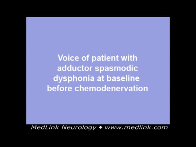
Laryngeal dystonia, previously referred to as spasmodic dysphonia, is a focal dystonia that affects the intrinsic laryngeal muscles (thyroarytenoid, lateral cricoarytenoid, interarytenoid, and posterior cricoarytenoid). The more prevalent subtype, adductor laryngeal dystonia, involves the muscles that close (adduct) the vocal cords—specifically the thyroarytenoid, lateral cricoarytenoid, and interarytenoid muscles—resulting in strained, strangled, and effortful speech with phonatory breaks. Less commonly, abductor laryngeal dystonia affects the posterior cricoarytenoid muscles, which are the only muscles that open (abduct) the vocal cords for breathing, leading to breathy speech and voiceless pauses.
A distinctive feature of laryngeal dystonia is its task-specific nature: symptoms typically occur during speech, whereas laryngeal function remains normal during other activities, such as swallowing, coughing, laughing, yawning, or singing. Diagnosis relies on patient history, an auditory-perceptual evaluation of the voice, and laryngeal endoscopy to rule out other disorders. Although several causative genes have been linked to some forms of laryngeal dystonia, it is most often idiopathic. The primary treatment involves electromyography-guided botulinum toxin injection into the affected intrinsic laryngeal muscles. Patients report significant improvements in vocal function, quality of life, and work productivity following successful treatment. Recent advancements in treatment include the use of the oral medication sodium oxybate, which has been reported to improve vocal symptoms in patients with alcohol-responsive laryngeal dystonia, and the injection of daxibotulinumtoxin A, a formulation of botulinum toxin that may have a longer duration of action in a subset of patients.
|
• Laryngeal dystonia is diagnosed clinically, based on history, perceptual voice evaluation, and laryngeal endoscopy. | |
|
• Adductor laryngeal dystonia typically results in strained, strangled, effortful speech with phonatory breaks on vowels. Abductor laryngeal dystonia generally causes breathy speech with voiceless pauses. Mixed laryngeal dystonia has characteristics of both. Vocal tremor is often coexistent. | |
|
• Laryngeal dystonia is rare, with females more likely affected than males. Adductor laryngeal dystonia is more common (> 80%) than the abductor or mixed forms. | |
|
• Laryngeal dystonia may be initially misdiagnosed as a functional or psychogenic voice disorder, and some patients suffer a delay in diagnosis of many years. | |
|
• EMG-guided botulinum toxin injections into the intrinsic laryngeal musculature have become the mainstay of treatment for laryngeal dystonia. | |
|
• Laryngeal dystonia is a lifelong condition. Successful management is facilitated by an interprofessional team that includes speech language pathologists, neurologists, and otolaryngologists. |
Dystonia is a neurologic hyperkinetic movement disorder characterized by sustained or repetitive involuntary muscle contractions. In primary dystonia (familial or sporadic), the dystonia is the only clinical abnormality (aside from occasional tremor), with acquired causes ruled out. In contrast, secondary dystonia results from identifiable causes, such as head injury, tardive dyskinesia (a drug side effect), or other neurologic diseases (eg, Wilson disease or Parkinson disease). Dystonias can also be classified based on the body regions affected: focal, segmental, or generalized.
Laryngeal dystonia is a focal dystonia resulting in task-specific, action-induced spasm of the vocal cords. Historically, Tiberius Claudius Drusus Nero Germanicus, who became emperor of Rome 41 AD, has been suspected to have laryngeal dystonia (154). It was first described by Traube in 1871 as a “nervous hoarseness” in a young girl and assigned the label of spastic dysphonia (193). The patient only spoke with great effort, and “the laryngoscopic examination revealed spastic closure of the vocal cord, whereby the left arytenoid cartilage shifted in front of the right one while probably also the vocal cords were particularly overlapping of each other” (168). Schnitzler may be the first one to suspect organic etiology, in 1895, in two patients with “cramping of the vocal cord and forced voice” (171), who also had synkinesis of facial muscles and abnormal movements of the arms and legs (91). Schnitzler termed the condition “aphonia spastica” or spastic dysphonia. Due to the lack of other coexisting neurologic deficits, the disorder continued to be considered psychogenic (67; 168; 11). A century later, Aronson observed the fluctuating vocal pattern characteristic of laryngeal dystonia and proposed the term “spasmodic” instead of “spastic” (08; 09). Robe and colleagues were the first to postulate that this disorder was related to the central nervous system (155). Dedo proved the neurologic etiology of laryngeal dystonia with the success of his recurrent laryngeal nerve transection procedure, which was a bold decision at a time when most of his contemporaries still believed in a psychiatric etiology (41).
The onset of laryngeal dystonia is insidious in most patients (84%), initially presenting as nonspecific hoarseness with gradual worsening of vocal quality over months to years (81; 07; 187). In a small proportion of patients, the onset is sudden (81). The most common inciting events identified by patients are stress, upper respiratory infection, pregnancy, and parturition (37; 05).
Laryngeal dystonia may be divided into several types, depending on the specific intrinsic laryngeal muscles that are most affected.
Adductor laryngeal dystonia. Adductor laryngeal dystonia is the most common subtype and accounts for over 80% of cases (24). The intrinsic laryngeal muscles that close (adduct) the vocal folds are the paired thyroarytenoid and lateral cricoarytenoid muscles and the single interarytenoid muscle. There can also be involvement of supraglottic musculature. Dystonic activity in these muscles cause a strained, strangled speaking pattern (111). When the vocal folds close so tightly during a muscular spasm that the airflow is completely interrupted, phonation is blocked, resulting in a “vocal break” – no phonation produced.
Abductor laryngeal dystonia occurs in 17% of cases (25). The paired posterior cricoarytenoid muscles are the sole abductors that open the glottis for breathing. In this subtype, the vocal folds have opening spasms, thus, leading to a low-volume breathy yet effortful speech pattern and breathy vocal breaks.
Mixed laryngeal dystonia is rarely diagnosed, although some experts posit that all laryngeal dystonia has a mixed component (35).
Vocal tremor may coexist with laryngeal dystonia in up to one third of patients (95), although this is more common in females (144).
Unusual variants include singing and respiratory dystonia. Singing laryngeal dystonia occurs when dystonic vocal spasms occur only with singing while conversational speech is spared (24). Respiratory laryngeal dystonia results in laryngeal adductor spasms during breathing. Patients with respiratory dystonia usually have fairly normal speech but report dyspnea and inspiratory stridor. The stridor abates during sleep, and they do not have hypoxia on clinical exam (191).
Fine wire electromyography has revealed that both the thyroarytenoid and the lateral cricoarytenoid muscles might be affected in adductor laryngeal dystonia, even if the thyroarytenoid is more predominant (93). The thyroarytenoid and lateral cricoarytenoid muscles are equally involved in laryngeal dystonia with tremor (93).
Laryngeal dystonia most commonly disrupts normal speech, particularly within the typical range of pitch, volume, and speed used in everyday conversation. Interestingly, activities such as laughing often produce brief, normal-sounding vocalizations, and tasks like whispering, screaming, yawning, or singing may temporarily improve or normalize the voice. These features reflect the task-specific nature of dystonia. Vocal spasms typically occur during speech but may be relieved during emotional vocalizations—such as laughing, crying, or shouting. Although speech is a learned behavior relying on specific neural circuits, emotional vocalization is an innate function governed by different, more primitive systems (Nieder and Mooney 2019).
As with dystonia in other areas of the body, some patients with laryngeal dystonia will report that a tactile or proprioceptive sensory trick (geste antagonist) can improve symptoms. These sensory tricks may include behaviors, such as touching the larynx during speech, chewing gum/putting an object between the teeth, or laying down. Fluency may also be improved by speaking in a higher pitch or using a “character voice.” Vacation often brings an improvement in symptoms whereas stress almost universally exacerbates dystonia and worsens vocal quality. Some patients report that sedatives or consumption of alcohol reduces symptoms, which is also noted in vocal tremor (90). Most patients report the voice worsens with speaking in public or on the telephone (77).
Relieving factor | Aggravating factor | |
Stress | — | 47.3% |
| ||
Laryngeal dystonia tends to emerge gradually in midlife and then reaches a plateau in terms of severity. Spontaneous remission has not been reported in laryngeal dystonia, which can be seen with cervical dystonia (116). Untreated or poorly responsive laryngeal dystonia is often linked to psychological distress, including a sense of handicap, low symptom control, functional disability, and increased anxiety and depression.
Successful treatment can lead to significant improvements in both physical and mental functioning. In 2022, the general prevalence of anxiety and depression symptoms among U.S. adults was 18.2% and 21.4%, respectively (190). However, in a cohort of 142 patients receiving botulinum toxin injections, those with laryngeal dystonia showed notably lower rates: 13.4% for anxiety and just 2.4% for depression (72). Laryngeal dystonia can have a substantial impact on occupational functioning (77). Patients report a mean 30% reduction in voice-related work productivity, which significantly improves following botulinum toxin treatment (133). These productivity impairments are primarily due to presenteeism rather than absenteeism (132). Additionally, individuals with laryngeal dystonia often report avoiding communication and seeking employment in roles with lower vocal demands (16).
The gold standard of treatment for laryngeal dystonia is EMG-guided botulinum toxin chemodenervation of the affected intrinsic laryngeal musculature. The duration of action in most individuals is between 2 to 6 months. Repeated treatment is needed, possibly lifelong. According to a national survey, most practitioners who provide botulinum toxin injections for laryngeal dystonia work at an academic care center (126). This may limit access to repetitive treatment and cause burden and loss of work productivity for those who live in more rural or remote areas.
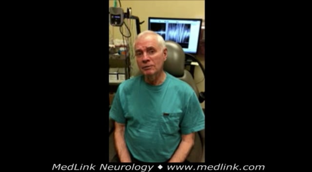
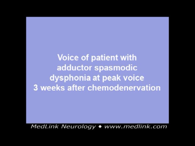
Case 1. The patient first noticed dysphonia in his late 40s. He was working in law enforcement on a hazardous undercover assignment. He began to have vocal difficulties that he attributed to laryngitis from occupational irritant exposure. His voice never recovered but continued to become more strained and effortful. About 10 years later, he saw a speech-language pathologist and reported that while on amusement park rides with his grandchildren, his voice was reproducibly normal. His speech-language pathologist diagnosed him with laryngeal dystonia. He did not seek treatment for another 5 years and was initially treated with 1 unit of botulinum toxin A to the bilateral thyroarytenoid muscles. He had significant breathiness and some difficulty with swallowing, and his dose was reduced. Currently, he receives an asymmetrical dose of 0.15 units of botulinum toxin A to one thyroarytenoid muscle and 0.25 units of botulinum toxin A to the contralateral thyroarytenoid muscle with vocal improvement from 40% to 50% normal function at baseline (when the effects of botulinum toxin have dissipated) to 80% to 90% of normal vocal function at the peak effect of the botulinum toxin injection with minimal breathiness or dysphagia. The Percent Normal Function scale (PNF or %NF) is an accepted functional scale to track vocal disability improvement during chemodenervation treatment for laryngeal dystonia; 100% indicates completely normal voice, and 0% indicates no functional voice (141).
Case 2. A 43-year-old female reported acute onset of voice changes in July 2016. She endorsed a strained effortful and “garbled” voice quality. She noted improvement in vocal quality with singing and with alcohol consumption. She was seen by her local ENT and referred to a speech-language pathologist and then further referred to the university for evaluation and management. As the vice president of her community college who frequently spoke to large groups, she had an extraordinary demand for voice use and quality in her vocational capacity. She was a nonsmoker and was otherwise healthy, with no other medical problems and no family history of neurologic disorders.
The patient was diagnosed with adductor laryngeal dystonia and initially treated with LEMG-guided botulinum toxin injections with a good effect. But after 2 years of treatment, she struggled with her injections causing too much initial breathiness and a shorter-than-desired duration of peak voicing. She was changed to an endoscopic approach to her injections and currently receives botulinum toxin injections to the bilateral thyroarytenoid and to the interarytenoid musculature. Her total dose is 2.5 units every 10 weeks of which she typically experiences 1 week of initial breathiness and mild dysphagia to liquids and 9 weeks of effect. She reports 4 weeks of peak voicing (90% to 100% of normal function), with a gradual decline to her baseline of 15% to 20% of normal function.
Classifying laryngeal dystonia as a medical disorder rather than psychiatric is attributed to Dedo after a recurrent laryngeal nerve resection (41). Nevertheless, the etiological and pathophysiological mechanisms underlying laryngeal dystonia are not known. Histological analysis of the recurrent laryngeal nerves in two patients who underwent recurrent laryngeal nerve resection revealed no apparent signs of either destruction or degeneration; however, the percentage of thin nerve fibers (diameter ranging from 5 to 10 microns) was higher than in normal controls (98). In another study, slight morphometric differences were found between the recurrent laryngeal nerve removed from patients with laryngeal dystonia and control recurrent laryngeal nerves in two groups, but these cannot explain causation of laryngeal dystonia (36).
Focal dystonias, such as laryngeal dystonia, are generally thought to be due to dysfunction in the basal ganglia. Supporting this view, secondary dystonia frequently results from lesions in basal ganglia structures, such as the putamen or globus pallidus. However, several findings challenge this localized model. Laryngeal dystonia is now understood as a disorder involving widespread brain network dysfunction, rather than being limited to abnormalities in the basal ganglia (59). Notably, primary dystonia lacks neurodegenerative changes, and lesions in brain areas outside the basal ganglia can also cause secondary dystonia. These insights support the emerging concept of dystonia as a neurofunctional disorder involving various aspects of the motor system circuitry, characterized by abnormal connectivity in the brain.
Brain imaging and structural findings. Structural brain imaging has revealed increased gray matter volume and cortical thickness in several regions involved in voice control, such as the laryngeal motor cortex, inferior frontal gyrus, temporal cortex, and cerebellum (179). Differences in brain structure have also been found between different types and causes of laryngeal dystonia, particularly in the superior corona radiata and arcuate fasciculus (20). Changes in cortical surface area have been observed in regions related to motor control, sensory processing, and auditory perception (99).
A meta-analysis by Kshatriya and colleagues confirmed that laryngeal dystonia involves widespread structural and functional abnormalities in several brain regions, including the basal ganglia, premotor cortex, parietal cortex, and insula (101). These findings reinforce the idea of laryngeal dystonia as a brain network disorder and suggest that functional MRI (fMRI) could be helpful for assessing brain activity during speech tasks. Haslinger and colleagues found that fMRI studies revealed reduced activity in the primary sensorimotor, premotor, and sensory association cortices during speech tasks in patients with laryngeal dystonia (65). Furthermore, Battistella and colleagues identified disrupted connectivity between regions in the frontoparietal and sensorimotor networks, with variations depending on laryngeal dystonia subtype and genetic background (14; 15).
Neurophysiological abnormalities. Neurophysiological studies support the presence of reduced inhibitory control in laryngeal dystonia. Cortical silent period, measured via transcranial magnetic stimulation, serves as a well-established marker of intracortical inhibition. A shortened cortical silent period reflects decreased cortical inhibition, even in muscles not directly involved in voice production.
Patients exhibit a shortened cortical silent period and abnormal blink reflex recovery, both indicative of impaired motor inhibition (161; 162; 186). Reduced cortical inhibition has been implicated in both motor and sensory systems in dystonia patients with adductor laryngeal dystonia (ADLD). Cortical silent periods recorded from the masseter and first dorsal interosseous muscles are significantly shortened compared to those in patients with muscle tension dysphonia and healthy controls. Cortical silent period also reveals differences between adductor spasmodic dysphonia and muscle tension dysphonia (162).
Neurophysiological studies further highlight key abnormalities in inhibitory control, sensorimotor integration, and brain plasticity. Electromyography in dystonia patients typically reveals co-contraction of agonist and antagonist muscles, prolonged muscle bursts, and overflow activity to unintended muscle groups.
EEG studies have shown excessive synchronization between somatosensory and premotor regions in patients with laryngeal dystonia, suggesting impaired regulation of sensory and motor signals (88).
Although this finding is not specific to laryngeal dystonia as similar reductions in cortical silent period have been reported in other focal dystonias, such as cervical dystonia, it reinforces the association between diminished cortical inhibition and dystonic pathophysiology. Whether this represents a causal factor or a marker of disease susceptibility remains unclear. The observation of reduced cortical silent periods in unaffected muscles supports the hypothesis of a more generalized dysfunction in GABAergic inhibitory networks.
Neurochemical alterations. Neurochemical studies have pointed to significant abnormalities in the dopamine and GABA systems in laryngeal dystonia. PET imaging has shown reduced dopamine release in the striatum during symptomatic speech, whereas dopamine release is increased during nonsymptomatic tasks, suggesting task-specific dysfunction. A study using [11C] raclopride found decreased dopaminergic transmission during symptomatic speech production in patients with laryngeal dystonia compared to controls, and increased dopaminergic activity during nonsymptomatic tasks, possibly reflecting compensatory adaptations in the nigrostriatal system (177).
In addition, reduced GABA activity in the inferior parietal cortex may contribute to the overactivity in motor pathways observed in laryngeal dystonia. This is further supported by the fact that over 50% of patients with laryngeal dystonia report symptomatic improvement following alcohol consumption, likely due to its effect as a GABA agonist (90). Although the precise mechanism remains unclear, the effect is postulated to result from modulation of GABAergic transmission. These chemical changes may play a role in making the brain’s movement circuits overactive in laryngeal dystonia.
The prevalence of primary laryngeal dystonia is estimated to be 5.9 per 100,000 (10). Laryngeal dystonia occurs more often in women than in men (25; 182; 02). The overall ratio ranges between 1.4 to 3.8 females to 1 male (25; 182). In one series, women made up to 79.3% of the population with laryngeal dystonia (02). In another series, laryngeal dystonia has a female preponderance (77.6%), with an average age of onset at 51 years (144). Males make up a larger percentage of the abductor laryngeal dystonia population than the adductor laryngeal dystonia population. Broken down into subgroups, the female-to-male ratio was 4.1:1 for adductor laryngeal dystonia and 2.2:1 for abductor laryngeal dystonia (172). Izdebski and colleagues reported that in a series of 200 patients with laryngeal dystonia, the age of onset was 41 years (+13.25 SD), with a range of 6 to 65 years for males, and 45.4 years (+13.3 SD), with a range of 7 to 78 years for females (81).
Factors associated with an increased risk of laryngeal dystonia, in small or isolated case-controlled studies, include a past history of mumps, blepharospasm, tremor, and intense occupational voice use (188); a personal history of cervical dystonia, sinus and throat illnesses, rubella, and dust exposure (189); a family history of voice disorders and tremor (25); an immediate family history of vocal tremor and meningitis; and an extended family history of head and neck tremor, ocular disease, and meningitis (189).
There are no known methods to prevent laryngeal dystonia.
The differential diagnosis of laryngeal dystonia is broad and includes both organic disorders and functional disorders. Table 2 lists some differential diagnoses of laryngeal dystonia. Essential voice tremor and muscle tension dysphonia can cause voice breaks; thus, they can form the most important differential diagnoses.
Vocal tremor, a condition acoustically characterized by low-frequency oscillations in voice, can occur in isolation or as a component of essential tremor. Essential tremor is a progressive action tremor and consists of involuntary and rhythmic movements at 4 to 12 Hz of one or more antagonistic muscles or muscle groups. It affects approximately 1% of all people and nearly 5% of those older than 65 years (110). Eighteen to 30% of patients with essential tremor have a vocal tremor (185). The movement disorder in vocal tremor is rhythmic rather than spasmodic. In addition to the intrinsic laryngeal muscles, it often involves pharyngeal and strap muscles. Lundy and colleagues found that unlike laryngeal dystonia, tremor is more often marked by fluctuations in frequency rather than just in intensity (114).
Muscle tension dysphonia. Muscle tension dysphonia, a functional dysphonia, may also mimic the strained voice quality of laryngeal dystonia (71). Evidence of consistent sound-specific phonatory breaks should raise suspicion of adductor spasmodic over muscle tension dysphonia. The hyperadduction of muscle tension dysphonia is generally sustained and is unlikely to be spasmodic. Although differences between muscle tension dysphonia and adductor laryngeal dystonia have been described on fiberoptic laryngoscopy, phonatory airflow measurement, and acoustic analysis, there is currently no single diagnostic test to differentiate these two disorders (159).
Careful auditory perceptual voice evaluation can help differentiate these diagnoses. One key difference is that neither vocal tremor nor muscle tension dysphonia demonstrate task specificity, whereas laryngeal dystonia does (present with talking as opposed to laughing and singing). The voice may deteriorate with stress in all conditions. Laryngeal endoscopy helps in the differential between laryngeal dystonia, vocal tremor, and muscle tension dysphonia; however, the diagnosis may be challenging in some cases. Even among experts, there can be notable variation when diagnosing patients with laryngeal dystonia, vocal tremor, or muscle tension dysphonia. In one study, international experts were asked to classify the vocal diagnosis of 178 patients across four different sites into 11 categories (112). The experts reviewed a portion of these patients’ video-laryngeal endoscopy and speech recordings, but due to a lack of diagnostic guidelines, there was poor interrater agreement regarding patient diagnoses.
In psychogenic dysphonia, another functional dysphonia, there are often several atypical characteristics, including loss of normal shouting, yawning, and laughing. Psychogenic dysphonia also does not present with tremor (107). In psychogenic dysphonia, the vocal symptoms are more often invariant across phonetic and most phonatory variables and not associated with sound prolongations or voice arrests (107). In one study, psychogenic speech and voice disorders were found to occur in 16.5% of 182 patients with psychogenic movement disorders. Among these patients, stuttering was the most common speech abnormality (n = 16, 53.3%), followed by speech arrests (n = 4, 13.3%), foreign accent syndrome (n = 2, 6.6%), hypophonia (n = 2, 6.6%), and dysphonia (n = 2, 6.6%) (11).
Vocal cord polyps or other mass lesions of the vocal cord can cause dysphonia characterized by strain and vocal breaks. These lesions would be visualized and diagnosed on laryngeal endoscopy.
A form of dysphonia similar to laryngeal dystonia has also been described in dominantly inherited ataxia with dentate calcification, currently assigned the name “spinocerebellar ataxia type 20” (94).
|
• Muscle tension dysphonia (159) |
Laryngeal dystonia may occur as an isolated dystonia or as part of generalized dystonia (123; 83; 66; 03; 122; 148). Yet, most patients with laryngeal dystonia present only with the vocal dysfunction, without neurologic manifestations in other areas of the body (25). Most commonly, laryngeal dystonia occurs sporadically, and there are no associated or underlying disorders. Laryngeal dystonia less frequently occurs in the setting of segmental or generalized dystonia. The reported prevalence of extralaryngeal dystonia in patients with laryngeal dystonia varies from 5% to 14% (61; 144). In one series that included 901 patients with vocal involvement, 82.5% had primary dystonia and 17.5% had secondary dystonia (25). In another series, 15.8% of patients with laryngeal dystonia had spread of dystonia, all of whom developed cervical dystonia (19). In another study, patients with cough, disordinate breathing, paroxysmal sneezing, and hiccups were found to have a higher incidence of extralaryngeal dystonia (147).
Several causative genes have been identified in familial dystonic syndromes that cause generalized dystonia, which have risk of components of laryngeal dystonia (44). Careful clinical characterization of the dystonic syndrome allows accurate phenotype-genotype correlation and may assist in identifying an underlying genetic diagnosis. Despite these familial dystonic syndromes having components of laryngeal dystonia, genetic screening of patients with laryngeal dystonia targeted at mutations in TOR1A, THAP1, and TUBB4 has a low diagnostic yield (62; 44).
|
Gene |
Gene function |
Disease |
Presentation |
Reference |
|
THAP1 |
Encodes a transcription factor that regulates endothelial cell proliferation |
DYT6 |
Dystonia affecting the cervical, cranial, and upper limb musculature, often with laryngeal involvement. |
(23; 201) |
|
TOR1A |
Encodes for a protein that is involved in cellular functions, such as protein folding, lipid metabolism, cytoskeletal organization, and nuclear polarity |
DYT1 |
Dystonia of the limbs, notably sparing the craniofacial muscles initially and then spreading and progressing to severe generalized dystonia. |
(54) |
|
TUBB4 |
Encodes for a neuronally expressed tubulin, and mutations lead to basal ganglia and cerebellum atrophy |
DYT4 |
Prominent spasmodic (“whispering”) dysphonia, which is distinct from abductor and adductor laryngeal dystonia, and associated craniocervical dystonia as well as a “hobby horse”–type gait. |
(108) |
|
ANO3 |
Encodes for calcium-gated chloride channels |
DYT24 |
Craniocervical dystonia, including laryngeal dystonia and mild upper limb dystonia, notably additionally with tremor. |
(183) |
|
GNAL |
Encodes a protein that mediated signaling within the olfactory epithelium |
DYT25 |
Cervical dystonia, something with head tremor and laryngeal dystonia, although isolated laryngeal dystonia/laryngeal dystonia has also been described, and generalized dystonia occurs in about 10% of cases. |
(12; 152) |
|
KMT2B |
Encodes a protein that methylates DNA and modifies the epigenome |
DYTKMT2B |
Progressive childhood-onset dystonia, with prominent cervical, cranial, and laryngeal dystonia. It is associated with typical facial features of an elongated face and bulbous nasal tip. |
(131) |
|
YY1 |
Yin Yang 1 protein, a transcription factor that plays a role in oligodendrocyte maturation and myelin gene expression |
YY1/THAP1 coregulation impairing DNA double stranded break repair |
Childhood-onset intellectual disability, behavioral abnormalities, oromandibular and laryngeal dystonia, speech and swallowing difficulties |
(55) |
Other reported associations include neuroleptic exposure, either immediately or as part of the tardive syndrome (199; 06), mitochondrial disease (149), valproic acid administration (with improvement after discontinuation) (142), central pontine myelinosis (173), amyotrophic lateral sclerosis (158), psychogenic dysphonia (167; 11), late-onset laryngeal dystonia with low arylsulphatase A (125), essential tremor (109), palatal myoclonus (50), hereditary spastic paraplegia type 7 (63), multiple sclerosis (47), or trauma (56).
Diagnosis is often delayed, possibly due to the unfamiliarity with the disease, and patients are frequently seen by multiple physicians before a definitive diagnosis is made. One study showed that patients had over a 4-year delay from initial presentation to a physician for vocal concerns to reaching a diagnosis of laryngeal dystonia (38). Another study from Japan showed that in 60% of patients, the diagnosis of laryngeal dystonia was delayed for more than 2 years (75).
Laryngeal dystonia is a clinical diagnosis. Eliciting a thorough neurologic and voice history with a targeted physical exam and laryngeal endoscopy is critical to achieve the correct diagnosis (130). For some individuals, the diagnosis is further confirmed by response to treatment, most often botulinum toxin chemodenervation. It is important to determine if there are other coexistent neurologic symptoms, such as tremor or dystonia of other body areas. It is also important to elicit a history of neuroleptic use or administration of medications used to treat nausea, such as prochlorperazine, promethazine, or metoclopramide.
• Amphetamines, cocaine | |
• Antiepileptics | |
• Antihistamines | |
• Antipsychotics | |
• Beta-adrenergic agents | |
• Caffeine | |
• Cimetidine | |
• Dopamine agonists | |
• Lithium | |
• MAO inhibitors | |
• Metoclopramide | |
• Oral contraceptives | |
(143) |
Patients often notice their symptoms after an illness, during a period of increased stress, or during a public speaking venue. Some individuals describe a gradual onset with symptoms noted only during periods of stress or increased speaking demands; others describe a sudden occurrence that may be related to an illness or life event. Many individuals remark that symptoms are exacerbated or more acutely perceived when speaking on the telephone. Speech may be better on awakening in the morning when relaxed and is worsened with fatigue or stress. Although the symptoms can wax and wane, there is almost always some sense of presence of the disorder. Certain words or combinations of words might be more difficult to say, depending on the type of laryngeal dystonia. Individuals may report that singing, laughing, yelling, and falsetto are relatively spared. Cough and swallow, along with other vegetative laryngeal tasks are preserved. Vocal tremor is frequently coexistent. A subset of patients may have alcohol-responsive dystonia and tremor in which symptoms are significantly reduced after an alcoholic beverage (90).
Onset of symptoms | ||
May be sudden | ||
Aggravating/alleviating factors | ||
Speaking | ||
Associated symptoms | ||
Odynophonia | ||
Character of symptoms | ||
Quality of voice | ||
Raspiness | ||
Flow | ||
Decreased breath support | ||
Control | ||
Loss of pitch control | ||
| ||
The primary modality to assess laryngeal dystonia is by listening to the patient during conversational speech and during elicited speech tasks with either vowel or voiceless consonant predominant sentences (86).
Adductor laryngeal dystonia phrases or sentences would include words with vowels or voiced sounds. Sentences that elicit strain or vocal breaks in individuals with adductor laryngeal dystonia include the following:
• “We eat eggs every evening.” | |
• “Ambling down Rainy Island Avenue.” | |
• Counting 80 to 85 |
When phonating a vowel, the vocal folds are brought together in adduction. Individuals with dystonia will hyper-adduct and create a strained, strangled sound. If the spasm is particularly strong, the sound will be cut off and a vocal break will occur.
Abductor laryngeal dystonia phrases or sentences would include words with vowels following a voiceless consonant (such as b, d, f, h, p, s or t). A breathiness is heard as a speaker transitions from a voiceless consonant to a voiced vowel sound. Sentences that elicit the breathy breaks in individuals with abductor laryngeal dystonia include the following:
• “The puppy bit the tape.” | |
• “Hammy hit the hammer hard.” | |
• “Taxi!” | |
• Counting from 60 to 65 |
For the voiceless consonant “s,” the fricative sound is created by placing the tongue against the teeth and palate and forcing air through the narrow space. The vocal folds remain open to power this air flow, and there is no contribution from vocal fold vibration. The vocal folds then close to allow the vowel sound that follows. For individuals with abductor laryngeal dystonia, the vocal folds will be delayed in closing due to abductor muscle hyperfunction. The excessively open glottis will then result in a breathy voice or breathy breaks.
Some clinicians have patients read the phonetically balanced “rainbow passage”:
When the sunlight strikes raindrops in the air, they act like a prism and form a rainbow. The rainbow is a division of white light into many beautiful colors. These take the shape of a long round arch, with its path high above, and its two ends apparently beyond the horizon. There is, according to legend, a boiling pot of gold at one end. People look, but no one ever finds it. When a man looks for something beyond his reach, his friends say he is looking for the pot of gold at the end of the rainbow… (53; 130). |
Some authors have documented a correlation between maximum phonation time and laryngeal dysphonia, which can be a simple office test to add to the armamentarium of evaluation. The average maximum phonation time in the adductor spasmodic dysphonia, abductor spasmodic dysphonia, and control groups was 25 seconds, 9 seconds, and 16 seconds, respectively. The positive predictive value of this test was 81.3%, negative predictive value 83.9%, with a sensitivity of 79.6% and specificity of 85.2% (85). For abductor laryngeal dystonia, the clinical sign of paroxysmal flaring of ala nasi during breathy breaks is also noted in many patients (129).
In addition to conversational speech, attention should be paid to a complete neurologic examination. Laryngeal dystonia usually presents without other neurologic findings, so any tremor, weakness, or cranial nerve neuropathy should prompt further neurologic evaluation. In the setting of associated clinical features, such as segmental dystonia, tremor, or gait disturbance, careful clinical characterization of the phenotype may provide clues to an underlying genetic dystonic syndrome (see etiology and pathogenesis).
Laryngeal endoscopy is a critical component of the physical exam for laryngeal dystonia. Adductor and abductor spasms can be visualized during connected speech, and other laryngeal lesions that cause hoarseness can be ruled out. Because laryngeal dystonia is a disorder of connected speech, transnasal rather than transoral laryngoscopy should be performed. Some authors have suggested a correlation between abductor spasmodic dysphonia and mixed spasmodic dysphonia with vocal fold sulcus (138).
Researchers have tried to identify modalities to diagnose laryngeal dystonia and differentiate it from muscle tension dysphonia, but these have not gained wide clinical use. The critical unmet need lies in identifying objective, quantitative clinical measures that are both specific and sensitive for diagnosing laryngeal dystonia and can reliably validate treatment outcomes over time.
One study suggested that the long-term average spectrum (LTAS), a tool that assesses the average amplitude spectrum across a selective frequency range and provides information on the spectral distribution of the speech signal over a period of time, may identify spectral noise differences between muscle tension dysphonia and adductor laryngeal dystonia in women (71). The use of neural network and support vector machine–based methods, in combination with a pattern recognition algorithm, has also been studied (169). Fine kinemetric analysis from high-speed digital imaging may assist in the clinical differentiation of adductor laryngeal dystonia and muscle tension dysphonia, although further studies are required (146).
In a study, researchers introduced a novel automated acoustic outcome measure, the spectral aggregate of the high-passed fundamental frequency contour (SAHfo), developed specifically for adductor laryngeal dystonia (120). This measure offers a preliminary yet promising objective and quantifiable method for detecting and monitoring the acoustic features, particularly laryngeal discontinuities, such as creak (nonmodal voice quality, vocal fry) and phonatory breaks (120)
The percentage of creak, as measured by an automated creak detector, may serve as a reliable quantitative marker for adductor laryngeal dystonia and helps to serve as a screening tool to differentiate adductor laryngeal dystonia from muscle tension dysphonia and control groups (119).
For cases in which the diagnosis is unclear or treatment efficacy has declined, electromyography can give some important information about the affected musculature. In a study of patients with adductor laryngeal dystonia, Yang and colleagues found there were increased amplitudes of the motor unit recruitment potentials and evoked potentials of the thyroarytenoid muscles (202). When combined with acoustic channels, there may be a delay from the onset of the electrical activity on EMG with the onset of the acoustic output in laryngeal dystonia (176), and this latency may be significantly related to the severity of adductor laryngeal dystonia (39).
Botulinum toxin injections are the mainstay of treatment for laryngeal dystonia. Blitzer and colleagues performed the first botulinum toxin treatment on a patient with spasmodic dysphonia in 1986, and their work was confirmed by a subsequent double-blind study (26; 195). The American Academy of Otolaryngology-Head and Neck Surgery as well as the American Academy of Neurology endorses botulinum toxin as primary therapy for spasmodic dysphonia.
Botulinum toxin is a neurotoxin produced by the bacterium Clostridium botulinum that prevents the release of acetylcholine from the nerve ending at the neuromuscular junction. The result is a partial paralysis of the injected muscle. The toxin is made of a heavy and a light chain. The heavy chain allows the neurotoxin to be endocytosed into the neuron. Once within the neuron, the light chain binds to the SNARE proteins. SNARE proteins allow acetylcholine-containing vesicles to fuse at the nerve terminal to exocytose acetylcholine.
There are eight different types of toxins produced by Clostridium botulinum (A, B, C1, C2, D, E, F, and G). Type A is the most potent, and type B is the second most potent.
Five different botulinum toxin products are available on the market. Four of these toxins are botulinum toxin type A and include Botox® (Allergan, Inc., Irvine, California, United States), Dysport® (Ipsen Ltd., Slough, Berkshire, United Kingdom), Xeomin® (Merz Pharmaceuticals, United States), and CBTXA (China). Myobloc®/Neurobloc® (Solstice Neurosciences, Inc., South San Francisco, California, United States) is botulinum toxin type B. In the United States, the available preparations include Botox®, Xeomin®, and Myobloc®/NeuroBloc®.
Neutralizing antibodies to botulinum toxin can develop. Injections of botulinum toxin type B can be safely and effectively used in patients with antibodies to type A (01).
DaxibotulinumtoxinA (DAXI) is a novel botulinum toxin type A (BoNTA) formulation. It has shown promising safety and efficacy for both aesthetic and therapeutic applications, including cervical dystonia and upper limb spasticity. DAXI is composed of a purified 150-kDa neurotoxin without complexing proteins and includes a unique stabilizing peptide (RTP004), which enhances binding of the neurotoxin to neuronal surfaces, improving stability and potentially making its effects last longer. In a report published by Marshall and Rosen, a 77-year-old patient was injected with DAXI in the TA-LCA complex and subsequently in the interarytenoid muscle (124). He reported an 80% better duration of benefit and a 57% improvement in symptoms than his best BtxA injection. Clinical trials are ongoing to establish the safety and efficacy of DAXI for the treatment of laryngeal dystonia.
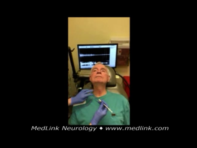
Techniques of laryngeal botulinum toxin injection. The most commonly employed technique for botulinum toxin injection in laryngeal dystonia is the EMG-guided percutaneous approach (134). Alternative methods include the “point-touch technique,” which relies on external anatomical landmarks (136), and injections under direct visualization via transoral, percutaneous, or transnasal approaches (57; 153). Some practitioners combine endoscopic visualization with EMG guidance (194).
A national survey of laryngologists demonstrated that 88% use EMG guidance, 9% rely on anatomic landmarks, and only 3% employ endoscopic guidance (174). Injection patterns vary and may be bilateral, unilateral, staggered, or use asymmetric dosing (127; 194; 197).
Target muscles. In adductor laryngeal dystonia, the thyroarytenoid muscle is most frequently targeted. Other intrinsic adductor muscles, such as the interarytenoid and lateral cricoarytenoid, may also be injected. For supraglottic involvement, injections into the false vocal folds have been utilized (181). In abductor laryngeal dystonia, the posterior cricoarytenoid muscle—the sole vocal fold abductor—is injected.
Thyroarytenoid muscle. A 25-gauge, Teflon-coated needle is inserted percutaneously just off midline (2–3 mm) through the space between the cricoid and thyroid cartilages, pointing towards the ipsilateral thyroarytenoid muscle. Bending the injection needle by 30 to 45 degrees can ease access to the muscle. The localization of the needle is verified by crisp motor unit potentials heard on EMG when the patient performs a long “/i/” (134; 32). The EMG activity is sustained during the phonation (68).
Lateral cricoarytenoid muscle. The EMG-guided percutaneous approach to the lateral cricoarytenoid muscle is similar to the EMG-guided percutaneous approach to the thyroarytenoid muscle. The needle is inserted further off midline in the cricothyroid space, at the junction of the cricoid cartilage and thyroid cartilage. After passing through the cricothyroid space, the needle is pointed towards the ipsilateral lateral cricoarytenoid muscle, and the location is confirmed with auditory feedback on the EMG machine during a sustained “/i/”. In this case, the EMG will show an increase and rapid drop-off in EMG activity, as the lateral cricoarytenoid muscle is responsible for the initial setting of the vocal cord position (68). A cadaver study showed that ultrasound-guided injection of the lateral cricoarytenoid muscle is feasible and precise (170).
Interarytenoid muscle. Injection is performed with EMG guidance, typically requiring topical laryngeal anesthesia. The needle is passed through the cricothyroid space and into the subglottis. A distinct air signal will be heard on the EMG when the needle is in the airway. The needle is then directed upwards towards the interarytenoid muscle. Insertional activity signal will be heard on the EMG machine when the muscle is entered, followed by recruitment activity during a sustained “/i/”.
False vocal fold injection. These are performed under direct endoscopic visualization without EMG. A flexible needle can be threaded through the working channel of a transnasal laryngoscope and the injection performed through this needle. However, this requires a large amount of liquid to prime the needle. Another approach is the peroral approach in which a flexible nasolaryngoscopy is performed to visualize the larynx. Then an orotracheal injector with a needle is passed perorally. The needle is visualized by the flexible laryngoscope and directed into the false vocal folds.
Lastly, a thyrohyoid approach can be used in which a 25-gauge needle is advanced through the thyrohyoid membrane and under direct visualization with a flexible nasolaryngoscope; the needle is directed into the false vocal fold.
In all approaches, the depth of injection is within the submucosal plane. This results in a bleb seen below the mucosa (181). Nasal anesthesia and decongestant are utilized during this procedure, in additional to laryngeal and airway anesthesia.
Posterior cricoarytenoid muscle (abductor laryngeal dystonia). The injection of botulinum toxin into the posterior cricoarytenoid muscle for abductor laryngeal dystonia is more challenging. Injection techniques usually include translaryngeal EMG guidance and laryngeal rotation (27), endoscopic guidance (156), and transcricoid rostrum (127), or a combination approach. As opposed to confirming the position of the needle with a sustained “/i/”, the position within the posterior cricoarytenoid muscle is confirmed with crisp motor unit potentials heard on EMG when the patient sniffs, which further abducts the vocal cords.
In the translaryngeal EMG-guided approach to injection of the posterior cricoarytenoid, the needle is passed through the skin overlying the cricothyroid membrane, through the airway, and through the posterior cricoid plate into the posterior cricoarytenoid muscle. Visualization with laryngoscopy is often utilized to improve the accuracy of the injection. As the mucosa of the airway is violated, topical anesthesia to the glottis and subglottis is typically required.
For the laryngeal rotation technique, the physician rotates the larynx to expose the posterior aspect of the thyroid cartilage. The needle is passed posterior to the thyroid lamina, along the lower half of the thyroid cartilage/superior border of the cricoid cartilage, until the needle hits the posterior surface of the cricoid. EMG auditory feedback is used to confirm the position.
Typically, unilateral posterior cricoarytenoid muscle injections are performed. A major concern for bilateral injections is bilateral vocal fold abductor weakness and the consequent narrowing of the airway for adequate respiration. Simultaneous bilateral posterior cricoarytenoid injections have also shown success in improving vocal outcome measures (92). Successful outcomes have also been reported using an initial unilateral posterior cricoarytenoid muscle injection, followed by a contralateral injection after 2 weeks, if necessary (24).
Botulinum toxin doses. Doses of botulinum toxin used for the treatment of laryngeal dystonia vary depending on the practitioner and the brand of toxin used. In the early literature, the doses of botulinum toxin (Botox®) into the thyroarytenoid muscle for adductor laryngeal dystonia ranged from 3.75 to 7.5 U for bilateral injections (34; 32; 33; 195) to 15 U for unilateral injections (134; 113). Up to 50 U per vocal cord has been used (84). Later literature and common practice have recommended the use of lower doses (30). In the authors’ experience, a chemodenervation-naive patient is started on botulinum toxin A at a dose of 0.5 to 1 unit in the bilateral thyroarytenoid muscle for the adductor laryngeal dystonia. This starting dose is based on Blitzer's original work (26). In one retrospective study of 126 patients, the mean dosage of onabotulinumtoxinA was 1.54 +/- 0.35 units per side (58).
Injection into the false vocal cords is typically at higher doses--anywhere from 5 to 10 units per false vocal fold (181).
The optimal dose of botulinum toxin varies between patients and does not appear to correlate with severity of laryngeal dystonia, age, or gender. In one study, BMI and overall health were correlated with a higher effective dose (203). Higher doses were required for symptom control in female patients with adductor laryngeal dystonia compared to males in one retrospective chart review, although the reasons for this observation are unclear (106).
Patients are warned about potential coughing with liquids and breathy vocal quality, which typically last no more than a week. They are then instructed to return when their voice breaks start to worsen. At that time, at the authors’ institution, they are asked to judge their best voice on a scale of 0% to 100%, with 100% being a completely normal voice and 0% being no voice at all. They are then asked about any initial side effects and how long those symptoms lasted. At that point, the decision is made to increase or decrease the botulinum toxin dose or change the injection interval.
There appears to be a lower rate of resistance with botulinum toxin in laryngeal dystonia compared with other dystonias, and this may be related to the low antigen challenge associated with the doses used (135).
Surgery. After recognition of the organic etiology of laryngeal dystonia, the development of recurrent laryngeal nerve resection confirmed the neurologic basis of the disease process. The first patient to undergo recurrent laryngeal nerve resection was a woman with a 17-year history of laryngeal dystonia who had been treated by more than 29 physicians from different specialties without improvement. Blocking of the recurrent nerve with lidocaine improved her spasm, whereas placebo injections did not improve the patient’s voice (41). After five repeated reproducible results with lidocaine, the patient underwent sectioning of the right recurrent laryngeal nerve with essentially normal voice within 1 week after the procedure, with the help of speech therapy.
Another less invasive procedure involved crushing the recurrent nerve, which initially appeared to be equally effective. Gross recurrent laryngeal nerve destruction is no longer a contemporary management for laryngeal dystonia, although this procedure is of great historical importance regarding the recognition that this disease has a neurologic and not psychiatric pathophysiology.
For adductor laryngeal dystonia, selective laryngeal adductor denervation-reinnervation (SLAD-R), first described in 1999 (18), involves selective denervation of the adductor branch of the recurrent laryngeal nerve. The distal nerve stumps are reinnervated with a nonlaryngeal nerve, generally the sternohyoid branches of the ansa cervicalis nerve (128).
Isshiki and colleagues offered an apparently mechanical solution to the problem of hyperadduction in adductor laryngeal dystonia with laryngeal framework surgery (80; 78). In this procedure, a lateralization laryngoplasty, or type II thyroplasty, is performed by means of a midline division and expansion of the thyroid cartilage.
Bilateral thyroarytenoid muscle myectomy is also used in the treatment of adductor laryngeal dystonia. Successful treatment of adductor laryngeal dystonia has also been reported in a small series of patients using selective lateral laser thyroarytenoid myotomy (74; 60). Sustained improvements in voice handicap index and GRBAS scale have been observed following endoscopic laser thyroarytenoid myoneurectomy (196; 60).
Surgical treatment for abductor laryngeal dystonia is more nuanced and has been less described in the literature. Bilateral vocal fold medialization, or type I thyroplasty, has been used by some.
Deep brain stimulation for adductor laryngeal dystonia is described for the treatment of laryngeal dystonia in a phase 1 clinical trial (69). A phase 2 clinical trial of deep brain stimulation for laryngeal dystonia is currently being conducted.
Other therapeutic modalities. Voice therapy has been reported to be helpful in conjunction with botulinum toxin treatment to retrain a long-term compensatory behavior superimposed on laryngeal dystonia (137), although this has not been consistent. Significant improvement with speech therapy may suggest an alternate diagnosis of muscle tension dysphonia (13). In a systematic review of 21 studies of voice therapy in patients with laryngeal dystonia, the adherence rate was quite low (about 60%) (121).
Although to a lesser degree than in the past, many patients with laryngeal dystonia are still subjected to psychiatric treatment. Psychotherapy is useful to help coping with social stresses, which can be considerable; the patient, however, needs to be made aware first that his or her condition is an organic disorder and to be treated accordingly either with botulinum toxin or surgery. Acupuncture has not been proven effective by expert raters, although Lee and colleagues found many patients reported subjective improvements in voice production (104).
Electrical and vibrotactile stimulation of the larynx has been explored as a neuromodulation strategy to address muscle spindle dysfunction and related proprioceptive and somatosensory abnormalities in laryngeal dystonia. Electrical stimulation of the thyroarytenoid muscle with a hook electrode was reported to improve symptoms in one small case series (150). Vibrotactile stimulation of the larynx is currently under research. Konczak and colleagues have demonstrated beneficial effects of vibrotactile stimulation to the skin over the larynx, self-administered by patients, with effects emerging within 15 to 20 minutes and lasting up to an hour or more (96).
An open-label study of sodium oxybate demonstrated reduced voice symptoms in alcohol-responsive laryngeal dystonia in those with and without coexistent voice tremor (160).
A phase IIb randomized, double-blind, placebo-controlled clinical trial demonstrated that a single 1.5 g dose of oral sodium oxybate significantly improves voice symptoms in patients with alcohol-responsive laryngeal dystonia, with effects seen across all laryngeal dystonia subtypes and symptom severities. The drug’s efficacy, which mimics alcohol’s GABAergic modulation, was not observed in alcohol nonresponsive patients, whose response was similar to placebo. Side effects, including dizziness, daytime sleepiness, and nausea, were mild, transient, and more frequent in alcohol-nonresponsive patients. The benefits appeared within 40 minutes, lasted about 5 hours, and did not lead to rebound symptoms, supporting its use on an as-needed basis. The safety profile was favorable, with no significant changes in cognition or vital signs. These findings suggest sodium oxybate may be a pathophysiologically relevant, noninvasive alternative for patients with alcohol-responsive laryngeal dystonia, particularly those unresponsive to or opting out of botulinum toxin injections. A phase III multicenter trial is recommended for further validation (180).
Botulinum toxin injections. As noted above, botulinum toxin injection is the mainstay of treatment for laryngeal dystonia, with positive outcomes. Treatment with botulinum toxin leads to a reduction in spasmodic contractions observed on video laryngoscopy (51). Yet, lower functional gain was noted in patients with dystonia in other body regions (118). A meta-analysis of 30, mostly single-blind studies, indicated moderate overall improvement as a result of botulinum toxin treatments (31). The Cochrane Collaboration Review noted that although only one study (195) out of the nearly 77 reports was double-blind and fulfilled their inclusion criteria, most of the studies had similarly positive effects (200). In one series of 131 patients, 67 of 574 injections (12%) were categorized as failures: failure was associated with injection by a new physician, the patient being a professional voice user, and a lack of breathiness in the voice after injection (204).
Improvement after injection with botulinum toxin for adductor laryngeal dystonia is realized an average of 2.4 days after injection; the average peak benefit occurs at 9 days post injection (28). For abductor laryngeal dystonia, the average onset of effect is 4.1 days, with a peak effect at 10 days.
For adductor laryngeal dystonia, the positive benefit of botulinum toxin injections lasts for approximately 15.1 weeks (28). The mean duration of benefit is 10.5 weeks for those with abductor laryngeal dystonia (28). Most insurance companies only allow for botulinum toxin injections every 3 months (four times per year). However, there is a considerable subset of patients who require short interval treatment to optimize their outcomes. In a study, 27.5% of patients required injections less than 90 days apart (short interval) while not showing an increased side effect profile (102). These patients were statistically younger than those who did not require short-interval botulinum toxin injections.
In the largest treatment series in 2010, patients with adductor laryngeal dystonia reported an improvement to 91.2% of normal with botulinum toxin injection (24). Treatment of abductor laryngeal dystonia was less satisfactory, with patients reporting an improvement to 70.3% of normal. Another study noted average subjective phonation improvement of 33% with adductor laryngeal dystonia (118). In another study examining outcomes of onabotulinumtoxinA, 88.1% of injection cycles for 328 patients with adductor laryngeal dystonia were noted to be maximally beneficial (145).
A meta-analysis showed significant improvement in several quality-of-life measures after treatment with botulinum toxin, including the Voice Handicap Index (VHI) and Voice-Related QoL scales (51; 52). Furthermore, patients on an established botulinum toxin injection routine have been shown to have high degrees of general and disease-specific self-efficacy, a concept that combines self-esteem and locus of control (73). Furthermore, the higher the self-efficacy, the lower the VHI and levels of anxiety and depression.
Possible complications from botulinum toxin injections into the thyroarytenoid musculature include breathiness, throat pain, and dysphagia. Initial functional decline following botulinum toxin injection was observed in 28.5% of cases (141). In a series of 1300 patients over 24 years of age, adductor injections were associated with a 25% risk of mild, transiently breathy voice and a 10% risk of transient coughing with drinking fluids. Breathy dysphonia has been reported to be as high as 50.9%, lasting on average 20 days (48). Local pain, bruising, and itch were reported in less than 1% of individuals (24). Rarely, bilateral abductor paralysis has been reported and is likely a result of toxin diffusion around the muscular process of the arytenoid to the posterior cricoarytenoid muscles (198).
There are some data to suggest that botulinum toxin is less effective in patients with concurrent vocal tremor (141). Patients with adductor laryngeal dystonia and associated vocal tremor may benefit from combined interarytenoid and thyroarytenoid muscle botulinum toxin injections (87). Botulinum toxin injections into the interarytenoid muscle have a 50% response rate in patients previously unresponsive to traditional injection sites (86).
Voice after supraglottic false vocal fold botulinum toxin injection for adductor laryngeal dystonia is significantly improved without the side effect of a period of a significantly breathy voice initially after injection (181).
Different protocols have been proposed for injecting botulinum toxin into the thyroarytenoid muscle, either unilaterally or bilaterally. Initial dosages of 1.25 units bilaterally of onabotulinumtoxinA resulted in a statistically significant shorter duration of breathiness without significant differences in clinical effectiveness or voice outcome in one study, compared with an initial dosage of 2.5 units bilaterally (157). Unilateral injection may result in fewer adverse events, such as breathiness or hoarseness (97; 21). Unilateral injections improve the performance ratio of strong voice interval divided by breathy voice interval (97). Upile and colleagues found that low-dose unilateral injections resulted in no significant difference in patient outcome in terms of voice duration, voice score, and complication rate when compared with bilateral injections (197). However, another study found that bilateral injections had a significantly higher proportion of patients than unilateral injections (36% vs. 33%, p=0.0228), with both greater than 3-month duration of effect and less than 2-week duration of side effects (46). Some studies have shown that the duration of benefit in men is significantly longer and with less swallowing difficulty after bilateral injections (117). Lee and colleagues found that alternating injections to the thyroarytenoid muscle was effective, with fewer side effects than bilateral injections, although the treatment effect was shorter (105). This was additionally supported by Stone and colleagues, who found that unilateral botulinum toxin injections reduce side effects without sacrificing improved voice outcomes, although with a shorter duration of improvement (184). Interestingly, they also studied bilateral injections with different doses to each side and found that this can extend the duration of effect, although with more side effects compared to a unilateral injection (184). Another study also found that the side effect profile was better for unilateral injections, but the authors concluded that bilateral injections had a better side effect/optimal effect profile (46).
Surgical outcomes for laryngeal dystonia. Recurrent laryngeal nerve sectioning initially showed remarkable therapeutic results. Dedo reported the results of sectioning of the recurrent laryngeal nerve in 34 patients (41). Short- and mid-term results were good, with a recurrence rate of 10% to 15%, respectively (82; 42; 43). Long-term results showed a gradual decline in benefit from the procedure. Crushing of the recurrent laryngeal nerve also from widespread recurrence of symptoms had a success rate of 13% at 3 years (22).
The initial reports of selective laryngeal adductor denervation-reinnervation (SLAD-R) presented favorable results in 19 of 21 patients (18). Only one patient underwent further treatment with botulinum toxin postoperatively. Positive results using expert and untrained judge’s perceptual evaluation of the voice were also reported in a smaller series (04). Additional studies have also reported favorable results after a mean follow-up time of 7.5 years (128). Functional reinnervation of the vocal cord adductors by ansa cervicalis has been observed 10 years after successful SLAD-R surgery (40). SLAD-R remains an important therapeutic option, particularly for patients who derive limited benefit from optimally administered botulinum toxin injections. However, it is essential to note that patient satisfaction with SLAD-R varies. Although some individuals report sustained improvement, others may experience only partial benefit or a recurrence of symptoms over time. As SLAD-R is irreversible, careful patient selection and preoperative counseling regarding both the potential benefits and the limitations of the procedure are critical.
Isshiki reported success in a small series with informal judgments regarding voice quality by both patients and physicians after type II thyroplasty (80; 78). A further small study using laryngeal framework surgery also resulted in significant acoustic and aerodynamic improvement in adductor laryngeal dystonia (166). Follow-up after 2 to 5 years revealed unsatisfactory outcomes in seven of 90 cases and were mainly attributed to inadequate technique and, in one case, an incorrect indication (163). Surgical mechanical faults appear to be the main cause of unsuccessful outcomes (79). Analyses of revision surgery cases indicated that the wings of titanium bridges would often break without patients displaying any signs, symptoms, or tissue damage. Based on the malfunctions detected and the analyses of failed devices, development of a new bridge with improved specifications is anticipated (164). Another long-term follow-up study (average of 41.3 months) following type II thyroplasty demonstrated sustained improvement in voice symptoms and quality of life (165).
One study found no difference in the postoperative Voice Handicap Index 10 score between patients who underwent bilateral thyroarytenoid muscle myectomy compared to those who received type II thyroplasty (140). Bilateral thyroarytenoid muscle myectomy appeared to improve strangulation, interruption, and tremor but worsened breathiness when compared to type II thyroplasty.
In one case series, six patients underwent vocal fold medialization via type 1 thyroplasty and demonstrated significant benefit on Voice Handicap Index (VHI) and Voice-Related Quality of Life (VR-QOL) measures. The four patients who had undergone the procedure at least 1 year prior to their survey reported a sustained benefit to their symptoms (45). One patient with staged bilateral posterior cricoarytenoid partial myoneurectomy demonstrated sustained benefit up to 8 years postoperatively (17).
Outcomes of voice therapy. With regards to voice therapy, one study did not demonstrate additional benefits in patients receiving voice therapy compared to those receiving botulinum toxin alone or those who received botulinum toxin with sham voice therapy (175). Further studies are required to confirm these results.
Deep brain stimulation. Deep brain stimulation is believed to work by changing the activity within abnormal motor networks in the brain, helping to normalize the overtly active motor responses seen in dystonia. Unlike the quick improvements seen with deep brain stimulation in conditions like Parkinson disease or essential tremor, its effects in laryngeal dystonia are usually delayed. Patients may need several weeks or months to experience noticeable benefits. This delayed response supports the conceptualization of dystonia as a disorder of sensorimotor integration, wherein reorganization and adaptation of motor circuitry require extended time following neuromodulation. Patient response to deep brain stimulation varies widely. Although some individuals show significant improvement, others experience only limited or no benefit. So far, no single factor—including the presence of the DYT1 gene mutation—has been proven to reliably predict treatment outcomes. Determining which patients with primary dystonia will benefit most from deep brain stimulation remains an important area for future research (143).
Deep brain stimulation of the globus pallidus internus (GPi) resulted in improvement of segmental dystonia, including laryngeal dystonia in one patient, with maintenance of symptom control at 10-year follow-up (70). Some patients with primary dystonia with a component of laryngeal dystonia had no benefit to their laryngeal dystonia with GPi stimulation (76). One patient with essential tremor and adductor laryngeal dystonia had sustained benefit to her laryngeal dystonia with bilateral ventral intermediate (Vim) nucleus of the thalamus stimulation (115). Another patient with essential tremor and coincident adductor laryngeal dystonia treated with unilateral thalamic deep brain stimulation experienced significant improvement in vocal dysfunction with isolated Vim and combined Vim and ventral oralis anterior (Voa) nucleus of the thalamus (151). Honey and Kruger have reported promising results with bilateral thalamic deep brain stimulation (100; 69).
A study highlighted the role of diffusion MRI in identifying individualized biomarkers of response to thalamic deep brain stimulation. The approach supports personalized deep brain stimulation strategies for every patient, especially using directional electrodes, and calls for further studies to validate these biomarkers (64).
Vibrotactile stimulation. Laryngeal vibrotactile stimulation temporarily improved symptoms in nine of 13 patients (89). This improvement was associated with a suppression of theta band power in the left somatosensory motor cortex, similar to what is seen during successful sensory tricks.
Konczak and colleagues conducted a single-group, 11-week randomized controlled crossover trial to evaluate the effects of vibrotactile stimulation to the skin over the larynx at two dosages—20 minutes once weekly versus three times weekly—self-administered by patients with adductor-type laryngeal dystonia at home over two 4-week treatment blocks (96). This produced acute improvements in voice quality measured as an increase in smoothed cepstral peak prominence by 0.41 dB and greater than 30% reduction in speech effort in about one third to one half of participants, with effects emerging within 15 to 20 minutes and lasting up to an hour or more for some. The trial also showed that vibrotactile stimulation provides additional benefit, even in patients already receiving botulinum toxin (BoNT) treatment, and can improve symptoms beyond the BoNT “golden period.” Although the effects were generally short-lived and no long-term cumulative benefits were confirmed, the findings support vibrotactile stimulation as a promising, low-cost, noninvasive adjunct home-based therapy for adductor laryngeal dystonia (96).
Pregnancy has been associated with the sudden onset of adductor laryngeal dystonia in small studies (05). Botulinum toxin injections are not given to women who are pregnant or breastfeeding.
Laryngeal spasms associated with laryngeal dystonia will relax under sedation or anesthetic; thus, there are no additional considerations for anesthesia in abductor laryngeal dystonia or adductor laryngeal dystonia.
All contributors' financial relationships have been reviewed and mitigated to ensure that this and every other article is free from commercial bias.
Tanya K Meyer MD
Dr. Meyer of the University Washington has no relevant financial relationships to disclose.
See ProfileShruti Dhingra MS DNB
Dr. Dhingra of the University of Washington has no relevant financial relationships to disclose.
See Profile
Robert Fekete MD
Dr. Fekete of New York Medical College received consultation fees from Acadia Pharmaceutical, Acorda, Adamas/Supernus Pharmaceuticals, Amneal/Impax, Kyowa Kirin, Lundbeck Inc., Neurocrine Inc., and Teva Pharmaceutical, Inc.
See ProfileNearly 3,000 illustrations, including video clips of neurologic disorders.
Every article is reviewed by our esteemed Editorial Board for accuracy and currency.
Full spectrum of neurology in 1,200 comprehensive articles.
Listen to MedLink on the go with Audio versions of each article.
MedLink, LLC
3525 Del Mar Heights Rd, Ste 304
San Diego, CA 92130-2122
Toll Free (U.S. + Canada): 800-452-2400
US Number: +1-619-640-4660
Support: service@medlink.com
Editor: editor@medlink.com
ISSN: 2831-9125
Sleep Disorders
Jul. 03, 2026
Movement Disorders
May. 24, 2026
Movement Disorders
May. 18, 2026
Movement Disorders
May. 18, 2026
Movement Disorders
May. 18, 2026
Movement Disorders
May. 18, 2026
General Child Neurology
Apr. 24, 2026
Movement Disorders
Apr. 12, 2026