Stroke & Vascular Disorders
Cerebral revascularization: surgical and endovascular approaches
Sep. 15, 2025
MedLink, LLC
3525 Del Mar Heights Rd, Ste 304
San Diego, CA 92130-2122
Toll Free (U.S. + Canada): 800-452-2400
US Number: +1-619-640-4660
Support: service@medlink.com
Editor: editor@medlink.com
ISSN: 2831-9125
Toll Free (U.S. + Canada): 800-452-2400
US Number: +1-619-640-4660
Support: service@medlink.com
Editor: editor@medlink.com
ISSN: 2831-9125
Nearly 3,000 illustrations, including video clips of neurologic disorders.
Every article is reviewed by our esteemed Editorial Board for accuracy and currency.
Full spectrum of neurology in 1,200 comprehensive articles.
Listen to MedLink on the go with Audio versions of each article.
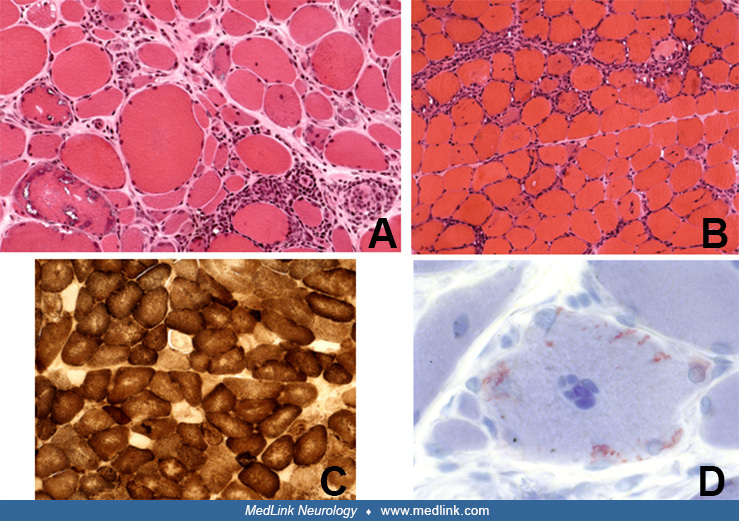
(A) Cross-sections of H&E-stained muscle biopsies demonstrate scattered inflammatory foci with lymphocytes invading or surrounding healthy-appearing muscle fibers combined with autophagic vacuoles with bluish-red material in areas not invaded by T cells and chronic myopathic features (increased connective tissue, atrophic and hypertrophic fibers). (B) In clinical inclusion body myositis, there is inflammation without vacuoles. (C) Characteristic in typical inclusion body myositis are COX-negative fibers and (D) crystal violet-positive congophilic amyloid deposits within the vacuoles. (Contributed by Dr. Marinos Dalakas.)

