Sleep Disorders
Sleeptalking
Jan. 18, 2025
MedLink, LLC
3525 Del Mar Heights Rd, Ste 304
San Diego, CA 92130-2122
Toll Free (U.S. + Canada): 800-452-2400
US Number: +1-619-640-4660
Support: service@medlink.com
Editor: editor@medlink.com
ISSN: 2831-9125
Toll Free (U.S. + Canada): 800-452-2400
US Number: +1-619-640-4660
Support: service@medlink.com
Editor: editor@medlink.com
ISSN: 2831-9125
Nearly 3,000 illustrations, including video clips of neurologic disorders.
Every article is reviewed by our esteemed Editorial Board for accuracy and currency.
Full spectrum of neurology in 1,200 comprehensive articles.
Listen to MedLink on the go with Audio versions of each article.
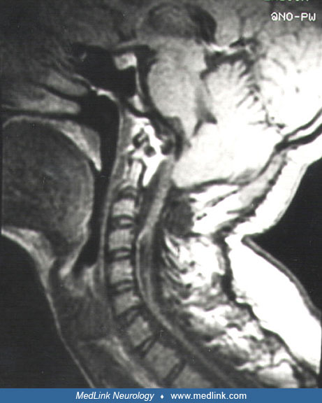
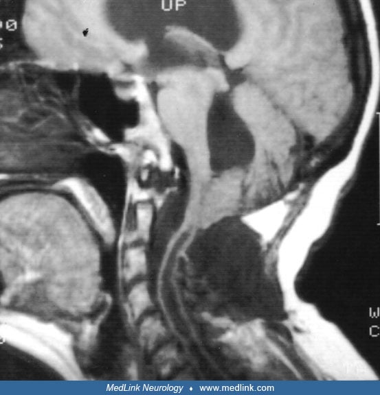
Sagittal brain MRI scan of a 40-year-old patient with a 10-year history of progressive syringomyelia. Examination: left Horner syndrome; lower motor neuron signs in upper limbs; spastic paraparesis; dissociated sensory loss in upper limbs, trunk, and face, with an onion-bulb distribution. There is a large cyst in the medulla with extension to the pons. (Contributed by Dr. Martín Nogués.)