Developmental Malformations
PEHO
Dec. 01, 2025
MedLink, LLC
3525 Del Mar Heights Rd, Ste 304
San Diego, CA 92130-2122
Toll Free (U.S. + Canada): 800-452-2400
US Number: +1-619-640-4660
Support: service@medlink.com
Editor: editor@medlink.com
ISSN: 2831-9125
Toll Free (U.S. + Canada): 800-452-2400
US Number: +1-619-640-4660
Support: service@medlink.com
Editor: editor@medlink.com
ISSN: 2831-9125
Nearly 3,000 illustrations, including video clips of neurologic disorders.
Every article is reviewed by our esteemed Editorial Board for accuracy and currency.
Full spectrum of neurology in 1,200 comprehensive articles.
Listen to MedLink on the go with Audio versions of each article.
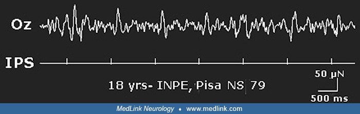
Visual evoked potentials at MO electrode are of a high amplitude. Overlapped is the schematic representation of average VEPs amplitude for 10 controls. Checks of 20 minute of arc; reversal rate of 1.7 Hz; contrast of 57%. MO = mid occipital electrode. (Contributed by Dr. Renzo Guerrini.)