Neuromuscular Disorders
Becker muscular dystrophy
Feb. 09, 2026
MedLink, LLC
3525 Del Mar Heights Rd, Ste 304
San Diego, CA 92130-2122
Toll Free (U.S. + Canada): 800-452-2400
US Number: +1-619-640-4660
Support: service@medlink.com
Editor: editor@medlink.com
ISSN: 2831-9125
Toll Free (U.S. + Canada): 800-452-2400
US Number: +1-619-640-4660
Support: service@medlink.com
Editor: editor@medlink.com
ISSN: 2831-9125
Nearly 3,000 illustrations, including video clips of neurologic disorders.
Every article is reviewed by our esteemed Editorial Board for accuracy and currency.
Full spectrum of neurology in 1,200 comprehensive articles.
Listen to MedLink on the go with Audio versions of each article.
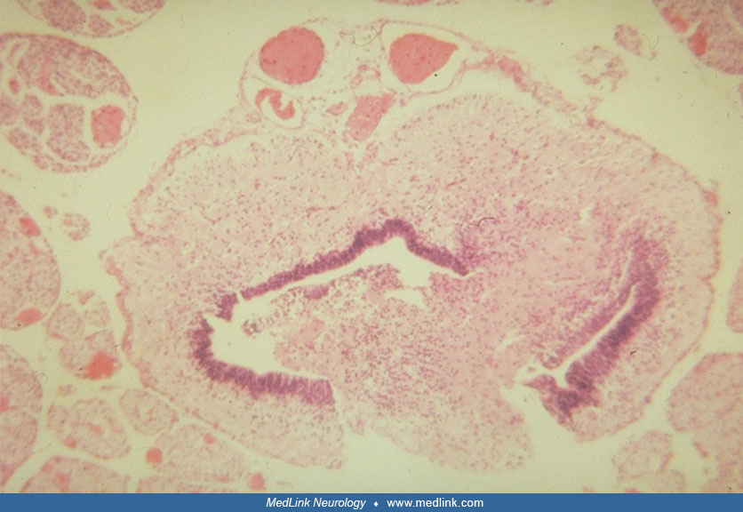
The ependyma of the central canal is discontinuous and divided into two portions with intervening neural tissue; the central canal is also much larger than expected at this age. Ventral horns are fused in the ventral midline and motor neurons are few. A well-formed dorsal median raphe separating the dorsal columns of the 2 sides is absent. (Hematoxylin-eosin; x40) (Used with permission. Sarnat HB. Cerebral dysgenesis. Embryology and clinical expression. New York: Oxford Univ Pr, 1992.)


