
Sleep Disorders
Sleep-related urologic dysfunction
Jul. 06, 2026
MedLink, LLC
3525 Del Mar Heights Rd, Ste 304
San Diego, CA 92130-2122
Toll Free (U.S. + Canada): 800-452-2400
US Number: +1-619-640-4660
Support: service@medlink.com
Editor: editor@medlink.com
ISSN: 2831-9125
Toll Free (U.S. + Canada): 800-452-2400
US Number: +1-619-640-4660
Support: service@medlink.com
Editor: editor@medlink.com
ISSN: 2831-9125
Worddefinition
At vero eos et accusamus et iusto odio dignissimos ducimus qui blanditiis praesentium voluptatum deleniti atque corrupti quos dolores et quas.
Cerebral arteriovenous malformations are congenital vascular malformations in the brain that are the underlying cause of 1% to 2% of all strokes, 4% of strokes in young adults, 9% of subarachnoid hemorrhages, and 4% of intracerebral hemorrhages (03; 50). Brain arteriovenous malformation detection rate in a random-effects meta-analysis was 1.31 per 100,000 person-years, and in the largest case series, the prevalence of brain arteriovenous malformation-associated aneurysms was 10% to 20% (26). With regards to intracerebral hemorrhage, there is a 9.8% per year risk of cerebral arteriovenous malformations hemorrhage in patients with intranidal aneurysms, which is higher than the overall population of patients with cerebral arteriovenous malformations (26).
Though mostly asymptomatic, these lesions come to clinical attention with a variety of neurologic presentations, including headaches, seizures, progressive neurologic deficits, or by incidental discovery (20). Short-term morbidity and mortality associated with arteriovenous malformations are typically low, although poor outcomes may occur in the long-term given the cumulative risk of hemorrhage. In determining the need for intervention, key morphologic and clinical characteristics, such as age, size, location, vascular features, and, most importantly, the estimated risk of hemorrhage, are considered (05). When published in 2013, the first large-scale randomized clinical trial comparing medical and interventional management of unruptured brain arteriovenous malformations, the ARUBA trial, showed that medical management alone remained superior to interventional therapy for the prevention of death or symptomatic stroke, even up to 5 years follow-up (49). Since that time, there has been burgeoning research into interventional therapy options, with both invasive and noninvasive treatment modalities, including surgical resection, embolization, and radiation therapy. Intervention for unruptured brain arteriovenous malformations remains controversial, though studies have suggested more promising functional outcomes and safety with intervention than initially established in the ARUBA trial (02). The purpose of this article is to discuss the clinical manifestations, pathophysiology, diagnosis, prognosis, and management of cerebral arteriovenous malformations.
|
• Cerebral arteriovenous malformations are congenital vascular malformations in the brain that result in direct connections between arteries and veins, without intervening capillary beds. | |
|
• Cerebral arteriovenous malformations are relatively uncommon, but a notable, cause of intracranial hemorrhage, especially in younger adults. | |
|
• Most cerebral arteriovenous malformations are asymptomatic and are discovered incidentally with neuroimaging. | |
|
• Cerebral arteriovenous malformations are reliably identified by CT and MR angiography, but conventional angiography remains the definitive diagnostic modality. | |
|
• Interventions for cerebral arteriovenous malformations include surgical resection, stereotactic radiotherapy, and endovascular embolization. | |
|
• Management decisions and risk prognosis differ between unruptured and ruptured cerebral arteriovenous malformations. |
Vascular malformations of the central nervous system (CNS) have been described in the literature dating back to the 19th century. With advances in imaging modalities and microsurgical techniques, the understanding of and treatment options for these lesions have expanded. Modern categorization and nomenclature of CNS vascular malformations was established by McCormick in 1966, using histopathologic features to classify each lesion as a venous angioma, cavernous malformation, capillary telangiectasia, or arteriovenous malformation. The Spetzler-Martin grading system (Table 1) for cerebral arteriovenous malformations takes into account major factors influencing the risk of surgical resection and hemorrhage (76). It is the most commonly used grading system, helping clinicians make treatment decisions and offering a standardized classification terminology. A supplementary scoring system was published in 2010 taking into account additional components: age at resection, hemorrhage before resection, and diffuseness of the arteriovenous malformations nidus. This supplementary grading scale (also known as the Lawton-Young Grading System) was found to be more accurate at predicting neurologic patient outcome than the Spetzler-Martin system alone and further clarified surgical risk stratification (51). It was further validated, even with a higher proportion of high-grade arteriovenous malformations; however, perforators play important role on the outcome (34). During arteriovenous malformation resection, there is significant difficulty with coagulation of these perforators, and these vessels can retract into the white matter. This is seen with large complex arteriovenous malformations, and the perforators that were studied were medial lenticulostriates, insular lenticulostriates, thalamoperforators, and brainstem perforators (34).
|
Characteristic |
Number of points assigned |
|
Size of arteriovenous malformation |
1 point |
|
Location |
0 points |
|
Pattern of venous drainage |
0 points |
|
| |
|
• Most cerebral arteriovenous malformations are asymptomatic and discovered incidentally. | |
|
• Neurologic presentations of cerebral arteriovenous malformations vary, including seizures, headaches, and hemorrhage. | |
|
• The annual hemorrhage rate of cerebral arteriovenous malformations ranges from 1% to 3% but varies based on size, location, and morphology. |
Most cerebral arteriovenous malformations (cAVMs) are asymptomatic and are discovered as an incidental radiographic finding. They are usually diagnosed between the first and fourth decade of life but have a bimodal peak: one in childhood and again at age 30 to 50 (28; 20). If symptoms are present, they vary based on age, size, vascular features, and lesion morphology location (48). Symptoms may include seizures, headaches, focal neurologic manifestations, or resultant hemorrhage. The most dreaded complication and presentation of cerebral arteriovenous malformations is intracranial hemorrhage (48; 20).
A large meta-analyses (n = 3923) estimating the natural history of cerebral arteriovenous malformations reported an annual hemorrhage rate of 3% (31). Earlier studies reported the average annual mortality in patients with untreated cerebral arteriovenous malformations from 0.7% to 1% (58; 08). Another patient-level meta-analysis published in 2014 found an overall annual rate of intracranial hemorrhage of 2% (42).
Due to the variability of locations in cerebral arteriovenous malformation-related hemorrhage, clinical presentation can vary considerably. Cerebral arteriovenous malformations are generally located in the brain parenchyma, resulting in parenchymal hemorrhages. However, secondary subarachnoid, intraventricular, or subdural extension may occur. More superficially located arteriovenous malformations may exhibit a pattern of hemorrhage that masquerades as aneurysmal or convexal subarachnoid hemorrhage. Moreover, arteriovenous malformations that have deep draining systems can lead to primary intraventricular hemorrhage. Hemorrhages may be clinically silent and can be incidentally discovered on subsequent imaging.
Numerous risk factors affect the risk of hemorrhage. According to one of the largest systematic reviews, the annual hemorrhage rate is 2.2% for unruptured arteriovenous malformations and 4.5% for ruptured arteriovenous malformations (31). Children were not at higher risk for subsequent hemorrhage after initial hemorrhage compared to adults (27). However, clinically silent hemorrhage visualized on neuroimaging is a risk factor for subsequent hemorrhage (33). Certain anatomical features, such as the presence of an associated aneurysm, exclusively deep venous drainage, and deep subcortical brain location, all confer an increased risk of hemorrhage (41; 15; 78; 16; 33). Additionally, certain angio-architectural features of brain arteriovenous malformations have been found to confer an increased risk of hemorrhage, for which other features provide protective benefit. For example, one study showed that a larger diameter of the nidus and the presence of venous drainage alterations were protective (with OR = 0.54 and 0.40, respectively), as compared to the presence of prenidal aneurysms, which increased the risk of hemorrhage (with OR = 4.69) (68). Arteriovenous malformation size does not appear to influence the risk of hemorrhage but does influence the surgical risk associated with treatment. During pregnancy and the puerperium period, there is an increased risk of intracerebral hemorrhage for women with arteriovenous malformations (45). Additional risk factors continue to be identified, such as variants of APOE E4 being associated with a 4-fold higher risk of intracerebral hemorrhage (64).
Brain arteriovenous malformations often present with seizures. Though the etiology is not entirely clear, mechanisms include increased venous back pressure related to outflow obstruction, periarteriovenous malformation nidus gliosis, and cortical irritation from prior hemorrhage (25; 28). The majority of seizures are simple partial or complex partial seizures; however, the rate of generalized tonic-clonic seizures ranges from 27% to 35% (59; 36). Superficially temporal or frontal lesion locations correlates with clinical seizure presentation (28). According to a prospective population-based study in Scotland (n = 229 patients) with arteriovenous malformations, the 5-year risk of first-ever seizure after presentation was higher when presenting with intracranial hemorrhage or focal neurologic deficit than for incidental arteriovenous malformations (23% vs. 8%). Moreover, 58% of these patients were at a higher risk of developing subsequent epilepsy (38).
Headaches can present as the initial symptom of cerebral arteriovenous malformations. As the incidence of arteriovenous malformation-associated headache is unknown, there was a study that 0.2% of patients who underwent neuroimaging for headache, in the setting of a normal neurologic examination, were found to have an arteriovenous malformation (48; 23). A risk factor for arteriovenous malformation-associated headache is occipital arteriovenous malformation location, and patients can have concurrent visual symptoms (eg, field cuts, blurring, scintillations, and diplopia). The following have been proposed as mechanisms underlying the pathophysiology of arteriovenous malformation-associated headache: increased intracranial pressure, steal phenomenon, and cortical spreading depression (CSD). Increased intracranial pressure activates the trigeminovascular system, and the high pressure can trigger a headache. Patients with a history of migraine have increased sensitivity to small increases in intracranial pressure and are more likely to experience headache (23). Hemodynamic changes, such as hypoperfusion, and mechanical stimulation secondary to arteriovenous malformation can activate cortical spreading depression and the trigeminovascular system (23). Arteriovenous malformation nidus can serve as an area of focus that triggers cortical spreading depression, which determines headache lateralization and aura.
Focal neurologic deficits can be seen in patients with cerebral arteriovenous malformations; however, the incidence is unknown. Its significance depends on the lesion’s location and adjacent structures. It has been hypothesized that a vascular steal syndrome may be related to focal deficits, as well as more obvious effects from hemorrhage or post ictal (48).
The prognosis of intracerebral hemorrhage due to arteriovenous malformations is more favorable than intracerebral hemorrhage without arteriovenous malformation (52). Annual rebleeding risk with arteriovenous malformation ranges from 0.9% to 4%. Mortality associated with the initial bleed is low, ranging from 1.5% to 9%. One study with arteriovenous malformation-related intracerebral hemorrhage (34 out of 342 total intracerebral hemorrhage patients) reported that patients with ruptured arteriovenous malformations had higher odds of ambulatory independence at discharge (OR 4.4) compared to patients with other causes of intracerebral hemorrhage (52). However, given the annual cumulative risk of hemorrhage with arteriovenous malformations, the long-term outlook tends to be less favorable. Recurrent hemorrhage is estimated to be around 1% in 4 to 7 years and 2% annually after the first year of hemorrhage. It is well known that with repeated hemorrhages, morbidity and mortality rise substantially (48).
Patients with intracerebral hemorrhage secondary to cerebral arteriovenous malformations rupture tend to be younger, with lower pre-stroke and admission blood pressures and higher admission Glasgow Coma Scale (GCS) scores. They are more likely to have lobar location of intracerebral hemorrhage compared to all patients with spontaneous intracerebral hemorrhage (50). Though cerebral amyloid angiopathy hemorrhage can also be lobar, this would tend to affect patients above the age of 60. The ruptured brain arteriovenous malformations prognostic (RAP) score was proposed to help to stratify the risk of poor long-term outcome after cerebral arteriovenous malformation rupture. The RAP score was a stronger predictor of a poor long-term outcome and inpatient mortality than the intracerebral hemorrhage score. For a RAP score 6 or higher, sensitivity and specificity for predicting poor outcome were 76.8% and 90.8%, respectively (73); however, more studies are needed for further validation of this score.
Other prognostic factors associated with the development of intracerebral hemorrhage in patients with arteriovenous malformations include older age, exclusively deep venous drainage and location, and associated arterial vasculature. The first validated risk prediction tool, the R2eD AVM score, was developed for hemorrhage risk stratification. The score looks at race (nonwhite vs. white), exclusive deep location, arteriovenous malformation size (small or large), venous drainage, and monoarterial feeding (1 vs. > 1 feeding artery) (24). If a patient scores a 6, indicating that all risk factors are present, there was a 78% median hemorrhage presentation risk (24). External validation of the R2eD AVM score was performed through a retrospective 10-year study in 122 patients with cerebral arteriovenous malformations and was found to be acceptable (07).
Patients presenting with progressively worsening neurologic deficits have the worst prognosis as these symptoms indicate a large arteriovenous malformation that is difficult to manage and treat. Additional tools have now been established, such as the VALE scoring system. The VALE system identified four risk factors (ventricular system involvement, venous aneurysm, deep location, and exclusively deep drainage) and serves as an additional tool to provide a tailored approach to identify high-risk individuals who may benefit from earlier intervention (13).
A 33-year-old right handed nurse is referred for recurrent headaches that have been increasing in frequency. These are described as episodic, retro-orbital throbbing with nausea, light and noise sensitivity, and occurring three to four times per month. They are occasionally preceded by a visual aura of “sparkling lights” that ascend in the peripheral visual field and dissipate. The patient takes over the counter analgesics with some relief, but headaches can last for hours or several days before dissipating. They have no other significant past medical history and are on no prescription medications. Their social and family history are otherwise unremarkable. On exam, vital signs are normal. There is no papilledema. Reflexes are brisk throughout. The remainder of the neurologic exam is otherwise normal. Their primary care provider ordered a brain MRI, which revealed a tangle of vessels centered in the region of the right posterior cingulate gyrus measuring 1.6 cm x 1.6 cm, predominantly fed by the right anterior cerebral artery with both superficial and deep venous drainage. There were no associated signs of recent or past hemorrhage.
A follow-up cerebral angiogram confirmed the morphology and drainage patterns, largely to the superior sagittal sinus, but also some deep drainage to the straight sinus and internal cerebral veins. There were no intranidal aneurysms.
After review of the imaging, this was determined to be a Spetzler-Martin grade 3 arteriovenous malformation (1 point for size less than 3 cm, 1 point for deep venous drainage, and 1 point for potentially eloquent location). Based on the results of the ARUBA clinical trial, medical management and observation of this unruptured arteriovenous malformation is associated with better outcomes compared to procedural intervention. Diagnosis of this patient’s headaches is consistent with migraine with aura, with the arteriovenous malformation likely representing an incidental finding.
Etiology. Arteriovenous malformations were traditionally considered sporadic congenital vascular malformations of the nervous system; however, there are reports of de novo arteriovenous malformation formation. Although dysregulated vasculogenesis may result in congenital appearance, it is now possible that this may occur in childhood or even adulthood (17). Some rare cases of familial arteriovenous malformations have been reported in the literature, but it is unclear if they reflect a clear genetic connection or pure coincidence (83).
Some genetic syndromes are associated with arteriovenous malformations, with the most common being hereditary hemorrhagic telangiectasias, also known as Osler-Weber-Rendu syndrome. Hereditary hemorrhagic telangiectasias is an autosomal dominant condition that involves haploinsufficiency of signaling genes for transforming growth factor beta (20). The presence of numerous cerebral arteriovenous malformations, otherwise uncommon, is highly predictive of hemorrhagic telangiectasias (06). The Curacao Criteria were developed to support a clinical diagnosis of hereditary hemorrhagic telangiectasias, including the presence of spontaneous recurrent epistaxis, multiple telangiectasias in characteristic sites of oral mucosa and appendages, arteriovenous malformations in any location, or a first-degree relative with hereditary hemorrhagic telangiectasias (Table 2). Pulmonary arteriovenous malformations in hereditary hemorrhagic telangiectasias may also be a source for extracardiac shunting and paradoxical emboli causing arterial strokes. Studies have shown that malformations in hereditary hemorrhagic telangiectasias develop as a result of KRAS-induced activation of the MAPK-ERK signaling pathway in brain endothelial cells (56). Ongoing work involving next generation sequencing is aiming to detect a molecular basis and pathogenic mechanisms involving angiogenesis as an etiology for sporadic cases of arteriovenous malformations (71).
|
Criterion |
Definition |
|
Epistaxis |
Spontaneous, recurrent |
|
Telangiectasias |
Multiple in typical sites including oral mucosa (lips, mouth), nose, ears, fingers |
|
Arteriovenous malformations |
Any location including: |
|
Family history |
First degree relative with HHT |
|
| |
A congenital nonhereditary neurocutaneous syndrome is Wyburn Mason syndrome, which is also known as racemose angioma. It presents with multiple arteriovenous malformations primarily affecting the face, retina, and CNS (typically midbrain) (74). Cerebrofacial arteriovenous metameric syndrome (CAMS) has been used as a classification system that describes where arteriovenous malformations are expressed in the cerebral, orbital, and facial region. Wyburn-Mason syndrome is designated as CAMS2, as it involves cortex and diencephalon, optic chiasma, optic nerve, retina, sphenoid, maxilla, and cheek (74).
Pathogenesis. Arteriovenous malformations have unique hemodynamics with direct connections between arteries and veins, without intervening capillary beds. These connections contain the nidus; a tangle of abnormal dilated channels that are neither arterial nor venous. Arterial blood is shunted through the nidus with elevated flow in both ends and pressure on the venous end, predisposing the vessel towards inflammation and thrombosis (20). The arterial supply or venous drainage may be composed of single or multiple vessels. Surrounding gliotic brain tissue is usually admixed with the vascular tangle, along with occasional micro or macrocalcifications (20).
In cerebral arteriovenous malformations, although draining veins are large and easily recognizable on imaging, the arteries feeding them can be small, sometimes referred to as “cryptic” (69). These feeding arteries have medial hypertrophy and endothelial thickening, narrowing their lumen, which can then become stenotic and even occluded (57). The arteries within the cerebral arteriovenous malformations are usually deficient in the muscularis layer. Arteries within the lesion can also be large, but lined with thin walls, due to poorly developed internal elastic lamina and media. Examination of adjacent and intralesional parenchyma reveals abnormal gliosis (47). Angiographic findings indicate that the arterial portions of arteriovenous malformations shunt blood directly toward the venous components. This rapid shunting of blood into large, dilated, draining veins increases blood flow, promoting the formation of aneurysms.
Arteriovenous malformations tend to grow slowly after initial formation. The feeding arteries and draining veins grow in caliber, recruiting adjacent vessels into its architecture. One theory as to why arteriovenous malformations grow is the inability of the abnormal vessel group to accommodate arterial pressure (47). Other important anatomic features associated with hemorrhagic presentation include the presence of drainage into the galenic system, venous outflow obstruction, and deep or infratentorial location (01).
Cerebral arteriovenous malformations can also cause small recurrent intracerebral hemorrhages. These bleeds cause loss of adjacent brain parenchyma as hematoma and necrotic tissue are reabsorbed. The space created by this process may allow room for an arteriovenous malformation to abut and grow. Pulsation of the arteriovenous malformation is also thought to slowly damage surrounding tissue (48). In addition, although endothelial dysfunction has been thought to be a contributing case of hemorrhage risk in cerebral arteriovenous malformations, neuroinflammation and hypoxia likely play a role in the progression of malformations. One study found that reactive astrocytes underwent changes promoting vascular instability, which in turn further promoted endothelial dysfunction, with COX-2 upregulated in the microenvironment of malformations, leading researchers to explore the potential role of COX-2 inhibitors as a treatment target (82).
|
• Cerebral arteriovenous malformations occur rarely, in about 0.1% of the population, but account for at least 1% to 2% of strokes and up to 9% of all nontraumatic subarachnoid hemorrhages. |
The overall incidence of brain arteriovenous malformations is about 1.10 to 1.42 cases per 100,000 (12). Ninety percent of cerebral arteriovenous malformations are supratentorial with the rest located in the posterior fossa (48). Brain arteriovenous malformations account for 1% to 2% of all strokes, 4% of strokes in young adults, 9% of subarachnoid hemorrhages, and 4% of intracerebral hemorrhages (03; 50). The estimated risk of hemorrhage in an untreated, unruptured arteriovenous malformation is 1% to 3% per year (12). With respect to ruptured arteriovenous malformations causing hemorrhage, this serves as a predictor for subsequent hemorrhage.
Morphologic risk factors associated with an increased risk of hemorrhage in arteriovenous malformations include venous drainage pattern, fewer draining veins, nidus location and size, presence of associated arterial aneurysms, or venous varices (12). As aforementioned, the most common genetic cause of cerebral arteriovenous malformations is autosomal dominant hereditary hemorrhagic telangiectasia. About 10% to 25% of patients with hereditary hemorrhagic telangiectasia will develop at least one cerebral arteriovenous malformation.
As cerebral arteriovenous malformations are thought to be due to sporadic genetic mutations, no information is available at this time for prevention. For family members of patients with hereditary hemorrhagic telangiectasias, screening is indicated, but otherwise, there is no role for routine screening of the general population (83). As with any autosomal dominant condition, genetic counseling is strongly encouraged for patients and family members with consideration of hereditary hemorrhagic telangiectasia.
Arteriovenous malformations fall under a large and heterogenous group of cerebral vascular malformations, including venous angiomas, cavernous angiomas (cavernomas), complex intracranial aneurysms, and capillary telangiectasias. Venous angiomas comprise a group of anomalous veins, usually separated from normal brain parenchyma. These vessels do not fill during the arterial phase of cerebral angiography, which differentiates them from arteriovenous malformations.
Cerebral cavernous malformations, also referred to as cavernous angiomas, cavernous hemangiomas, or cavernomas, are well-encapsulated masses of sinusoidal vessels separate from brain parenchyma. These lesions are not connected to the normal cerebral circulation and do not opacify during the arterial phase of cerebral angiography, differentiating them from arteriovenous malformations. Cavernous angiomas are often sporadic or may be associated with familial cases (syndromes of cerebral cavernous malformations including CCM1, CCM2, and CCM3 genetic loci) (22; 43; 84).
Capillary telangiectasias are small lesions in which capillaries are separated from each other by normal brain parenchyma commonly located in the pons and brainstem. These lesions are multiple and quite small compared to arteriovenous malformations, and they do not fill during contrast angiography (10).
Dural arteriovenous fistulas are defined as vascular abnormalities in which arteries arise from branches of the carotid or vertebral arteries and drain directly into the dural leaflets of the venous sinuses (89). These tend to be supratentorial, although the most common location is the transverse-sigmoid sinus junction. Etiology can be secondary to dural sinus thrombosis, trauma, infection, or prior craniotomy (89). These are distinguished from arteriovenous malformations, as there is no parenchymal nidus, and the arterial supply involves the dura mater (65).
Cerebral proliferative angiopathy (CPA) is a rare and progressive vasculopathy characterized by normal brain parenchyma interspersed with abnormal vascular channels. Cerebral proliferative angiopathy may be confused with cerebral arteriovenous malformations, but there are angio-morphological features that help distinguish them. Distinguishing features of cerebral proliferative angiopathy include an absence of dominant feeders or flow-related aneurysms, proximal stenoses on the feeding arteries, extensive transdural supply to both healthy and pathological tissues, large size, presence of capillary angioectasia, and moderately enlarged veins. On perfusion-weighted MRI, there is increased blood volume within the nidus, with an increased mean transit time of flow indicative of capillary and venous ectasia and an area of hypoperfusion. In arteriovenous malformations, mean transit time is decreased, and the perinidal areas are not as severely hypoperfused as in cerebral proliferative angiopathy (32).
Moyamoya syndrome is characterized by the development of extensive lenticulostriate collaterals due to progressive steno-occlusive disease of the distal internal carotid arteries. It may be acquired as a result of severe intracranial arterial disease (eg, carotid atherosclerosis, sickle cell disease, antiphospholipid antibody syndrome, and thyroid disease) or genetically mediated (ie, Moyamoya disease) with a bimodal age of distribution in children and young adults. It is distinguished from cerebral arteriovenous malformations with delayed perfusion and transit time, including angiographic filling of the small vessel collateral network (ie, moyamoya meaning “puff of smoke”) and does not involve any direct arteriovenous filling. Like cerebral arteriovenous malformations, moyamoya may be a secondary cause of intraparenchymal hemorrhage due to rupture of friable lenticulostriate vessels.
In patients who undergo CT angiography for evaluation of secondary intracerebral hemorrhage, there are vascular mimics that can present with the spot sign. In a retrospective chart review of 96 intracerebral hemorrhage patients (between January 2002 and May 2007) who underwent CT angiography, spot sign mimics, which were defined as 1 to 2 mm foci of enhancement within the parenchymal hematoma, were identified with micro-arteriovenous malformation and moyamoya (29). The patients were also younger at the time of presentation. In the patient with moyamoya, there was an enhancing focus within the inferior Basal Ganglia/Nucleus accumbens that was found to be a pseudoaneurysm arising from the lenticulostriate artery (29). The study also demonstrated that the presence of a vessel extending from the brain or ventricular surface into the hematoma suggests an underlying vascular formation.
A developmental venous anomaly is characterized by a cluster of radially arranged medullary venous channels that converge and drain into a collecting vein, resulting in the typical “caput medusa” appearance on imaging (54). In the evaluation of developmental venous anomaly through cerebral angiography, there is a normal arterial and capillary phase along with a wedge-shaped collection of dilated medullary veins that drain into an enlarged collection of veins in the late venous phase. Atypical features such as intraparenchymal hemorrhage or ischemia adjacent to a developmental venous anomaly should prompt further investigation to distinguish it from an arteriovenous malformation (54). Though controversial with regards to naming, arteriovenous malformations can drain into a large developmental venous anomaly and, thus, have been called “venous predominant parenchymal” arteriovenous malformations. Developmental venous anomalies are also often adjacent to a cavernous hemangioma.
|
• Cerebral arteriovenous malformations are reliably identified by CT and MR angiography. | |
|
• Digital subtraction angiography remains the gold standard for diagnosis. |
Cerebral arteriovenous malformations are typically diagnosed by neuroimaging and are most often incidental findings. Arteriovenous malformations are reliably identified by CT and MRI, including angiographic imaging (CTA and MRA). Digital subtraction angiography is used for a definitive diagnosis (20).
Noncontrast CT is limited in detecting arteriovenous malformations, but it can show certain characteristics supporting a diagnosis. Enlarged or calcified vessels along the margin of the hemorrhage or regions of increased density around the nidus, indicating an underlying vascular anomaly, can be seen (18). The overwhelming majority of patients with indeterminate results undergo CT angiography. Advances in adapting temporal coding into CT angiography techniques now permit better delineation of arterial, nidal, and venous components (20).
Advances in MR and MR angiography technology now make it comparable to CT and CTA in terms of accuracy of detecting arteriovenous malformations in intracerebral hemorrhage. Both CT and MR imaging provide information about the arteriovenous malformation as well as adjacent brain tissue, which is essential in determining treatment plans (20).
Adjunctive imaging studies, such as transcranial color duplex and regular Doppler, have been studied extensively and can help analyze the hemodynamic changes occurring pre- and postnidus, and can be used to assess for changes following treatment (09).
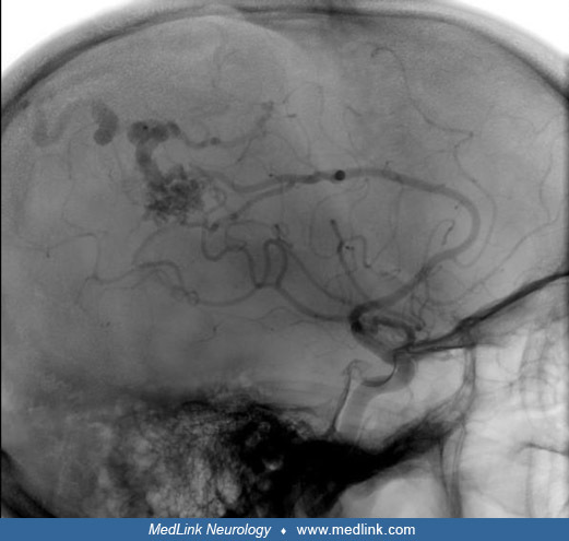
The gold standard for diagnosing arteriovenous malformations remains digital subtraction angiography (DSA), which is often pursued after CT or MR angiography identifies a suspicious lesion. Digital subtraction angiography is additionally used to help with treatment planning and follow-up.
|
• Arteriovenous malformation treatment aims to completely eliminate the nidus and the shunt. | |
|
• The primary treatment modalities are surgical excision, stereotactic radiosurgery, and endovascular embolization. | |
|
• Multimodality approaches are often used in a stage-wise process. | |
|
• The ARUBA trial compared conservative (nonsurgical) management to surgical intervention in unruptured cerebral arteriovenous malformations and found better outcomes (prevention of stroke or death) with conservative management at 5 years follow-up. |
The goal of arteriovenous malformation treatment is completely eliminating the nidus and arteriovenous malformation shunt, along with reducing morbidity and mortality associated with the natural course of these lesions. Following diagnosis and evaluation of an arteriovenous malformation, three treatment modalities are accepted: microsurgery or macro-surgery, stereotactic radiosurgery, and endovascular embolization. Endovascular embolization can also be performed before microsurgery and stereotactic radiosurgery to reduce bleeding risks and nidal volumes (20).
Intervention is indicated for ruptured arteriovenous malformations. Cerebral arteriovenous malformations with angiographic features suggesting an increased risk of recurrent hemorrhage, such as an associated aneurysm, are treated acutely, whereas other arteriovenous malformations are generally treated 4 to 6 weeks after the hemorrhage, as absorption of the hematoma and resolution of any surrounding edema improve access to the arteriovenous malformation for future intervention (05).
Surgical excision. Open surgical excision has a well-established history of use in treating arteriovenous malformations and offers the best chance for immediate cure (05). However, the surgery is often complicated and requires complex planning with angiographic review of the microarchitecture. Surgical risks associated with surgery include intraoperative hemorrhage and damage to adjacent cerebral and vascular structures. Successful surgery completely removes the rupture-prone malformed vessels and obliterates feeding and draining vessels. This modality results in immediate elimination of hemorrhage risk and complete eradication of the nidus. Introducing surgical adjuncts, such as functional MRI and diffusion tensor imaging-based tractography, as well as stereotactic neuro-navigational systems, allows for more accurate intervention and minimization of postoperative morbidity risk (20). New surgical techniques continue to be explored, such as use of exoscope during microsurgical resection to provide improved field of view and magnification of up to 24 times has been proposed along with use of indocyanine green video angiography (ICG-VA) to assist in observing the hemodynamics (81).
As discussed, the Spetzler-Martin scale is commonly used to establish surgical intervention risk. Arteriovenous malformations of larger size, deep venous drainage patterns, and near eloquent cortex have higher surgical risks and may require adjunctive presurgical interventions. The ARUBA trial results applied to Spetzler-Martin grade I and II arteriovenous malformations, and there is speculation that the high rate of morbidity and mortality in the study may have been due to a low rate of patients who received microsurgery (37). With regards to grade I and II arteriovenous malformations, it is unclear whether the results from the ARUBA trial can be applied to these particular classifications with respect to microsurgical treatment (37). In a study of 977 arteriovenous malformation patients, 155 ARUBA-eligible patients with Spetzler-Martin grades I and II arteriovenous malformations underwent microsurgical resection (86). Though complete obliteration was 99.2%, early disabling effects and permanent disabling deficits occurred in 9.3% and 3.4%, respectively.
Grade III arteriovenous malformation is associated with an operative morbidity risk of 16% due to significant variability in eloquence, size, and venous drainage (37). In a study of 174 resected cerebral grade III arteriovenous malformations, the Spetzler-Martin grading scale was modified to better assess outcomes (44). The modification was described as the following: III- (S1V1E1), III (S2V1E0), III+ (S2V0E1), and III* (S3E0V0); (S: size, V: deep venous drainage, E: eloquence). The operative morbidity risk was found to be III-, III, and III+ was 2.9%, 7.1%, and 14.8%, respectively. Surgery was favorable for both III- and III. In addition, poor outcomes were associated with perirolandic arteriovenous malformations, which demonstrates how eloquence can significantly affect operative morbidity risk.
Surgical intervention of grade IV and V arteriovenous malformations is associated with significant operative risk. Minor deficit risk was 20%, and major deficit risk was 7% and 12%, respectively (37). Preoperative stereotactic radiosurgery or embolization should be considered to decrease the operative risk. It was found that preoperative radiation for high-grade arteriovenous malformations reduced intraoperative blood loss and reduced hospital stay. Regarding embolization, an approach was developed to target high-flow feeding vessels for selective occlusion and avoid nidal penetration (72; 37). With reduced arteriovenous malformation, the patient could transition to treatment with microsurgery or stereotactic radiosurgery. It was found that there was a reduction in morbidity to less than 4% compared to approximately 8% with standard nidal occlusion, though there needs to be further validation (72; 37).
Stereotactic radiosurgery. Stereotactic radiosurgery is another treatment modality for arteriovenous malformations. It is particularly helpful for small lesions (less than 3 cm max diameter with volume less than 12 cm3), lesions near the eloquent cortex that could be damaged by surgery, or lesions deemed too risky due to anatomical or medical reasons (21). Targeted radiation is delivered over a period of years. Patients who undergo successful radiosurgery are protected from future hemorrhage once complete obliteration is achieved. Therefore, patients still undergoing radiotherapy, called the latency period, are still at risk of hemorrhage until the treatment is complete (62). Recommended radiation fraction doses and delivery schedules vary in the literature, as do the reported success rates with stereotactic radiosurgery. Radiation doses and arteriovenous malformation volumes can predict obliteration rates (40). Radiosurgery alone is efficacious in obliterating smaller arteriovenous malformations, whereas larger lesions require additional therapies. A scoring system used for predicting outcomes of arteriovenous malformation stereotactic radiosurgery is the modified radiosurgery-based arteriovenous malformation score. This score looks at nidus volume, age, and nidus location as a calculation (0.1 x nidus volume+ 0.02 x age + 0.5 x nidus location). The Virginia Radiosurgery AVM scale is a modified scoring, which incorporates prior hemorrhage rather than patient age, and a favorable outcome is defined as obliteration without post-stereotactic radiosurgery hemorrhage or permanent symptomatic radiation-induced complication (12).
Neurologic function after stereotactic radiosurgery appears preserved or improved in most patients with arteriovenous malformations (79). The most frequent complication post-stereotactic radiosurgery for arteriovenous malformation is radiation-induced changes (RICs) (12). These can manifest between 6 and 18 months after stereotactic radiosurgery as perinidal T2 weighted hyperintensities on MRI. Although most radiation-induced changes are asymptomatic and transient, 10% of all stereotactic radiosurgery-treated patients with arteriovenous malformation develop focal neurologic deficits, seizures, and headaches. Three percent of patients can have permanent neurologic deterioration related to radiation-induced changes (12). Risk factors associated with the development of radiation-induced changes include lack of prior arteriovenous malformation hemorrhage, repeat stereotactic radiosurgery, and deep arteriovenous malformation location. Other factors that may contribute to complications from stereotactic surgery include lesion location, target volume, and radiation dose to surrounding normal tissue (20). Cyst formation occurs in 1% to 3% of patients treated with stereotactic radiosurgery for arteriovenous malformation and can develop 6.5 years after intervention (12). It is thought to occur due to the formation of frail telangiectatic perinidal vessels that are susceptible to rupture, which promotes serum and protein exudation, edema accumulation, and eventual cyst formation. Risk factors for cyst development include high radiosurgical dose, large nidus volume, and lobar nidus location. About 70% of post-stereotactic radiosurgery cysts are asymptomatic, though surgical intervention may be necessary if patients become symptomatic or the cyst continues to enlarge (12).
Endovascular embolization. Endovascular treatment modalities utilize microcatheters to deliver microparticles or embolizing agents to obliterate feeding vessels in the target lesion (67). Though endovascular therapy has been used in addition to microsurgery and stereotactic radiosurgery, further advances in catheter technology and endovascular techniques have allowed for complete arteriovenous malformation obliteration at a rate of 13% to 57.1% (37). However, in the literature, embolization with curative intent as a stand-alone intervention continues to have mixed outcomes. One prospective cohort study comparing stand-alone endovascular therapy with medical management in unruptured arteriovenous malformations found that intervention with embolization was inferior to medical management in preventing the risk of long-term intracranial bleeding and death but did suggest a possible benefit to more targeted embolization techniques (14). Arteriovenous malformation characteristics to be considered are lesions less than 3 cm, noneloquent location, and fewer arterial pedicles. A prospective randomized trial comparing transvenous versus transarterial embolization showed that transvenous embolization may be more effective in terms of angiographic results at 3 to 6 months (63).
Palliative endovascular treatment may also be used to reduce vascular steal symptoms. Morbidity and mortality rates for attempted surgical resection alone of grade V arteriovenous malformations approach 50%, but this risk is significantly reduced with staged presurgical embolization (67). Liquid embolic agents, such as ethylene-vinyl alcohol copolymer (Onyx) and n-butyl cyanoacrylate, have demonstrated better vessel occlusion and polymerization kinetics (37). Endovascular surgery itself carries risks and should be used when it can significantly reduce the overall risk. Possible complications include new neurologic deficits, intraoperative and postembolization hemorrhage, and subsequent ischemic stroke (20). Predictive scores regarding procedural morbidity and mortality have been developed, such as the Buffalo score, Puerto Rico score, and arteriovenous malformation embocure score, though these have yielded mixed results on predictive efficacy (37).
Multimodality treatment strategies are often utilized for large or complex lesions. Combining different techniques reduces the risk of subsequent interventions, lowering overall treatment risk. Using the Spetzler-Martin grading scale as a guide, endovascular embolization can be followed by surgery, or it can be followed by stereotactic radiosurgery. The intention of multimodality intervention is complete lesion obliteration as partial obliteration increases hemorrhage risk (55). Several institutions have reported applying this model with radiographic obliteration rates of 38% to 83% and permanent neurologic morbidity rates of 4% to 14% (20). A case-control study reviewed patients who underwent embolization prior to stereotactic radiosurgery compared to stereotactic radiosurgery only and found no statistical differences in obliteration rates or complications. When undergoing multiple treatments, the risk of complications from each individual procedure should still be considered, as higher rates of new neurologic deficit in staged endovascular and surgical therapy was found compared to surgery alone (35).
The benefit of staged embolization before surgery for large arteriovenous malformations has been established (77; 61), but not all combination therapies are beneficial. Andrade-Souza and colleagues showed significantly decreased obliteration rate with embolization before radiosurgery compared to radiosurgery alone (04). In another retrospective study, embolization before radiosurgery did not lead to an improved rate of total obliteration nor reduction in delayed hemorrhage risk (39). However, a multicenter study of patients who underwent stereotactic radiosurgery with or without prior embolization found a trend towards reduced incidence of primary outcomes (nonfatal hemorrhagic stroke or death) in the combined embolization and stereotactic radiosurgery when compared to stereotactic radiosurgery alone in large volume (> 10 mL) arteriovenous malformations (46). Given conflicting evidence, an individualized approach and discussion of risks and benefits must be taken.
Conservative medical management (observation). There remains a paucity of randomized clinical trials comparing medical (conservative) versus surgical (interventional surgery, microsurgery, or endovascular) in unruptured cerebral arteriovenous malformations. Although new trials are now underway, for years the ARUBA trial (A Randomized trial of Unruptured Brain Arteriovenous malformations) was the only randomized clinical trial that compared medical (conservative) versus surgical (50). Patients with unruptured cerebral arteriovenous malformations were assigned to medical versus interventional treatment, including surgery, radiotherapy, or endovascular therapy. In 2013, after collecting outcome data on 223 patients followed for a mean of 33 months, randomization was halted early. Composite rates of symptomatic stroke (ischemic and hemorrhagic) and death were higher in the interventional compared to medical treatment group (31% vs. 10%). Rates of neurologic disability were also lower in conservatively treated patients. Deaths occurred in 2% of patients in both groups. Criticism surrounding the study includes the variable nonstandardized approaches used in the interventional treatment arm, a small sample size, and a relatively short follow-up period. This was remedied with a 5-year follow-up study finally done, which found that after a mean follow-up of 50 months, medical management still remained superior to medical management with interventional therapy for the prevention of death or symptomatic stroke in patients with an unruptured cerebral arteriovenous malformation (49). However, studies have continued to call into question the generalizability and validity of the ARUBA trial results, and unruptured brain arteriovenous malformations management remains a controversial topic (02). The TOBAS trial is an ongoing prospective multicenter, randomized controlled trial, and registry also aimed at investigating the best management for brain arteriovenous malformations, including both unruptured and ruptured (70). The trial includes both randomized controlled trials and a registry of conservatively managed patients with the primary outcome of the randomized controlled trial being death or disabling stroke at 10 years.
Monoclonal antibody. Bevacizumab, which is a monoclonal antibody against VEGF, has been used in the treatment of perilesional edema related to radiation necrosis. A single-arm pilot study sought to test bevacizumab for treating cerebral arteriovenous malformations (53). Patients with large brain arteriovenous malformations that were deemed unsuitable for invasive treatment were included in the study. Although bevacizumab was well tolerated, there were no radiographic changes over the 52-week study period. However, this study was underpowered due to difficulty with funding and patient recruitment.
A case report in 2012 evaluated the use of bevacizumab to control perilesional edema refractory to corticosteroid therapy after Gamma Knife surgery for intracranial arteriovenous malformation (85). Significant adverse effects to consider are the following: seizure (6%), fatigue (3.6%), hypertension (8.3%), neutropenia, diarrhea, and pneumonia (1.2%). With regards to neurosurgical complications, bevacizumab has also been associated with worse wound healing if given prior to the procedure (85). Of note, a significant adverse effect of bevacizumab is intravascular hemorrhage. Therefore, this use of medication should be weighed against the patient’s risk of hemorrhage secondary to their underlying arteriovenous malformation. Patients must be closely followed with imaging and neurologic examinations.
Other therapies. Therapeutic investigations have targeted inflammatory markers such as matrix metalloproteinases. Tetracyclines, such as doxycycline and minocycline, have been investigated to inhibit nonspecific inhibitors that can increase vascular stability and reduce risk of spontaneous hemorrhage. However, there is no evidence of clinical efficacy or hemorrhagic risk reduction in patients (60).
Thalidomide and lenalidomide have been explored as a potential therapy through an increase in PDGFB levels, which could strengthen vessel walls and reduce intracerebral hemorrhage possibility (60).
Rapamycin, an mTOR inhibitor that regulates cellular proliferation and survival, has been used as an off-label treatment for medical management of large superficial arteriovenous malformations. The mechanism of action involves forming a complex with intracellular FK binding proteins (eg, FKPB12), which inhibits mTOR and arrests the G1 phase of the cell cycle and, thus, stops blood vessel formation (30). Duration of treatment ranged from 3 to 24.5 months, with the median duration being 12 months across cases. In this particular study, the maximum dose was 40 mg, and levels were maintained between 3 to 20 ng/mL (30). Adverse reactions included hyperlipidemia, minor opportunistic infection, and oral mucositis and oral ulcers.
Bone marrow/monocyte transfusion has been explored as a potential therapy to reduce the severity of brain arteriovenous malformations. In mouse models, bone marrow-derived cells participate in VEGF-stimulated brain angiogenesis, and injection of normal human monocytes can rescue defective vessel formation and heart function (60).
The KRAS mutation has also been a target of potential treatment. As most brain arteriovenous malformations have been found to have somatic KRAS mutations, one study identified lovastatin to be a possible pharmacological treatment option. Using human umbilical vein samples, the KRAS mutation was found to induce endothelial-mesenchymal transition (EndMT) through activation of the ERK-TGF-β/ BMP-SMAD4 signaling pathway. This study then demonstrated that lovastatin inhibits the EndMT by suppressing TGF-β/BMP pathway activation and SMAD4 acetylation (87). Further exploration of this novel treatment modality is needed.
Data from case reports and series showed that the overall recurrence of arteriovenous malformations was 2.7% in adult patients, with an average age of recurrence at 17.8 years and overall mean time from the obliteration of original arteriovenous malformation to recurrence being 4.22 years (75).
Patients are at risk for developing arteriovenous malformation-associated epilepsy. Through an interventional approach, the risk of seizures can be lowered by the removal of epileptogenic foci. However, there can also be an increased risk of seizures because of surgery, endovascular embolization, and stereotactic radiosurgery (38). A prospective, population-based, observational cohort study analyzed arteriovenous malformation treatment versus conservative management on the risk of a first seizure, the risk of epilepsy, and the chances of achieving 2-year seizure freedom (38). There was no difference in the 5-year risk of seizures with arteriovenous malformation treatment or conservative management. In patients who present with intracerebral hemorrhage secondary to arteriovenous malformation, the increased risk of seizures was secondary to larger hematoma volume. The risk of seizure following arteriovenous malformation treatment for unruptured incidental arteriovenous malformations was higher than with conservative management, though this was not statistically significant. Hence, this may be consistent with arteriovenous malformation treatment, which independently worsened short-term outcomes for adults with unruptured arteriovenous malformations.
There has been more literature discussing the association between cerebral arteriovenous malformations and pregnancy. Previously, it has been speculated that fluctuating hormone levels in pregnancy may play a role in arteriovenous malformation-related intracranial hemorrhage during and after pregnancy. However, a cohort cross-over design evaluated women who underwent pregnancy and delivery from the healthcare cost and utilization project state inpatient databases for California (2005 to 2011), Florida (2005 to 2014), and New York (2005 to 2014). Five hundred sixty-eight women out of 4,022,811 had an arteriovenous malformation. Intracerebral hemorrhage rates during pregnancy and puerperium were 6355.4 and 14.4 per 100,000 person-years for women with and without arteriovenous malformation, respectively (45). It was found that in women with a cerebral arteriovenous malformation, the risk of intracranial hemorrhage increased 3.27-fold during pregnancy and puerperium compared with nonpregnant period (45). This increase in hemorrhage during pregnancy was also reported in a 2023 meta-analysis that saw an 11% risk in pregnant females versus 6.7% in nonpregnant females (19). The risk of rupture of a previously unruptured arteriovenous malformation in pregnant women has been estimated to be 3.5% per year versus 3.1% in nonpregnant women (88). In a cohort cross-over analysis, the relative risk for intracerebral hemorrhage was over 3-fold associated with pregnancy and delivery in women with arteriovenous malformation (45).
During embolization of an intracranial arteriovenous malformation, general anesthesia is typically used. Patients undergo endotracheal intubation along with invasive arterial blood pressure monitoring. Intraoperative neurophysiological monitoring though somatosensory evoked potential helps to determine the adequacy of perfusion through the vessels in which the embolization catheters are situated (80). Propofol-based total intravenous anesthesia can improve the somatosensory evoked potential signs, and neuromuscular blockade agents are used to prevent reflex patient movement.
It is important to monitor for normal perfusion pressure breakthrough after arteriovenous malformation embolization. As areas of the brain that were previously underperfused may have higher perfusion pressures after treatment, this can lead to cerebral edema or hemorrhage, so blood pressure must be carefully monitored (80).
Once the arteriovenous malformation has been occluded, patients should be weaned off anesthesia and extubation for neurologic assessment. They should be monitored in an ICU that will allow for frequent neurologic checks. Patients may report smelling an odor of rotten eggs several days after onyx embolization as dimethyl sulfoxide is released in their breath (80). They should be counseled that this is a normal phenomenon.
COVID-19. COVID-19 has been associated with both ischemic stroke and venous thrombosis in the setting of endothelial dysfunction, thus causing a prothrombotic state and systemic inflammation. Further elucidation into this mechanism shows that in the setting of SARS-CoV-2 infection, the inflammatory stimuli can cause parenchymal cells, perivascular cells, and monocytes to release tissue factor (TF) into the bloodstream, which form complexes with factor VIIa, which then activate coagulation pathways to generate thrombin (11). Although COVID-19 is not associated with development of arteriovenous malformation, a case report noted spontaneous arteriovenous malformation thrombosis due to progressive venous drainage thrombosis 2 weeks after COVID-19 pneumonia (66). There are two mechanisms to note: arterial and venous occlusion. With regards to arterial occlusion, it can be secondary to arteriovenous malformation hemorrhage, associated edema mass effect, thromboembolic events, or atherosclerotic occlusion (66). In arteriovenous malformation thrombosis due to venous occlusion, this can be due to hematoma mass effect, hypercoagulable state, or venous stenosis (66).
All contributors' financial relationships have been reviewed and mitigated to ensure that this and every other article is free from commercial bias.
Jimmy Suh MD RPNI
Dr. Suh of the Medical University of South Carolina received a consulting fee from Siemens.
See ProfileElisabeth Ertel MD
Dr. Ertel of the Medical University of South Carolina has no relevant financial relationships to disclose.
See Profile
Steven R Levine MD
Dr. Levine of the SUNY Health Science Center at Brooklyn has no relevant financial relationships to disclose.
See ProfileNearly 3,000 illustrations, including video clips of neurologic disorders.
Every article is reviewed by our esteemed Editorial Board for accuracy and currency.
Full spectrum of neurology in 1,200 comprehensive articles.
Listen to MedLink on the go with Audio versions of each article.
MedLink, LLC
3525 Del Mar Heights Rd, Ste 304
San Diego, CA 92130-2122
Toll Free (U.S. + Canada): 800-452-2400
US Number: +1-619-640-4660
Support: service@medlink.com
Editor: editor@medlink.com
ISSN: 2831-9125
Sleep Disorders
Jul. 06, 2026
Sleep Disorders
Jul. 05, 2026
General Child Neurology
Jun. 24, 2026
General Child Neurology
Jun. 10, 2026
Epilepsy & Seizures
Jun. 02, 2026
General Neurology
May. 13, 2026
General Child Neurology
May. 12, 2026
Epilepsy & Seizures
May. 08, 2026