


Epilepsy & Seizures
Driving and epilepsy
Jul. 08, 2026
MedLink, LLC
3525 Del Mar Heights Rd, Ste 304
San Diego, CA 92130-2122
Toll Free (U.S. + Canada): 800-452-2400
US Number: +1-619-640-4660
Support: service@medlink.com
Editor: editor@medlink.com
ISSN: 2831-9125
Toll Free (U.S. + Canada): 800-452-2400
US Number: +1-619-640-4660
Support: service@medlink.com
Editor: editor@medlink.com
ISSN: 2831-9125
Worddefinition
At vero eos et accusamus et iusto odio dignissimos ducimus qui blanditiis praesentium voluptatum deleniti atque corrupti quos dolores et quas.
Since the first EEG recordings by Hans Berger in the 1920s, the electroencephalogram (EEG) has remained a pivotal diagnostic tool analyzing the brain’s 3-dimensional electrophysiological activity in a 2-dimensional format for interpretation. EEG graphically displays voltage differences between two sites of the brain to represent intrinsic function recorded over time. Because surface electrodes are routinely used to record EEG, the scalp, skull, and meninges serve as barriers to influence the EEG; therefore, features of the cortical waveforms may be altered. To ensure the accuracy of the recording where very low amplitude electrophysiological potentials are encountered, the technical aspects of recording are important to understand and maintain.
Interpreting EEG necessitates a baseline understanding of electrophysiology, biological aspects of recording, and physical principles to achieve accurate pattern recognition. Adherence to the 10 to 20 international system of electrode placement, principles underlying instrumentation and electrical safety, standardized digital recording, and applying crucial recording concepts throughout the performance of the EEG are a few important technical requirements and incorporate advances in technology over time. However, the technical challenge of recording interpretable EEG with little or no artifact has remained the same, though artifact reduction and elimination algorithms have helped limit “interference” with interpretation. Importantly, EEG must be run by an experienced and knowledgeable technologist capable of supervising the technical aspects of the recording, from electrode application to visual display for interpretation by a physician, as well as storing and archiving the data. In this article, the authors discuss important technical aspects of EEG that may influence the recording quality and, as a result, impact the clinical application of scalp EEG.
|
• The EEG procedure begins with electrode placement using the international 10-20 system. Instrumentation includes all components that connect the patient to the EEG machine. With each component of a recording system, there is potential to encounter interference or noise from outside sources that produce artifactual signals capable of obscuring brain-generated waveforms. | |
|
• The electrodes are a part of the recording system that are connected by leads to the EEG machine. Electrodes are the weakest link in ensuring the integrity of the EEG recording due to technical limitations predisposing to artifact. Silver-silver chloride and gold electrodes are applied after debridement of the skin surface using an adhesive paste as an electrical conduit (30). Interelectrode impedances up to 10 kilo-ohms (kOhms) are acceptable, but optimal recording still requires balanced impedances (18). New electrode technologies, including dry electrodes that do not require gel, electrode caps, and rapid EEG devices to reduce the delay in obtaining an EEG in acute neurologic settings, eliminate the need for trained EEG technologists to secure individual electrodes and save timely delays in recording (23). | |
|
• The pins on the tip of the electrodes connect the electrode lead wires to the jack box. Electrode leads have a site-specific location on the jack box, reflecting the anatomical location where the electrode is attached to the head. The jack box is connected via an insulated input cable to the EEG machine, where amplification of the waveforms occurs. The EEG machine can arrange waveforms for visual display in a montage. The electrodes are compared by the differential amplifier to be arranged as pairs (bipolar) or as single (monopolar or referential) measurements. The montage then configures them in a “map,” which typically displays them for visual analysis using a left-over-right array for visual analysis (01). | |
|
• Amplifiers are designed to increase the sensitivity of waveform detections recorded from the brain and filter the frequencies recorded. The EEG machine has a low-frequency filter (high pass), a high-frequency filter (low pass), and a 60-Hz notch filter. Standard settings of 7 µV/mm sensitivity, 70 Hz high-frequency filter, and 0.5 Hz or 1 Hz low-frequency filter should be the initial parameters of recording EEG. | |
|
• Sampling rates are based on the Nyquist-Shannon theorem. The minimum sampling rate should be at least 256 samples per second; however, 512 hertz (Hz) is preferred to prevent “aliasing” high frequencies that may be falsely displayed as lower frequencies using high-resolution computer screens (05). | |
|
• The risks associated with performing an EEG are very low, but the potential for adverse electrical safety exists if the ground is faulty or if there is more than one ground used. In this case, electricity could inadvertently be conducted through the patient and produce bodily harm. In addition, fire hazards represent a rare electrical concern if high oxygen levels are in use while operating the EEG machinery. Rarely, cutaneous infections, metal allergy, or focal inflammatory responses may result from electrode contact with the scalp. |
The first known tracing in animals of fluctuating cortical potentials that constitute the EEG was performed by Richard Caton. He was a physician and physiologist from Liverpool, England, who in 1875 used a galvanometer to discover the electrical nature of the brain in rabbits and monkeys (33). In 1924, a German neuropsychiatrist, Hans Berger, performed the first EEG recording in humans using a Siemens double-coil galvanometer and published his first report of a human 3-minute EEG recording in 1929 recognizing the alpha rhythm. Berger could not observe the recording live; instead, he had to develop the paper form of EEG, similar to a photograph to visualize the recording (06). Berger continued to advance in his attempts to record EEGs as technology improved. His colleagues at the Institute of Brain Research in Berlin made significant improvements in the quality of the EEG with the help of JF Tonnies, who was an engineer at the institute. From the initial 1-channel EEG, Tonnies is credited with developing the first ink-writing oscillograph, which he called the neurograph. He went on to develop the first differential amplifier as well as the concepts of impedance and volume conduction (33). In the late 1920s, electrical engineer Harry Nyquist determined that the number of independent pulses that could be put through a telegraph transmitting the number of channels over a unit of time was limited when it was less than twice the bandwidth of the channel (05). In the 1940s, Claude Shannon expanded on the Nyquist theorem to incorporate the sampling rate that became known as the Nyquist-Shannon theorem. Current sampling rates in digital equipment are based on the Nyquist-Shannon sampling theorem to prevent “aliasing” of the EEG data and false representation of the brain signal caused by a lower sampling frequency than the actual waveform. Nearly all proprietary EEG systems now provide 512 Hz sampling rates to ensure adequate representation of the wide range of frequencies in the EEG.
By the 1950s, EEG technology started to become invasive, and the use of invasive electrodes and the exploration of deep intracerebral regions began. In the 1980s, data collection and analysis developments allowed the EEG to be digitalized and recorded on videotape. The first-generation commercial digital EEG systems were introduced in the 1990s (33). Over the years, computerized networking enabled remote EEG reading and simultaneous video recording of the patients, making continuous EEG (cEEG) a reality (25; 22). As manual review and interpretation of cEEG became increasingly labor-intensive, methods were developed to assist in rapid and accurate EEG interpretation. In the last 2 decades, complex algorithms enabling quantitative EEG analyses, such as wavelet analysis and Fourier analysis, were developed to improve the display of the EEG signal (10). Automated spectral analysis was introduced to study spectral arrays using the Fast Fourier Transform to generate a spectrogram, a color plot providing the temporal dispersion of the EEG frequency spectrum separated by the power contained in independent components for analysis (22). These methods have improved the ease and diagnostic power of EEG by displaying quantitative frequency representation in trends, especially in intensive care units (29).
Terminologies. Understanding the technical aspects of recording EEG requires a foundation of terminology encompassing its instrumentation. The following is a glossary of terms that are used to facilitate the concepts involved in recording EEG.
Aliasing. Aliasing is a signal-processing term. Aliasing occurs when a system is measured at an insufficient sampling rate and creates a frequency misrepresentation of the recorded activity.
Amplifier. An amplifier is an electronic device that can increase the power of a signal. An amplifier functions by using electric power from a power source to increase the amplitude of the voltage or current signal. The amplifier gain is the ratio of the output signal to the input signal.
Analog to digital converter (AD converter). Analog to digital converter is a system that converts an analog signal into a digital signal.
Artifacts. Artifacts are noncerebral signals that often contaminate the recordings in both temporal and spectral domains within a wide frequency band. Internal sources of artifacts may be due to physiological activities of the subject (eg, ECG, EMG or muscle artifacts, EOG) and its movement. External sources of artifacts are environmental interferences, recording equipment, electrode pop, and cable movement. In addition, some artifacts appear focal, whereas others appear diffusely.
Bandwidth. Bandwidth refers to a frequency range.
Bipolar recordings. Bipolar recordings are carried out where active electrode pairs are compared to record the difference between each pair.
Calibration. Calibration is the comparison of measurement values delivered by a device under test with those of a calibration standard of known accuracy. A mechanical calibration is performed at the beginning and end of each recording to test the accuracy of the amplifiers.
Capacitance. Capacitance is the ability to store an electrical charge.
Channel. Channel refers to the output of an amplifier that displays electrical information.
Common-mode rejection. Common-mode rejection refers to the characteristic of differential amplification where a signal that is the same in the two amplifier inputs is “rejected” or not recorded (there is no potential difference). Common-mode rejection ratio relates to the ratio of signal to noise.
Chart drive. Chart drive refers to the motion component of the EEG.
Derivation. Derivation refers to recording from an electrode pair with the output displayed in one recording channel.
Electrode. Electrode is a solid electric conductor through which an electric current enters or exits an electrolytic cell or other medium. The electrode is considered to be the first component in a series of instruments involved in recording EEG.
Electrode test. Electrode test is the test applied to electrodes to assess impedance.
Epileptiform discharges. Epileptiform discharges are the EEG patterns of spikes and sharp waves associated with an increased risk for developing seizures and epilepsy.
Filter. Filter refers to particular circuits within the amplifiers that attenuate frequencies or frequency bands.
Gain. Gain is the ratio of the output signal to the input signal, or the amount of magnification being used to amplify or increase the voltage of a signal.
Galvanometer. A galvanometer is an electromechanical instrument for detecting and measuring electric current.
High-frequency filter. High-frequency filters reduce the sensitivity of the EEG to high frequencies. It can be adjusted by a stepped control available on all EEG machines that perform digital recording.
Impedance. Impedance is the measurement of resistance to an alternating electrical current. To obtain a record with minimal electrical noise, the impedance of the scalp electrodes should be under 10 kOhms.
Input I. Input refers to the first of two electrode inputs measured by the EEG differential amplifier.
Input II. Input II refers to the second of two electrode inputs measured by the EEG differential amplifier.
Interictal. Interictal refers to the period between seizures, or convulsions characteristic of an epilepsy disorder.
Low-frequency filter. A low-frequency filter reduces the sensitivity of the EEG to record frequencies that are lower than the cut-off filter setting.
Jack box. A jack box is the electrode board receiving each pin of the electrodes. Electrodes are plugged into the jack box to pre-amplify and convert an analog signal to a digital format. Each site is labeled with the electrode’s anatomical name and is also configured as a head diagram to alleviate confusion about where to plug in the electrodes.
Nyquist-Shannon theorem. The Nyquist-Shannon theorem states that the sampling rate must be at least twice the highest analog frequency component.
Ground. Ground is the reference point in an electrical circuit from which voltages are measured.
Montage. The montage is a standardized arrangement of selected pairs of electrode channels displayed in a “map” of the brain’s electrical activity chains for review. The most common montage is the A-P (anterior-posterior) longitudinal bipolar montage (aka the “double banana”). This is because the electrode configuration has the appearance of two bananas laid front to back over each of the brain hemispheres when the electrodes are connected. In a bipolar montage, neighboring electrodes may be paired in a chain to form an array either anterior to posterior (longitudinal bipolar) or side to side (transverse bipolar). The ACNS recommends that a standard EEG recording should contain at least one longitudinal bipolar montage, one transverse bipolar montage, and one referential montage.
Notch filter. A notch filter is a circuit that filters out narrow band frequencies, for example, a 60 Hz (or 50 Hz) signal. This is particularly important when recording in intensive care unit settings where various electrical equipment is used.
Oscillograph. An oscillograph is a device that records the waveforms of changing currents, voltages, or any other quantity that can be translated into electric energy, such as sound waves.
Phase reversal. Phase reversal refers to the principle means of electrophysiological localization using bipolar recording. The phase reversal reflects the maximal amplitude electrophysiological phenomenon of interest (eg, spikes or sharp waves) where waveforms “point” towards each other in adjacent channels “reversing” their deflection in the up and down directions.
Polarity. Polarity refers to negative, positive, or neutral values and relates to the polarity convention, which dictates that an upward deflection is surface-negative and a downward deflection is surface-positive.
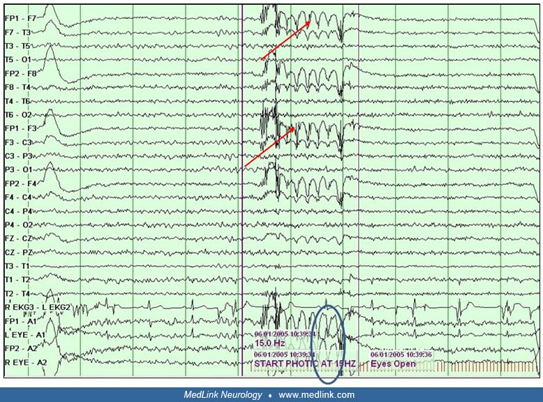
Reactivity. Reactivity refers to an alteration of EEG activity by external sensory stimuli.
Reference electrode. Reference electrode refers to an electrode that is electrically inactive relative to the active electrode site. This reference electrode remains consistent compared to electrodes in the montage. Examples of a reference electrode include ipsilateral ear/mastoid or the vertex location comparing a single electrode to all others in the electrode array. In a referential recording, the activity from the active electrode is compared to the reference to produce a potential difference where absolute amplitude remains the site of maximal electrophysiological involvement.
Resistance. Resistance is opposition to direct electrical current (DC) flow.
Sensitivity. Sensitivity refers to the ratio of input voltage to output recorded in a channel of the EEG recording.
Nearly 3,000 illustrations, including video clips of neurologic disorders.
Every article is reviewed by our esteemed Editorial Board for accuracy and currency.
Full spectrum of neurology in 1,200 comprehensive articles.
Listen to MedLink on the go with Audio versions of each article.
MedLink, LLC
3525 Del Mar Heights Rd, Ste 304
San Diego, CA 92130-2122
Toll Free (U.S. + Canada): 800-452-2400
US Number: +1-619-640-4660
Support: service@medlink.com
Editor: editor@medlink.com
ISSN: 2831-9125