


Neuro-Oncology
Mesenchymal non-meningothelial tumors [Brief]
Jan. 16, 2024
MedLink®, LLC
3525 Del Mar Heights Rd, Ste 304
San Diego, CA 92130-2122
Toll Free (U.S. + Canada): 800-452-2400
US Number: +1-619-640-4660
Support: service@medlink.com
Editor: editor@medlink.com
ISSN: 2831-9125
Toll Free (U.S. + Canada): 800-452-2400
US Number: +1-619-640-4660
Support: service@medlink.com
Editor: editor@medlink.com
ISSN: 2831-9125
Nearly 3,000 illustrations, including video clips of neurologic disorders.
Every article is reviewed by our esteemed Editorial Board for accuracy and currency.
Full spectrum of neurology in 1,200 comprehensive articles.
Listen to MedLink on the go with Audio versions of each article.
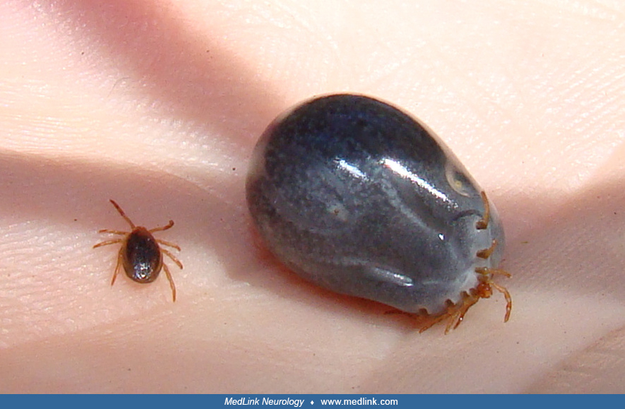
Axial T1-weighted MRI of the brain at the level of the medulla shows a normal appearance of the medulla and the cerebellum. However, on closer inspection, there is a small, isointense area (arrow) seen attached to the skin in the occipital region on the right side. (Source: Sarwar Z, Osborne AF, Shah C, Rathore MH. Neuroimaging in tick paralysis: looking outside the box. Infect Dis Rep 2022;14(6):837-40. Creative Commons Attribution 4.0 International [CC BY 4.0] license, creativecommons.org/licenses/by/4.0.)