Epilepsy & Seizures
Familial focal epilepsy with variable foci
Jul. 06, 2022
MedLink®, LLC
3525 Del Mar Heights Rd, Ste 304
San Diego, CA 92130-2122
Toll Free (U.S. + Canada): 800-452-2400
US Number: +1-619-640-4660
Support: service@medlink.com
Editor: editor@medlink.com
ISSN: 2831-9125
Toll Free (U.S. + Canada): 800-452-2400
US Number: +1-619-640-4660
Support: service@medlink.com
Editor: editor@medlink.com
ISSN: 2831-9125
Nearly 3,000 illustrations, including video clips of neurologic disorders.
Every article is reviewed by our esteemed Editorial Board for accuracy and currency.
Full spectrum of neurology in 1,200 comprehensive articles.
Listen to MedLink on the go with Audio versions of each article.
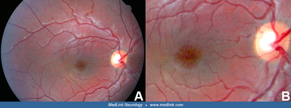
(A) The macula of juvenile retinoschisis patients always presents with a classic foveal schisis or spoke-wheel appearance of the macula. Of these patients, 50% will also have peripheral areas of retinoschisis. (B) This is a magnified view of the fundus of photo A. (Contributed by Dr. James Walters.)