Neuropharmacology & Neurotherapeutics
Drug-induced neuropathies
Dec. 06, 2025
MedLink, LLC
3525 Del Mar Heights Rd, Ste 304
San Diego, CA 92130-2122
Toll Free (U.S. + Canada): 800-452-2400
US Number: +1-619-640-4660
Support: service@medlink.com
Editor: editor@medlink.com
ISSN: 2831-9125
Toll Free (U.S. + Canada): 800-452-2400
US Number: +1-619-640-4660
Support: service@medlink.com
Editor: editor@medlink.com
ISSN: 2831-9125
Nearly 3,000 illustrations, including video clips of neurologic disorders.
Every article is reviewed by our esteemed Editorial Board for accuracy and currency.
Full spectrum of neurology in 1,200 comprehensive articles.
Listen to MedLink on the go with Audio versions of each article.
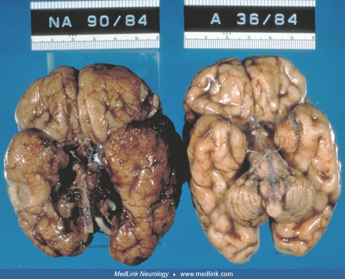
Basal views of brain of 2 neonates at autopsy, after 2 weeks of formalin fixation. The one on the left (NA90/84) has global cerebellar hypoplasia. The one on the right (A36/84) is a normal, age-matched control. The brainstems and cerebral hemispheres appear externally normal in both. There is a thin layer of subarachnoid blood in the leptomeninges of A36/84, giving it a darker appearance; this is, though, unrelated to the cerebellar hypoplasia. Note the difference in the lateral extension of the cerebellar hemispheres in the two brains. (Contributed by Dr. Harvey Sarnat.)

