Fabry disease
Dec. 02, 2025
MedLink, LLC
3525 Del Mar Heights Rd, Ste 304
San Diego, CA 92130-2122
Toll Free (U.S. + Canada): 800-452-2400
US Number: +1-619-640-4660
Support: service@medlink.com
Editor: editor@medlink.com
ISSN: 2831-9125
Toll Free (U.S. + Canada): 800-452-2400
US Number: +1-619-640-4660
Support: service@medlink.com
Editor: editor@medlink.com
ISSN: 2831-9125
Nearly 3,000 illustrations, including video clips of neurologic disorders.
Every article is reviewed by our esteemed Editorial Board for accuracy and currency.
Full spectrum of neurology in 1,200 comprehensive articles.
Listen to MedLink on the go with Audio versions of each article.
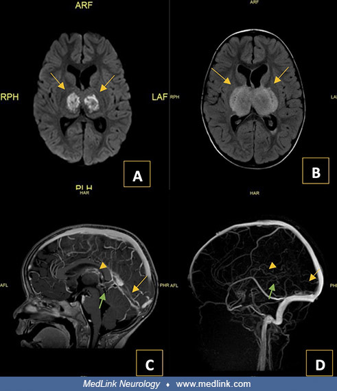
MRI and MRV brain. (A) Axial DWI showing restricted diffusion in the bilateral thalami (arrows), indicative of cytotoxic edema and suggestive of infarction. (B) Axial T2 FLAIR image demonstrating bilateral thalamic hyperintensities (arrows), involving the posterior limb of the internal capsule, consistent with vasogenic edema. (C) T1 gadolinium-weighted sagittal sequence showing intraluminal filling defects, confirming thrombosis in the internal cerebral vein (arrowhead), vein of Galen (green arrow), and straight sinus (yellow arrow). (D) Venogram (time-of-flight) demonstrating absence of flow-related signal within the internal cerebral vein (arrowhead), vein of Galen (green arrow), and straight sinus (yellow arrow). (Contributed by Dr. Mahendranath D Moharir.)