Neuropharmacology & Neurotherapeutics
Nusinersen
Apr. 23, 2026
MedLink, LLC
3525 Del Mar Heights Rd, Ste 304
San Diego, CA 92130-2122
Toll Free (U.S. + Canada): 800-452-2400
US Number: +1-619-640-4660
Support: service@medlink.com
Editor: editor@medlink.com
ISSN: 2831-9125
Toll Free (U.S. + Canada): 800-452-2400
US Number: +1-619-640-4660
Support: service@medlink.com
Editor: editor@medlink.com
ISSN: 2831-9125
Nearly 3,000 illustrations, including video clips of neurologic disorders.
Every article is reviewed by our esteemed Editorial Board for accuracy and currency.
Full spectrum of neurology in 1,200 comprehensive articles.
Listen to MedLink on the go with Audio versions of each article.
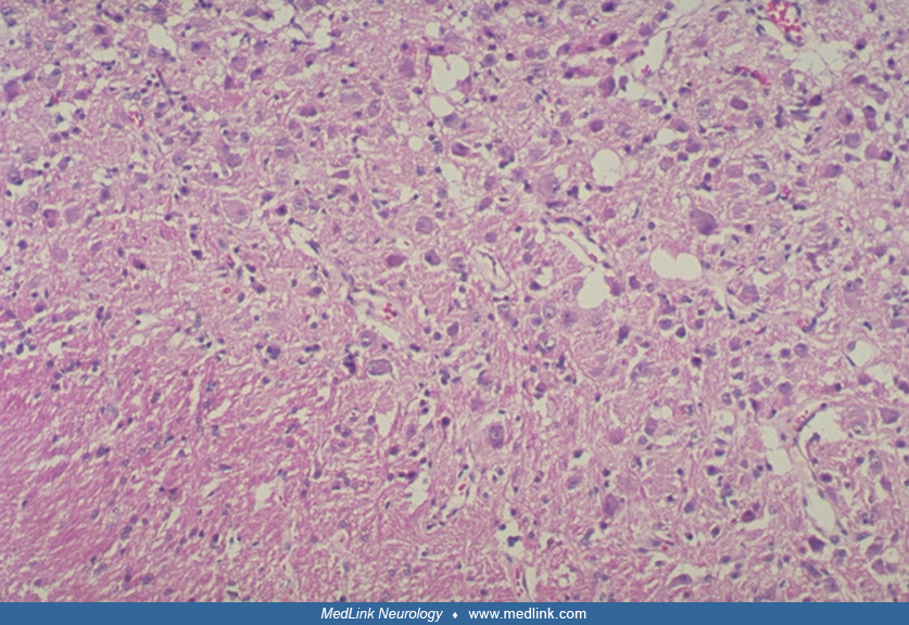
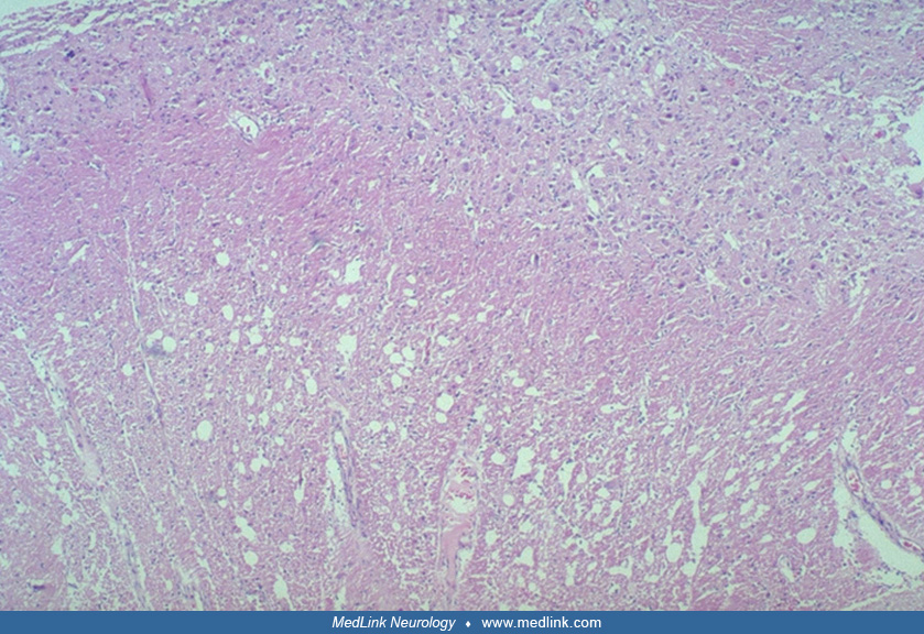
The lesion of the cerebellar cortex at low magnification shows enlarged, vacuolated molecular layer and hypertrophic cells replacing the Purkinje and granule cell layers. This architecture gives the lesion the gross appearance, both by imaging and pathologically, of thickened folia. (Hematoxylin-eosin; x100) (Contributed by Dr. Laura Hair.)