Neuro-Oncology
Pediatric-type diffuse high-grade gliomas (excluding diffuse midline gliomas)
Aug. 26, 2025
MedLink, LLC
3525 Del Mar Heights Rd, Ste 304
San Diego, CA 92130-2122
Toll Free (U.S. + Canada): 800-452-2400
US Number: +1-619-640-4660
Support: service@medlink.com
Editor: editor@medlink.com
ISSN: 2831-9125
Toll Free (U.S. + Canada): 800-452-2400
US Number: +1-619-640-4660
Support: service@medlink.com
Editor: editor@medlink.com
ISSN: 2831-9125
Nearly 3,000 illustrations, including video clips of neurologic disorders.
Every article is reviewed by our esteemed Editorial Board for accuracy and currency.
Full spectrum of neurology in 1,200 comprehensive articles.
Listen to MedLink on the go with Audio versions of each article.
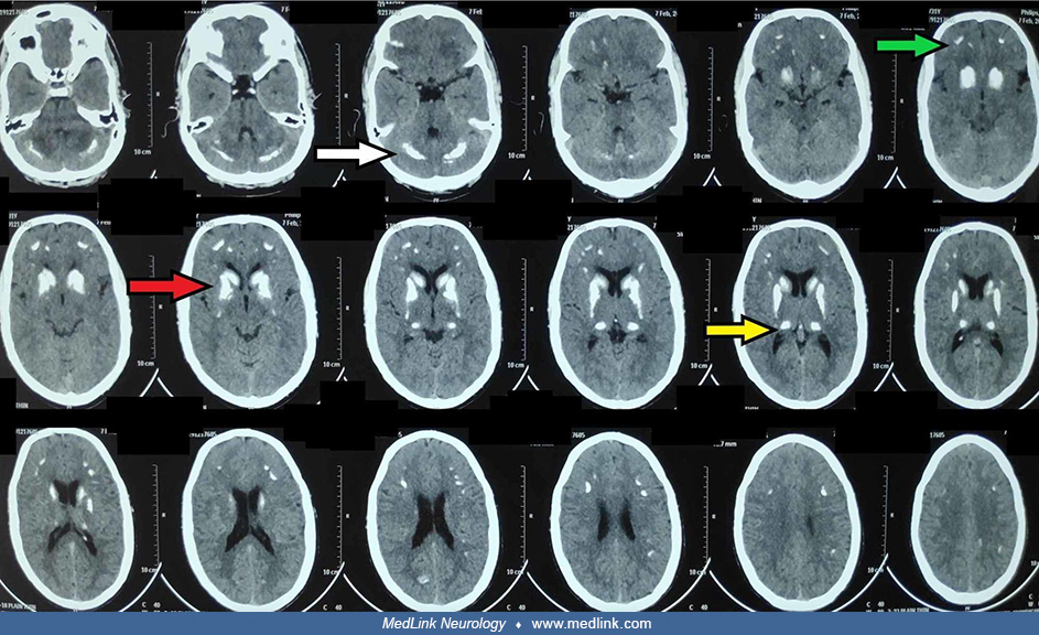
A noncontrast brain CT scan showing bilateral, symmetrical calcifications in the basal ganglia, thalami, corona radiata, and dentate nuclei. (A) Axial, (B) coronal, and (C) sagittal images showing bilateral calcifications of the basal ganglia and thalami (red arrows). (D) Axial image showing bilateral calcifications of the corona radiata (red arrows). (E) Axial and (F) coronal images showing bilateral calcifications of the dentate nuclei (red arrows). (From: Mohammed YE, Gebreselassie KH, Sesega EH, Wolie AA, Ahmed BJ. Recurrent seizures in an adolescent female with extensive intracranial calcifications: a case report of Fahr's syndrome secondary to hypoparathyroidism. Cureus 2025;17[2]:e78780. Creative Commons Attribution 4.0 International [CC BY 4.0] license, creativecommons.org/licenses/by/4.0.)