Stroke & Vascular Disorders
Hyperhomocysteinemia
Mar. 31, 2025
MedLink, LLC
3525 Del Mar Heights Rd, Ste 304
San Diego, CA 92130-2122
Toll Free (U.S. + Canada): 800-452-2400
US Number: +1-619-640-4660
Support: service@medlink.com
Editor: editor@medlink.com
ISSN: 2831-9125
Toll Free (U.S. + Canada): 800-452-2400
US Number: +1-619-640-4660
Support: service@medlink.com
Editor: editor@medlink.com
ISSN: 2831-9125
Nearly 3,000 illustrations, including video clips of neurologic disorders.
Every article is reviewed by our esteemed Editorial Board for accuracy and currency.
Full spectrum of neurology in 1,200 comprehensive articles.
Listen to MedLink on the go with Audio versions of each article.
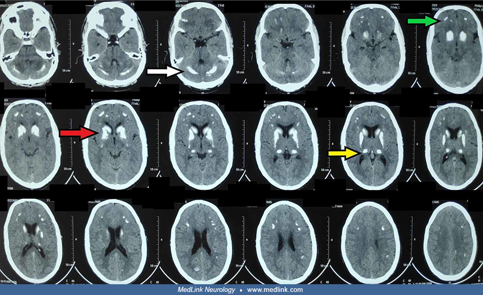
Axial brain sections on a susceptibility-weighted MRI sequence showing bilateral symmetrical calcification in a 31-year-old man with hypoparathyroidism and hypocalcemia. The calcification is evident as “blooming,” predominantly in bilateral basal ganglia (red arrow) and thalami (yellow arrow). Smaller lesions are also seen in the frontal cortex (green arrow) and the cerebellum (white arrow). (From: Gaba S, Singla M, Gupta M, Dua A, Gaba N. From psychiatry to neurology and endocrinology: a case of hypoparathyroidism. Cureus 2020;12(9):e10666. Creative Commons Attribution License CC-BY 4.0.)