Peripheral Neuropathies
Hexacarbon neuropathy
May. 07, 2025
MedLink, LLC
3525 Del Mar Heights Rd, Ste 304
San Diego, CA 92130-2122
Toll Free (U.S. + Canada): 800-452-2400
US Number: +1-619-640-4660
Support: service@medlink.com
Editor: editor@medlink.com
ISSN: 2831-9125
Toll Free (U.S. + Canada): 800-452-2400
US Number: +1-619-640-4660
Support: service@medlink.com
Editor: editor@medlink.com
ISSN: 2831-9125
Nearly 3,000 illustrations, including video clips of neurologic disorders.
Every article is reviewed by our esteemed Editorial Board for accuracy and currency.
Full spectrum of neurology in 1,200 comprehensive articles.
Listen to MedLink on the go with Audio versions of each article.
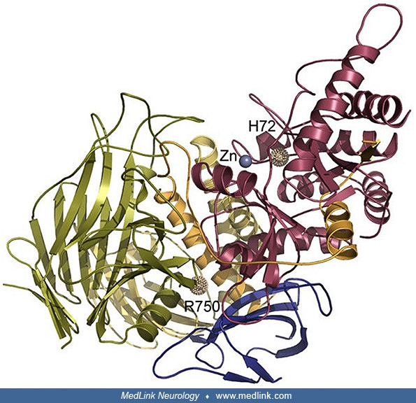
Peptides are colored a-red, b-orange, c-yellow, d-green, and e-blue. The active site is denoted by a Zn2+ ion. Two mutant sites are displayed, demonstrating the effect of mutations c.215A>T: p.H72L affecting Zn2+ coordination in the actives site (group 1 mutation) and the prevalent mutation c.2248C>T: p.R750W, which is likely affecting peptide e-d interaction (group 2 mutation). (Created by Dr. P. Heikinheimo, University of Helsinki, Finland. It has been prepared with the program PyMol. Source: Malm D, Nilssen O. Alpha-mannosidosis. Orphanet J Rare Dis 2008;3:21. Creative Commons Attribution License. http://creativecommons.org/licenses/by/2.0. Enlarged and edited by Douglas J Lanska MD MS MSPH to improve sharpness and contrast.)