Epilepsy & Seizures
Self-limited (familial) infantile epilepsy
May. 08, 2026
MedLink, LLC
3525 Del Mar Heights Rd, Ste 304
San Diego, CA 92130-2122
Toll Free (U.S. + Canada): 800-452-2400
US Number: +1-619-640-4660
Support: service@medlink.com
Editor: editor@medlink.com
ISSN: 2831-9125
Toll Free (U.S. + Canada): 800-452-2400
US Number: +1-619-640-4660
Support: service@medlink.com
Editor: editor@medlink.com
ISSN: 2831-9125
Nearly 3,000 illustrations, including video clips of neurologic disorders.
Every article is reviewed by our esteemed Editorial Board for accuracy and currency.
Full spectrum of neurology in 1,200 comprehensive articles.
Listen to MedLink on the go with Audio versions of each article.
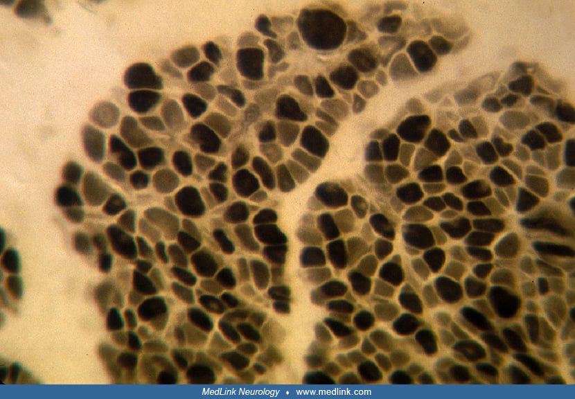
In transverse section, myofibers are smaller than expected, with an average diameter of 5 to 12.5µm (normal mean 15µm). About 20% of fibers have central nuclei or clear central spaces if the section is between nuclei, but no myofiber degeneration or necrosis are seen. X250. (Reproduced with permission from: Sarnat HB, O’Connor T, Byrne PA. Clinical effects of myotonic dystrophy on pregnancy and the neonate. Arch Neurol 1976;33:459-65. Copyrighted 1976, American Medical Assoc.)