Headache & Pain
Fibromyalgia
Feb. 16, 2026
MedLink, LLC
3525 Del Mar Heights Rd, Ste 304
San Diego, CA 92130-2122
Toll Free (U.S. + Canada): 800-452-2400
US Number: +1-619-640-4660
Support: service@medlink.com
Editor: editor@medlink.com
ISSN: 2831-9125
Toll Free (U.S. + Canada): 800-452-2400
US Number: +1-619-640-4660
Support: service@medlink.com
Editor: editor@medlink.com
ISSN: 2831-9125
Nearly 3,000 illustrations, including video clips of neurologic disorders.
Every article is reviewed by our esteemed Editorial Board for accuracy and currency.
Full spectrum of neurology in 1,200 comprehensive articles.
Listen to MedLink on the go with Audio versions of each article.
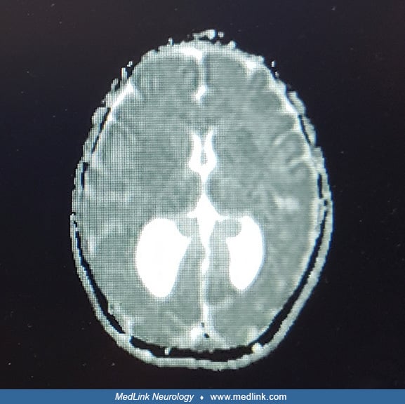
(A) Diagnosis of hydrocephalus was made in utero by ultrasonography. (B and C) Coronal sections of ultrasonography showing significant dilatation of occipital horns with vertical but normal in size front horns. Choroid plexi are normal. (D) Parasagittal section shows the dilated temporal and occipital horns (left); the roof of the ventricle is thin due to the absence of the corpus callosum. (E and F) Axial CT confirms dilatation of the (E) occipital horns and (F) temporal horns of the lateral ventricles and absence of the corpus callosum. (From: Sarnat HB. Cerebral dysgenesis. Embryology and clinical expression. New York: Oxford University Press, 1992a:238.)