
Stroke & Vascular Disorders
Fusiform and dolichoectatic aneurysms
May. 03, 2026
MedLink, LLC
3525 Del Mar Heights Rd, Ste 304
San Diego, CA 92130-2122
Toll Free (U.S. + Canada): 800-452-2400
US Number: +1-619-640-4660
Support: service@medlink.com
Editor: editor@medlink.com
ISSN: 2831-9125
Toll Free (U.S. + Canada): 800-452-2400
US Number: +1-619-640-4660
Support: service@medlink.com
Editor: editor@medlink.com
ISSN: 2831-9125
Worddefinition
At vero eos et accusamus et iusto odio dignissimos ducimus qui blanditiis praesentium voluptatum deleniti atque corrupti quos dolores et quas.
Aortic atherosclerotic plaques are recognized as significant contributors to cerebral infarction, with occurrences both spontaneous and iatrogenic, particularly during perioperative periods. The presence of large and complex atheromas increases the risk of cerebral infarction. Management strategies include addressing modifiable risk factors and considering antiplatelet therapy or anticoagulation. However, the effectiveness of these interventions awaits clarification through further studies to establish clear treatment protocols.
|
• Aortic arch atherosclerosis is a known cause of ischemic stroke. | |
|
• Plaques measuring 4 mm or greater and lipid rich core carry the highest risk of stroke, as compared to calcified plaques. | |
|
• Stroke often tends to be right hemispheric due to the location of plaque within the aorta. | |
|
• Specific guidelines regarding antiplatelet versus anticoagulation therapy are still underway; however, the usual treatments for ischemic stroke, such as antithrombotic therapy, statin therapy, and lifestyle changes, are recommended. |
Stroke is the fifth leading cause of death in the United States. Of the 795,000 strokes occurring annually in the United States, 87% are ischemic stroke and 10% are hemorrhagic stroke (105).
Atherosclerosis is a diffuse systemic vascular disorder affecting large and medium-sized arteries, causing patchy intimal plaques known as atheromas.
Aortic atheromatous plaques were first identified as a possible cause of stroke in the early 1990s, when transesophageal echocardiographic examination of three patients with cryptogenic stroke to identify potential cardiac sources demonstrated the presence of "large, protrusive plaques. . . with mobile projections that moved freely with the blood flow" (107). This was followed by a larger study in 1991 that demonstrated a higher incidence of embolism when aortic plaques had mobile elements (48). The association of aortic atheroma and stroke was first described by Amarenco and colleagues in a landmark autopsy study of 500 patients with cerebrovascular and other neurologic diseases. The prevalence of ulcerated plaques is 26% in patients with cerebrovascular diseases compared to 5% among patients with other neurologic diseases. Ulcerated plaques were present in 61% of cryptogenic cerebral infarcts compared to 22% with a known cause (06). Plaques that were at least 4 mm in thickness were found to be an independent risk factor for ischemic stroke (04; 70; 69).
The underlying mechanisms involved include: (1) thromboembolism, the detachment of thrombus overlying an unstable plaque, which typically presents acutely with ischemia; and (2) cholesterol crystal embolism, (atheroembolism), the dislodgement of cholesterol crystals from a ruptured plaque into small distal arteries, which tends to follow a subacute course and involves both mechanical obstruction and a local inflammatory response (Kronzon and Saric 2010; 88).
Prognosis with regard to vascular events, subsequent stroke, or mortality is a function of plaque (size and morphology) and other associated comorbidities, including atrial fibrillation and atherosclerosis in other vascular beds.
Plaque size. In the French Study of Aortic Plaques in Stroke, the recurrent stroke rate and vascular event rate were 12% and 26%, respectively, in the group with plaques greater than 4 mm, compared to 2.8% and 6% in those with plaques less than 1 mm, with a relative risk of recurrent brain infarction of 3.8 (95% CI 1.8–7.8) for plaques greater than or equal to 4 mm (32). A separate case-control study of first ischemic stroke reported an odds ratio of 13.8 for aortic arch plaques greater than 4 mm compared to controls (04). Mortality was significantly higher in the group with plaques greater than 4 mm compared to smaller plaque groups. These findings have been extended in more recent work: in a community cohort of 934 stroke-free participants followed for a mean of 11.3 years, large aortic arch plaques (≥ 4 mm) were independently associated with significantly increased long-term risk of cardiovascular events even in the statin era, with statin use found to modify this association (123). Additionally, aortic arch plaque thickness has been independently associated with white matter hyperintensity volume on brain MRI in stroke-free elderly participants, extending the prognostic implications of aortic plaque beyond overt stroke to subclinical cerebrovascular disease (106).
Plaque morphology. The highest risk for embolic stroke is found when aortic atheromas have mobile components, are hypoechoic (reflecting a lipid-rich core), or are ulcerated (101). In a long-term follow-up study of 1570 patients undergoing transesophageal echocardiogram over a mean of 8.7 years, ulceration independently predicted reduced survival and mobile plaque independently predicted higher embolic event rates, confirming the prognostic importance of morphological features beyond plaque size alone (46). In a prospective multicenter study of over 1000 acute ischemic stroke patients, those with embolic stroke of undetermined source had significantly thicker aortic arch plaques and a significantly higher proportion of ulcerated or protruding plaques compared to those with a determined stroke cause, supporting the causal role of high-risk plaque morphology in cryptogenic stroke (15).
Atrial fibrillation. The risk of stroke in patients with atrial fibrillation is significantly increased when associated with aortic atheroma. The Stroke Prevention in Atrial Fibrillation (SPAF) Investigators Committee on Echocardiography examined outcomes in 382 patients with high-risk nonvalvular atrial fibrillation who underwent transesophageal echocardiography. In the 35% of patients with complex aortic atheroma (greater than 4 mm, mobile, or ulcerated), the risk of stroke during 1 year ranged from 12% to 20%, compared to only 1.2% in patients without aortic atheroma (94). Data confirm that this coexistence is common and clinically important: complex aortic arch plaques were present in approximately 39% of stroke patients with nonvalvular atrial fibrillation, with prevalence rising sharply in those aged over 75 years (97). Furthermore, in a consecutive series of embolic stroke patients, those with both atrial fibrillation and coexisting aortic arch atheroma greater than or equal to 4 mm had the highest rates of short-term stroke recurrence and poor functional outcome at 3 months, demonstrating that the two conditions confer additive embolic risk (08). This discrepancy in stroke risk suggests a potential role of aortic atheromas in the mechanism of stroke even in patients with atrial fibrillation.
Coexisting hypercoagulable states. Thromboembolism appears to be much more frequent than atheroembolism in patients with severe aortic arch plaques, with a frequency reported to be as high as 33% at 1 year, compared with only 0.7% for atheroembolism. It is, therefore, conceivable that the coexistence of a hypercoagulable state in a patient with aortic arch plaque may increase the likelihood of superimposed thrombus formation and further enhance the embolic potential of the plaque. The APRIS study demonstrated a role for coagulation activation in the stroke mechanism on the basis of finding elevated prothrombin fragment F1.2, which is an indicator of thrombin generation in stroke patients with large aortic plaques; this subgroup also had significantly higher rates of recurrent stroke and death (23). In the PICSS/WARSS trial, large aortic plaques (≥ 4 mm) were present in 19.6% of 516 stroke patients and were associated with an approximately 2.5-fold increased adjusted risk of recurrent stroke or death at 2-year follow-up, independently of treatment assignment to warfarin or aspirin (24).
Atherosclerosis in other vascular bed. Aortic arch atherosclerosis frequently coexists with disease in other vascular territories, and this systemic distribution has important prognostic and clinical implications. In a study of cardiac patients undergoing transesophageal echocardiography, ascending aortic atherosclerotic plaques were present in 62.9% of the overall population, rising to 82.8% in those with carotid atherosclerotic plaques; there was also a stepwise increase in the prevalence of carotid plaques with increasing severity of aortic atherosclerosis, consistent with a shared systemic disease process (47). More broadly, supracardiac atherosclerosis — encompassing aortic arch and nonstenotic carotid plaques — has been identified as a substantially underestimated etiology in embolic stroke of undetermined source, with imaging features of high-risk plaque warranting active consideration in the stroke workup of this population (78). Patients with severe aortic stenosis showed a 47% prevalence of severe, complex aortic plaque compared to 9% in controls; nonobstructive aortic valve calcification correlated with aortic plaque in 86% of cases versus 30% in controls (119). Hypertension in individuals with aortic plaque may reflect underlying renal artery stenosis, and screening for this should be considered in such patients (84). Severe thoracic aortic plaque has also been associated with abdominal aortic aneurysm, suggesting a role for abdominal aortic aneurysm screening in patients with significant thoracic plaque (108).
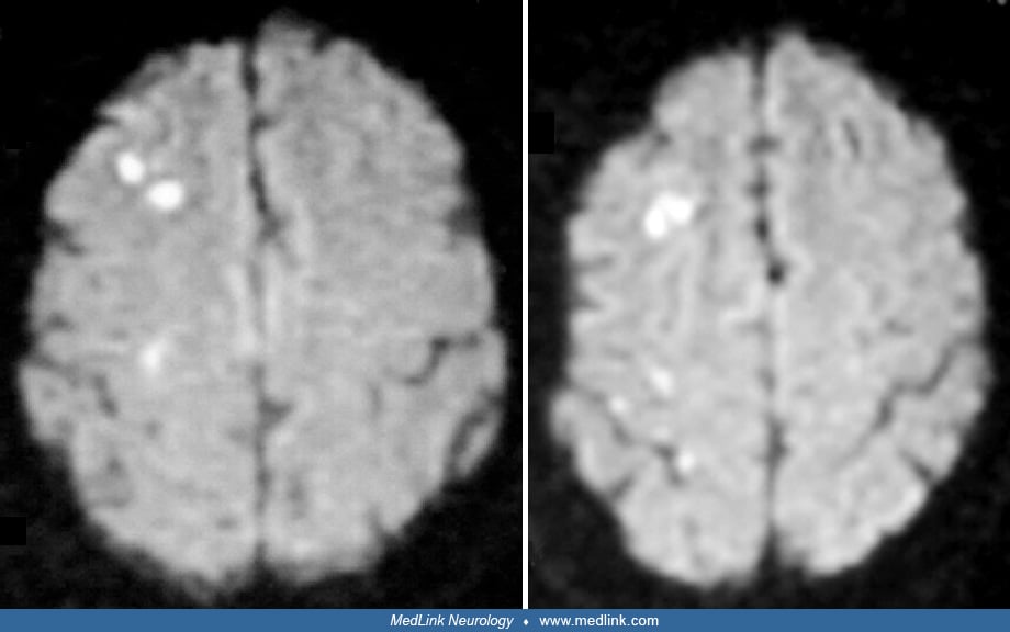
A 70-year-old, right-handed man with no prior medical history presented to the emergency room with a history of recurrent, transient left arm numbness. He had also experienced two transient episodes of left homonymous visual field deficits in the preceding week. He smoked two packs of cigarettes per day for at least 50 years.
Examination showed a blood pressure of 152/70 mm Hg, with a regular heart rate at 60 beats per minute. Language and memory were intact. Visual field examination was unremarkable. Motor examination showed a left pronator drift with reduced rapid alternating movements but intact strength on formal muscle testing. Gait and cerebellar examinations were normal, and there was no sensory loss.
ECG showed normal sinus rhythm. CT of the head without contrast was normal. MRI of the brain demonstrated several small white matter lesions in the deep right middle cerebral artery/anterior cerebral artery watershed arterial territories on diffusion-weighted imaging and FLAIR sequences.
MR angiography of the intracranial vessels showed no hemodynamically significant stenosis. Carotid duplex ultrasound showed minimal atherosclerotic disease bilaterally in both the proximal internal and distal common carotid arteries. Transthoracic echocardiogram was normal. Transesophageal echocardiography revealed a large 1.2 to 1.5 cm plaque in the ascending aortic arch without ulceration or mobility.
Atherosclerotic changes of the aorta occur as early as during the third decade of life and precede those of the coronary, cerebrovascular, and peripheral arteries. Plaque initiation and progression is a complex process initiated locally and modulated by various systemic, hemodynamic, and biological factors (126). Wall shear stress, defined as the frictional force exerted by the flowing blood on the vessel wall, plays a pivotal role in atherosclerosis. Low wall shear stress has been implicated in the initiation of plaque formation (19). Conversely, increased shear stress has an atheroprotective effect. The mechanical effect of lower shear stress alters gene expression in the endothelial cells triggering an inflammatory cascade and increased expression of adhesion molecules (113). LDL influx is high in areas of low wall shear stress. Accumulated LDL undergoes oxidation and initiates an inflammatory response by activating monocytes (through the expression of monocyte chemoattractant protein) and differentiation of monocyte to macrophage (33; 75). Macrophages, in turn, engulf modified LDL forming cholesterol laden foam cells and also secrete cytokines IL-1 and TNK-alpha, stimulating expression of adhesive molecules and, in turn, recruiting more monocytes, thus, propagating the cycle of inflammation (38; 11). The secretion of growth factors and metalloproteinases by the macrophages also results in cell proliferation and matrix degeneration, making the plaque unstable and prone to rupture (110).
The protective effect of HDL stems from its ability to promote an efflux of cholesterol from foam cells (71) and the inhibition of oxidative modification of LDL (64) by transporting antioxidants like paraoxonase (65) and apolipoproteins Apo-A1 and Apo-A2, which have intrinsic antioxidant properties (34). HDLs also inhibit cytokine and C-reactive protein–induced expression of adhesion molecules by endothelial cells (121; 116).
The causal relationship between characteristic features of aortic atheromas and stroke is now well established. Various characteristics of plaque size and thickness, morphology, location, and progression have been studied.
Grading of aortic plaques (plaque size/thickness). The thickness of atheromas has a direct correlation with the risk of embolic vascular events. Davila-Roman and colleagues, in a long-term follow-up study of 1957 patients who underwent intraoperative epiaortic ultrasound assessment of the ascending aorta during cardiac surgery, demonstrated that neurologic event rates increased progressively across grades of atherosclerosis severity, with patients in the severe atherosclerosis group having significantly higher long-term neurologic event rates than those with mild or moderate disease (21). At present, no universal grading system exists, with some authors describing plaques 5 mm or greater in thickness as complex plaques (49; 103; 111) and others using a thickness of 4 mm as the criterion (31).
In 1996, a classification of aortic plaques based on plaque morphology as visualized by transesophageal echocardiography was proposed by Montgomery and colleagues (72).
|
Grade |
Morphology |
|
I |
Normal intima or no/minimal intimal thickening |
|
II |
Extensive intimal thickening without protruding atheroma |
|
III |
Sessile atheroma protruding less than 5 mm in thickness into the aortic lumen |
|
IV |
Sessile (complex) atheroma protruding plaque 5+ mm in thickness or more into the aortic lumen |
|
V |
Mobile protruding atheroma, irrespective of thickness |
Not all studies have confirmed an independent association between aortic plaque and cerebrovascular risk. An analysis from the Stroke Prevention: Assessment of Risk in a Community (SPARC) study found that complex aortic plaques (greater than 4 mm or mobile) did not significantly increase the risk of cerebrovascular events after adjusting for age, sex, and other clinical risk factors (68). Importantly, however, the SPARC study examined a general population cohort without prior stroke, whereas studies demonstrating significantly increased risk, including the French Study of Aortic Plaques in Stroke and the APRIS study enrolled patients who had already suffered stroke or transient ischemic attack. This distinction is clinically important: aortic plaque burden may function as a marker of generalized atherosclerosis in the general population, while carrying direct embolic significance in the context of established cerebrovascular disease.
Definition of aortic plaques. Unstable or complex plaques are more frequently echolucent, heterogeneous, and noncalcified with or without mobile elements, and they are more prone to rupture. Mobile elements of the plaque are often referred to as aortic debris, superimposed thrombi, or complex plaque (23). Plaques with mobile elements are classified as complex, regardless of the plaque thickness. A nomenclature has been proposed for plaques in the coronary circulation. “Culprit” plaques refer to those causing the acute vascular event whereas “vulnerable” plaques refer to those plaques that have the potential to transform into culprit plaques. This classification aims to define plaques based on their potential to undergo acute change, which is dependent on morphology and plaque composition (73). A similar classification for aortic arch plaques would be of use in predicting future vascular events.
Plaque progression, such as the fibrous cap thickness, amount of lipid core, and the degree of active inflammation, play a role in determining plaque vulnerability.
A follow-up transesophageal echocardiographic study of patients with aortic atheroma over a period of 12 months reported an overall stability in the disease process, with close to 66% having the same atheroma grade at the end of the study period. Although none of the patients with atheromas smaller than 5 mm developed mobile lesions, 62% of those with more severe grades developed new mobile lesions. However, 70% of mobile lesions identified at baseline evaluation resolved spontaneously. Whether this is due to embolism of the mobile fragment or due to the healing process is uncertain. Therefore, though the overall disease process is stable, individual lesions are unpredictable and dynamic, demonstrating progression and regression over time (72).
Aging of the vasculature results in increased expression of proinflammatory transcription factors like nuclear factor (NF)-kappaB and decreased protective factors like hypoxia inducible factors, resulting in increased production of inflammatory cytokines (125). In addition, microbiological infection of the vessel wall triggers inflammation by direct cytopathogenicity, bystander effects, epitope spreading, and molecular mimicry (63).
Further progression of the disease process is also influenced by systemic factors like cigarette smoking, hypercholesterolemia, diabetes mellitus, and genetic influences. With further progression of the disease process, vascular smooth muscle cells and myofibroblasts migrate and deposit extracellular matrix, forming the fibrous cap. The oxygen-deprived central core contains extracellular lipids, cholesterol esters, and sometimes calcifications (14; 86). As the atherosclerotic process encroaches into the aortic lumen, the luminal diameter is preserved with the vessel wall undergoing outward remodeling. Thus, a normal lumen contour during an angiogram could be masking a severe atherosclerotic process (81). Vulnerable plaques typically have a thin fibrous cap, a larger lipid core, an increased number of inflammatory cells, and increased neoangiogenesis. The central role of inflammation has always raised the possibility of using inflammatory markers to detect disease progression. C-reactive protein has been shown to play an important role by decreasing eNOS-mediated dilatation in vivo. Maternal levels of C-reactive protein have been shown to predict atherosclerosis in children of hypercholesterolemic mothers (61). Serum amyloid A has been shown to increase proteoglycan synthesis, leading to increased LDL proteoglycan-binding affinity (120).
The potent amalgamation of inflammation, oxidized LDL, and vascular ageing results in plaque vulnerability to rupture. Local expression of matrix metalloproteinases and collagenase combined with smooth muscle apoptosis predispose to plaque rupture. Plaque rupture occurs at the plaque shoulder where mechanical strain is maximum, resulting in exposure of procoagulant factors like tissue factor (66). This activates the coagulation cascade, leading to superimposed thrombus formation and systemic embolism.
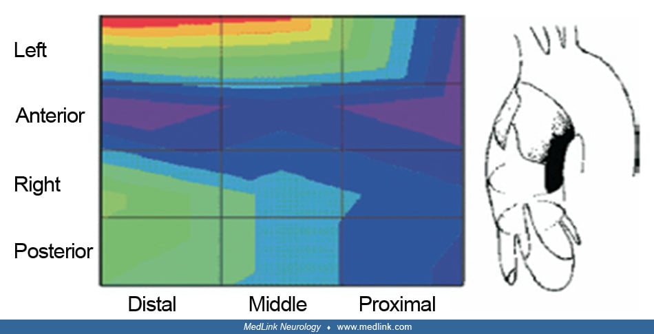
Plaque distribution. Distribution of clinically significant atherosclerotic plaques on the aorta varies depending on the location. The portion of the aorta upstream to the take-off of the left subclavian artery has the highest potential for atheroembolism leading to stroke. The ascending aorta is the least involved segment of the thoracic aorta in the atherosclerotic process (Barbut and Gold 1996; 82; 02). This proximal-to-distal gradient could be a reflection of the hemodynamics peculiar to the aorta. If complex plaques (greater than 4 mm) are found in the proximal thoracic aorta rather than the descending aorta, the odds of stroke increase from 1.5 to 13.8 (04).
Anomalies of the aortic arch are not infrequent and are associated with increased stroke events during carotid stenting procedures. This is due to increased technical difficulties secondary to distorted aortic arch and branching vessel anatomy (29). In the most frequent aortic arch variant, the left common carotid and innominate arteries share a common origin from the aortic arch. Rarely, the left common carotid may take origin directly from the innominate artery and is the second most common variant. Both variants are referred to as the bovine aortic arch, which is, in fact, a misnomer. The aortic arch in cattle has a single brachiocephalic trunk arising from the arch, which gives rise to both subclavian arteries and a bicarotid trunk (58).
Studies of embolic stroke following heart surgery have contributed significantly to understanding the role of aortic plaques. Aortic manipulation during coronary artery bypass grafting is considered to be one of the main causes of embolic stroke in the postoperative period. Patients with atheromatous change of the ascending aorta have increased incidence of postoperative stroke and a decreased 5-year stroke-free survival (114). Aortic debris collected from coronary catheters after advancement into the ascending aorta show signs of plaque disruption in up to 55% of plaques, with large lumen catheters having increased incidence of aortic debris (26).
More than 50% of neurologic events following cardiac catheterization occur in the posterior circulation (62), indicative of a predisposition of the posterior circulation to vascular insults. Early postoperative stroke is defined as stroke occurring within the first 24 hours of surgery or when the patient wakes up from anesthesia with a neurologic deficit. Delayed stroke refers to a new deficit after an initial normal recovery up to 30 days postoperatively. Aortic atherosclerosis can cause both types of stroke, being more significant in early strokes (42). Early postoperative stroke may present with multiple infarcts, secondary to a shower of emboli from aortic manipulation, whereas delayed strokes usually have single territory infarcts associated with fibrillation episodes (10).
Further studies have shown that involvement of the distal ascending aorta increases the stroke risk 5-fold. This is also the site where surgeons commonly place the aortic clamp. Another independent predictor of postoperative stroke is involvement of the middle lateral segment of the ascending aorta (115). This could be due to the altered hemodynamics secondary to the aortic arch anatomy, with higher blood flow velocities in the lateral aspect. Thus, the lesions in the lesser curvature of the ascending aorta are associated with higher stroke risk. Nevertheless, plaques from the descending aorta have been shown to embolize to the left subclavian artery during periods of retrograde blood flow, causing infarctions in the posterior circulation (40). In a study of patients with acute stroke by transesophageal echocardiogram and 3D MRI, retrograde flow was observed in as much as 31.7% of patients (40) with the average retrograde flow being 26 mm. The capacity of this flow to transport debris from plaques in the descending aorta to the left subclavian artery, leading to infarcts in the posterior circulation, cannot be overlooked (114).
Pathophysiology. Embolism from the aortic arch can be either due to thromboemboli from aortic mural thrombi secondary to plaque disruption with superimposed thrombosis or due to atheroemboli leading to cholesterol embolization. The more significant of these in relation to stroke appears to be thromboemboli. The frequency of thromboembolic episodes was 33% at a mean follow-up duration of 14 months in 42 patients with protruding aortic plaques (109). The incidence of emboli has a direct correlation with the grading of atheroma. One in three atheromas greater than 5 mm in thickness cause systemic emboli (92). This association was also demonstrated in a study of 335 patients undergoing transesophageal echocardiogram with the odds ratio for embolism increasing from 4 to 9.7 for grade 2 and 3 atheromas respectively (70). In another study, complex aortic plaques were more likely to be associated with small cortical infarctions than large artery atherosclerosis and cardioembolic etiologies. In addition, a subcortical pattern was more common in those with complex aortic plaque than those with cardioembolic mechanisms (51).
The prevalence of ulcerated plaques is three times higher in cases of cryptogenic stroke, compared to patients with known cause (06). In another study of 49 patients with cryptogenic stroke, transesophageal echocardiogram showed a high incidence of thoracic aortic atheroma of 46.9%, with microembolic signals being detected by transcranial Doppler in three of 10 patients with complex plaques (18). The prevalence rate of aortic plaques in a community screening of random individuals over 45 years of age for stroke prevention approached 51% (7.6% being complex plaques) (02). In another study, the prevalence of aortic arch plaques was greater in patients with severe aortic stenosis as compared to age- and gender-matched control subjects (74% vs. 41%) (96). Patients with significant intracranial atherosclerotic disease are more likely to have aortic arch atherosclerosis than those without (60.9% vs. 49.0%) (44). In a population-based cohort, aortic arch atherosclerosis was associated with an increased burden of white matter disease on MRI in people without known clinical stroke (106).
Ethnic differences are also present, with whites having a higher plaque burden and increased complex plaques compared to African Americans even though they had less severe arterial hypertension and diabetes mellitus (37). Another study demonstrated increased plaque burdens found in Jewish patients as compared to Arabic patients, but no differences were noted in the distribution of plaques by location or complexity (100). Gender differences have also been shown to exist. The established aortic plaque thickness of 4 mm for increased stroke risk may not hold true for both men and women. Plaque thickness between 3 and 3.9 mm was also associated with increased stroke risk in women (25). The reason for this remains unclear, with the influence of varied lipid profiles and inflammatory markers playing a possible role.
Traditional risk factors for atherosclerosis such as age, hypertension, dyslipidemia, and smoking have been associated with aortic atheroma (01; 68). Tribouilloy and colleagues reported that plasma homocysteine correlates with severity of thoracic aortic atherosclerosis, whereas Sen and colleagues demonstrated that hyperhomocysteinemia is an independent predictor of aortic atheroma progression in stroke and transient ischemic attack patients (104; 91; 90). In a cross-sectional study of 145 stroke-free participants, leukocyte count correlated with aortic arch plaque thickness consistent with the inflammatory component of atherosclerosis (28).
|
• Intraoperative cardiac surgery: Begin with the aorta when placed in the esophagus has made intraoperative transesophageal echocardiogram; escalate to epiaortic ultrasound if transesophageal echocardiogram shows significant plaque or if the patient has prior cerebrovascular accident, transient ischemic attack, or peripheral vascular disease. Epiaortic scanning is the only modality that eliminates the ascending aorta blind spot. | |
|
• Stroke workup/cryptogenic stroke: transesophageal echocardiogram remains first-line. CT (with cardiac gating) is a strong noninvasive alternative. MRI can combine aortic, cerebral, and carotid assessment in one session and is preferred when transesophageal echocardiogram is contraindicated. | |
|
• Plaque characterization/vulnerability assessment: MRI provides the best tissue differentiation (lipid vs. fibrous vs. cellular). OCT and IVUS are superior for luminal characterization when catheterization is already planned. IV-MRI and molecular imaging are research tools with high future potential. | |
|
• Screening high-risk patients: Non-contrast CT is rapid and accessible. FDG-PET/CT may detect early precalcification lesions and active inflammation but is not yet in routine practice. | |
|
• Transesophageal echocardiogram contraindicated: CT angiography or cardiac MRI are the preferred alternatives. |
Transesophageal echocardiography. Transesophageal echocardiogram has traditionally been the preferred modality for assessment of the thoracic aorta, with the proximity of the transducer to the aorta enabling high-accuracy measurement and grading of atheroma and detection of mobile elements. It is capable of predicting plaque composition, including calcification and has a demonstrated negative predictive value of 100% for the ascending aorta in the absence of plaque in the visualized regions (52). This reflects the normal distribution of plaques, in which the ascending aorta is the least involved segment.
Interrater reliability is good, particularly for plaques that of 4 mm or greater (41; 124; 117). Significant findings on transesophageal echocardiogram in cardiac surgery patients are often an indication for an epiaortic ultrasound to assess the segments not visualized on routine transesophageal echocardiogram. Newer 3D transesophageal echocardiogram technology allows reconstruction of images to predict plaque burden and localization (09).
The principal limitation of transesophageal echocardiogram is its inability to visualize the distal ascending aorta and the proximal aortic arch, regions that are important predictors of stroke due to the interposition of the tracheal air column. Up to 42% of the ascending aorta not being visualized may not be seen, leading to underestimation of the true plaque burden (53). Additional disadvantages include its semi-invasive nature and the requirement for conscious sedation.
Epiaortic ultrasound. Epiaortic scanning is an intraoperative technique that allows complete evaluation of the aorta, eliminating the blind spot inherent to transesophageal echocardiogram. It has consistently been shown to be superior to both transesophageal echocardiogram (22) and preoperative CT (13) for detecting high-risk aortic lesions. By guiding further surgical strategy, epiaortic scanning has been associated with a significant decrease in the incidence of postoperative cerebrovascular events, prompting some surgeons to advocate for its use as a standard of care in cardiac surgery patients (128).
Current guidelines recommend epiaortic scanning primarily for patients “at high risk for embolic stroke,” including those with a history of cerebrovascular or peripheral vascular disease and those with evidence of aortic atherosclerosis on other imaging modalities such as TEE, MRI, CT, or chest radiograph. Lesions greater than 3 mm in thickness, or mobile elements and their location, are reported to the surgeon who decides to guide further management (36).
The principal limitation is that epiaortic scanning can only be used in open-chest cardiac procedures. Its availability is decreasing as more surgeries transition to minimally invasive approaches.
Computed tomography. The major limitation of conventional CT has been respiratory and cardiac motion artefact. Multidetector CT, combined with ECG-gated cardiac cycle synchronization, largely addresses these issues and increases spatial and temporal resolution (85). In a prospective study of ischemic stroke patients, cardiac cycle-gated CT (CGCT) was found to be more effective than transesophageal echocardiography in detecting atheromas of 4 mm, whereas atheromas larger than 4 mm were detected with similar efficacy by both investigative modalities. CGCT also demonstrated higher sensitivity for calcification, and morphological features such as ulceration and protrusion appear were better visualized by CGCT (45), with good inter- and intraobserver reliability (79).
Spiral CT can detect 89% of protruding aortic atheromas seen on transesophageal echocardiogram and rules out disease in 82% of patients with no lesions on transesophageal echocardiogram. Ultrafast techniques allow the study to be completed in a single breath hold. Non-contrast CT has been proposed as a rapid screening tool for protruding aortic atheromas (102), and multislice CT can detect greater than 4 mm in almost half of patients presenting with transient ischemic attack (67).
The major pitfall of CT is its inability to detect non-calcified lesions, including aortic mural thrombi, and to identify mobile lesions with high embolic potential. Contrast enhancement may help detect noncalcified plaques or thrombi in the ascending aorta (102). Additional concerns include contrast-induced nephropathy and radiation-induced diseases such as malignancy and accelerated atherosclerosis.
Magnetic resonance imaging. MRI has improved understanding of atherosclerotic plaque physiology through its ability to differentiate between lipid, fibrous, and cellular plaque components, making it valuable in predicting plaque change and enabling serial evaluation of plaque progression or regression (118). A technique described by Fayad and colleagues overcame earlier limitations from respiratory and blood-flow artefact, with aortic plaque assessment demonstrating good correlation with transesophageal echocardiogram findings (30). A 3D MRI protocol has been developed that detects more high-risk plaques than transesophageal echocardiogram (39), and in high-risk patients, cardiac MRI can be combined with MRI of the brain and carotid and vertebral circulation in a single examination, which is particularly useful in the assessment of multiple infarcts (87).
MRI is a valuable tool in patients with contraindications to transesophageal echocardiogram and in the evaluation of cryptogenic stroke. Contraindications include implanted defibrillators, pacemakers, and cochlear implants, whereas patient factors such as claustrophobia and inability to maintain breath hold or regular heart rhythm may also limit its use.
Transesophageal MRI (TEMRI) combines the aortic wall proximity of transesophageal echocardiogram with the tissue characterization capabilities of MRI, using an MRI coil placed in the esophagus adjacent to the descending aorta. Its ability to visualize the entire aortic wall thickness combined with an improved signal-to-noise ratio makes it a promising future tool; however, routine use requires further validation and is limited by its semi-invasiveness and cost (93).
Positron emission tomography. FDG-PET detects glucose uptake in the aortic wall secondary to glycolytic activity, with increased uptake indicating active inflammation. The normal aortic wall has negligible glycolytic activity, and FDG uptake has also been shown to increase with age, correlating with the rise in vascular inflammation over time (17). As a standalone modality, PET is limited by its inability to precisely localize sites of increased uptake (12); however, combining its inflammatory imaging capability with the anatomical detail of CT or MRI theoretically enables localization of vulnerable plaques.
A study by Bural and colleagues demonstrated the feasibility of using FDG-PET to assess aortic arch atherosclerotic burden by multiplying standard uptake volume by aortic wall volume derived from CT, providing a relatively noninvasive screening protocol for high-risk patients (16). A retrospective study of 85 consecutive cancer patients found that younger patients had isolated FDG uptake without calcification, whereas older patients had both increased uptake and calcification, suggesting that PET may detect early inflammatory lesions that later progress to calcification with vascular aging (99).
At present, further large-scale studies are required to establish the clinical role of PET in aortic plaque imaging. Its use remains primarily investigational.
Intravascular ultrasound. Intravascular ultrasound is a catheter-based research tool capable of characterizing plaque composition, though it has limited sensitivity for lipid-rich cores. Newer methods such as integrated backscatter and elastography improve this: assessment of the radiofrequency envelope of the plaque with integrated backscatter enhances detection of the lipid-rich core, whereas elastography combines intravascular ultrasound with radiofrequency measurements to detect regions of the increased vessel wall strain indicative of lipid-rich plaques (27).
Aortic wall stiffness, measurable by intravascular ultrasound through plaque distensibility, has been associated with stroke risk in transesophageal echocardiography studies (95), and intravascular ultrasound-based stiffness measurement may, therefore, serve as an additional stroke risk predictor. A study of cardiac patients demonstrated the effectiveness of intravascular ultrasound in the assessing aortic atherosclerosis (98), though its clinical role in routine aortic assessment remains limited given the invasiveness of the procedure.
Optical coherence tomography. Optical coherence tomography is analogous to intravascular ultrasound but uses infrared light rather than ultrasound. It measures backscattered optical echoes from the region of interest, and its superior resolution compared to ultrasound facilitates better identification of lipid-rich plaques and intimal hyperplasia. Optical coherence tomography has also proved equivalent, if not superior, to intravascular ultrasound in identifying the different plaque components, with studies demonstrating good inter- and intraobserver reliability and high sensitivity for lipid-rich plaques (122).
In vivo use is limited by the need to clear blood in the imaging field, requiring saline injection or proximal balloon occlusion, which is a limitation shared with angioscopy. Only the luminal surface can be analyzed, restricting assessment of deeper plaque layers. Spectroscopy, particularly near-infrared spectroscopy, offers complementary chemical composition analysis and may prove synergistic with intravascular ultrasound and optical coherence tomography in identifying plaque progression. Thermography can detect local pockets of elevated temperature caused by inflammatory changes, providing another tool for identifying vulnerable plaques (43).
Intravascular MRI. Intravascular MRI uses intravascular coils to achieve higher plaque resolution than conventional external MRI. The coil does not need to be placed directly within the aorta, as adjacent structures such as veins can be used. There is a 74% agreement with histopathology for necrotic core grading (20), suggesting a potential role in detecting vulnerable plaques. Inter- and intraobserver concordance is superior to that of intravascular ultrasound (57), and gadolinium enhancement during intravascular IV-MRI has been shown to better identify the fibrous constituents that determine plaque stability, which could be used to assess the response to plaque-stabilizing therapies (56).
Incorporating the magnet and coil within the same scanner eliminates the need for an external scanner, and the technique produces a color-coded image of the plaque composition rather than purely anatomical detail (43). However, IV-MRI remains an early-stage research tool requiring further clinical validation.
Molecular plaque imaging. Molecular plaque imaging uses plaque-specific MRI contrast agents to identify vulnerable and culprit plaques noninvasively. Ultra-small particles of iron oxide (USPIO) are avidly phagocytosed by activated macrophages, homing within the aortic wall. Gadolinium-tagged nanoparticles that are targeted to attached fibrin can detect thrombi in situ (60).
Although most evidence to date has focused on the coronary circulation, the shared pathophysiology of atherosclerosis throughout the vasculature means these techniques are plausibly applicable to the thoracic aorta. Molecular imaging holds particular promise for the rapid identification of culprit plaques in patients presenting with acute stroke, as well as for follow-up evaluation of vulnerable plaques over time. Clinical translation remains at an early stage, pending further validation and regulatory development of contrast agents.
Antiplatelet versus anticoagulation. There are no proven guidelines for secondary prevention in patients with stroke and aortic arch atheromas. Prior clinical trials were not found to be significant and were, therefore, inconclusive.
Thus far, the only randomized trial to be completed is the Aortic Arch Related Cerebral Hazard (ARCH) trial, which compared warfarin with a target INR of 2 to 3 versus a combination of aspirin 75 to 150 mg daily and clopidogrel 75 mg daily in patients with stroke with aortic arch atheromas greater than 4 mm and no other source (05). The primary outcome was stroke, myocardial infarction, peripheral embolism, and death. The study was stopped prematurely because enrollment lagged, and there was insufficient funding to continue the trial. At the time of study termination, 349 patients had been randomized, 172 to aspirin and clopidogrel, and 177 to warfarin. Median follow-up was 3.4 years with the primary endpoint occurring in 7.6% of dual antiplatelet and 11.3% of anticoagulation patients (p=0.5). Major hemorrhage occurred in 2.3% and 3.4% of these patients, respectively. Because the study was underpowered relative to its initial plan of enrolling between 744 and 1488 patients, the authors concluded that there was insufficient information to make a final conclusion, and the trial could only help generate additional hypotheses.
In the NAVIGATE-ESUS trial, an exploratory analysis found no difference in the rate of recurrent stroke in a subgroup of patients with complex aortic arch atherosclerosis assigned to rivaroxaban versus aspirin (four vs. four) (77). The annualized rate of recurrent stroke was higher overall in those with complex versus noncomplex versus no aortic arch atherosclerosis (7.2% vs. 4.2% vs. 5.6%, respectively).
Though statins have not been tested specifically in patients with concomitant stroke and aortic arch atheroma, current evidence supports their use in these patients.
The American College of Chest Physicians/American Heart Association aortic disease guidelines recommend antiplatelet therapy, anticoagulant, or both in patients with aortic atherosclerotic disease and concomitant coronary artery disease (CAD) or peripheral artery disease (PAD). One study has shown that the combination of ACE inhibitors and aspirin is more effective in reducing the inflammatory markers in carotid atheromas compared to single-agent therapy (89). Whether this combination is of benefit for aortic atheroma progression or stabilization is uncertain. Further trials are needed to establish effective treatments of aortic atheroma.
Statins. Lipid-lowering agents, such as high-intensity statins, have been recommended for secondary prevention, atheroma stabilization, and, in turn, reducing the risk of emboli irrespective of LDL levels due to the additional pleiotropic effects of statins as well as their ability to stabilize and regress plaque progression. 2021 AHA stroke prevention guidelines recommend goal of less than 70 mg/dL to prevent recurrent stroke in patients with a stroke or transient ischemic attack and evidence of an aortic arch atheroma. The Stroke Prevention by Aggressive Reduction of LDL (SPARCL) trial has suggested that aggressive reduction of LDL levels by statins can significantly reduce both primary and recurrent stroke risk, irrespective of the stroke subtype (03). Also, a more than 50% reduction in baseline LDL levels was associated with a significantly decreased stroke occurrence (07). One study demonstrated that combination therapy of atorvastatin plus etidronate for 12 months reduced thickness of abdominal and thoracic aortic plaques by 12% to 14%, and combination therapy was more effective than either treatment alone (50).
Surgical management. Thoracic aortic replacement surgeries for atherosclerosis are usually performed only in patients undergoing cardiac surgery for other indications like coronary artery bypass grafting or valve replacement. Severe atheroma detected preoperatively or intraoperatively by epiaortic scanning often leads to ascending aortic replacement to reduce embolic risk. Some authors have advocated surgical management of aortic atheromas, with high embolic potential as the most effective therapy despite maximal medical management. In 2002, a patient with recurrent cerebral emboli from friable aortic atheromas refractory to medical therapy was successfully treated by aortic arch replacement with a Dacron graft. No further emboli were detectable at 1-year follow-up by transcranial Doppler (55). Some authors have advocated for total arch replacement using specialized techniques to reduce stroke risk (80). Despite recent evidence of some improvement, there is still limited long-term survival after ascending aortic replacement for atherosclerosis, 3-year survival at 74% and 55-year survival at 68%. At this time, surgical management is reserved for cases refractory to medical therapy (127).
Prevention of stroke following cardiac surgery. Patients who experience stroke as a complication of cardiac surgery have a fivefold higher risk of in-hospital mortality and morbidity compared with those without stroke (54). Periprocedural stroke most commonly results from thromboembolism, accounting for nearly 70% of cases, although hypoperfusion may also contribute. Preventive strategies, therefore, focus on identifying high-risk aortic atherosclerosis and minimizing aortic manipulation in the presence of severe plaques. Preoperative assessment of the aortic arch. Multimodal imaging, including preoperative CT and transesophageal echocardiography (TEE), along with intraoperative epiaortic ultrasonography and digital palpation plays a key role in detecting high-risk plaques and guiding surgical planning. These assessments allow modification of aortic clamping and cannulation sites, which has been shown to reduce postoperative stroke risk (59; 35). Among available modalities, epiaortic ultrasonography provides superior visualization of the ascending aorta and is considered the gold standard for evaluating aortic atheroma and guiding cannulation strategies. Protocols combining preoperative CT with intraoperative epiaortic ultrasound have proven effective in identifying patients at high embolic risk (74). When severe atherosclerosis is present, avoiding aortic manipulation through a no-touch anaortic off-pump CABG technique can further reduce stroke incidence (112). Alternative cannulation strategies, such as axillary artery cannulation, provide antegrade flow while avoiding manipulation of a diseased aorta and may be preferable to femoral access. In addition, plaque characteristics may help stratify risk; aortic plaque density measured as grayscale median (GSM) on B-mode ultrasound during TEE reflects plaque composition, with lower values indicating higher lipid content and vulnerability. A GSM of less than 100 in plaques larger than 3 mm has been associated with a significantly increased postoperative stroke risk and may identify patients who could benefit from off-pump coronary artery bypass grafting (76).
All contributors' financial relationships have been reviewed and mitigated to ensure that this and every other article is free from commercial bias.
Varsha Muddasani MD
Dr. Muddasani of University of Texas Health Science Center at Houston has no relevant financial relationships to disclose.
See Profile
Aparna M Prabhu MD MRCP
Dr. Prabhu of Thomas Jefferson University and Jefferson Einstein Medical Center has no relevant financial relationships to disclose.
See Profile
Steven R Levine MD
Dr. Levine of the SUNY Health Science Center at Brooklyn has no relevant financial relationships to disclose.
See ProfileNearly 3,000 illustrations, including video clips of neurologic disorders.
Every article is reviewed by our esteemed Editorial Board for accuracy and currency.
Full spectrum of neurology in 1,200 comprehensive articles.
Listen to MedLink on the go with Audio versions of each article.
MedLink, LLC
3525 Del Mar Heights Rd, Ste 304
San Diego, CA 92130-2122
Toll Free (U.S. + Canada): 800-452-2400
US Number: +1-619-640-4660
Support: service@medlink.com
Editor: editor@medlink.com
ISSN: 2831-9125
Stroke & Vascular Disorders
May. 03, 2026
Stroke & Vascular Disorders
May. 03, 2026
Stroke & Vascular Disorders
Mar. 10, 2026
Stroke & Vascular Disorders
Mar. 10, 2026
Stroke & Vascular Disorders
Mar. 10, 2026
Stroke & Vascular Disorders
Mar. 10, 2026
Stroke & Vascular Disorders
Mar. 10, 2026
Stroke & Vascular Disorders
Mar. 10, 2026