


Stroke & Vascular Disorders
Medical complications of stroke
May. 03, 2026
MedLink, LLC
3525 Del Mar Heights Rd, Ste 304
San Diego, CA 92130-2122
Toll Free (U.S. + Canada): 800-452-2400
US Number: +1-619-640-4660
Support: service@medlink.com
Editor: editor@medlink.com
ISSN: 2831-9125
Toll Free (U.S. + Canada): 800-452-2400
US Number: +1-619-640-4660
Support: service@medlink.com
Editor: editor@medlink.com
ISSN: 2831-9125
Worddefinition
At vero eos et accusamus et iusto odio dignissimos ducimus qui blanditiis praesentium voluptatum deleniti atque corrupti quos dolores et quas.
Fusiform and dolichoectatic aneurysms are subtypes of non-saccular aneurysms characterized by circumferential dilatation and elongation of an intracranial artery without a discrete neck. They should be distinguished from dolichoectasia, a broader arteriopathy characterized by arterial enlargement and tortuosity beyond normal anatomical limits that often reflects a milder or earlier stage of disease. Fusiform and dolichoectatic aneurysms may occur in association with hereditary conditions, connective tissue disorders, and infectious or inflammatory processes, although most cases are sporadic. Clinically, they may be detected incidentally or present with ischemic stroke, intracranial hemorrhage, or compression of adjacent neural structures. Management is challenging due to their complex morphology and variable natural history and requires individualized multidisciplinary decision making. Microsurgical approaches may be appropriate in selected cases, and advances in endovascular therapy, including flow diversion and stent-assisted coiling, have expanded treatment options. This article summarizes current knowledge on the etiopathogenesis, clinical presentation, diagnosis, and management of fusiform and dolichoectatic intracranial aneurysms, with dolichoectasia considered a related but distinct arteriopathy.
|
• Fusiform and dolichoectatic (non-saccular) aneurysms are elongated dilatations of intracranial arteries without a defined neck. Dolichoectasia represents a broader arteriopathy characterized by diffuse arterial enlargement and tortuosity beyond normal anatomical limits. | |
|
• Fusiform and dolichoectatic aneurysms can present with ischemic strokes, intracranial hemorrhage, or symptoms related to compression of cranial nerves or brain structures. | |
|
• The abnormal dilation of the blood vessel results from the fragmentation of the internal elastic lamina, atrophy of the smooth muscle layer, and connective tissue hyalinization. | |
|
• Management of these aneurysms is challenging, but individualized surgical and endovascular approaches, selectively applied to symptomatic or larger aneurysms, can be effective in preventing growth or rupture. | |
|
• Application of advanced endovascular techniques, including flow-directing stents and stent-assisted coiling, has increasingly allowed successful treatment of complex non-saccular aneurysms. |
An aneurysm is a pathologic, localized dilatation of a blood vessel. Saccular aneurysms have a shared inflow and outflow at a defined neck, whereas non-saccular aneurysms are characterized by arterial dilatation exceeding 1.5 times the normal vessel diameter, absence of a discrete neck, and separate inflow and outflow segments along the vessel (20). Non-saccular aneurysms are classically divided into acute dissecting aneurysms and chronic fusiform or dolichoectatic forms (46). Acute dissecting aneurysms will not be addressed in this article.
Non-saccular aneurysms may be classified by morphology as fusiform (from Latin fusus, for spindle) or dolichoectatic (Greek dolikhos meaning long and ektasis meaning distention of a tubular structure). Fusiform aneurysms refer to segmental, circumferential dilatation of a vessel, typically over a short segment with a spindle-like configuration. In contrast, dolichoectatic aneurysms involve diffuse elongation and tortuosity of an arterial segment with uniform enlargement of its circumference. A transitional subtype has also been described, characterized by focal dilatation combined with elongation and displacement (20). The term “atherosclerotic aneurysm” has historically been used to describe some of these vascular lesions. However, it should be avoided given the uncertain causal relationship between atherosclerosis and fusiform or dolichoectatic aneurysms.
Dolichoectasia is a distinct entity characterized by increased diameter and tortuosity of intracranial arteries beyond normal anatomical limits, typically with uniform enlargement of the vessel circumference (16). Dolichoectasia, also referred to as dilatative arteriopathy, represents an intracranial arteriopathy rather than a true aneurysmal vascular malformation. Although the terms “fusiform aneurysm,” “dolichoectatic aneurysm,” and “dolichoectasia” may overlap conceptually, they are not interchangeable and should be distinguished based on their differing morphological features and clinical implications.
The earliest description of abnormally dilated and elongated intracranial vessels is attributed to Giovanni Battista Morgagni in his 1761 text “De sedibus, et causis morborum per anatomen indagatis libri quinque” (45). A more specific characterization of these lesions emerged in the twentieth century with Wells’ description of a basilar fusiform aneurysm in 1922 (67), followed by Moniz’s first angiographic demonstration of a dolichoectatic aneurysm in 1934 (44). Dandy had previously described these vascular lesions in 11 instances in the vertebrobasilar circulation and in six instances in the internal carotid artery circulation (11). In 1954, Greitz and Lofstedt reported a series of five patients with basilar artery ectasia, highlighting predominantly compressive and ischemic presentations (25). Since these foundational observations, subsequent studies have substantially expanded the understanding of the pathophysiology, clinical spectrum, and management of these complex vascular lesions.
|
• Fusiform and dolichoectatic aneurysms may be incidental or present with ischemia, hemorrhage, or mass effect. | |
|
• Ischemic stroke is the leading cause of morbidity and mortality, whereas hemorrhage is the least common presentation, unlike saccular aneurysms. | |
|
• Mass effect is frequent and may involve cranial nerves (especially V and VII), brainstem compression, or ventricular obstruction with hydrocephalus. |
Intracranial non-saccular aneurysms occur in both the vertebrobasilar and internal carotid circulations. Fusiform aneurysms are more frequently observed in the internal carotid and middle cerebral artery territories, whereas dolichoectatic aneurysms predominantly involve the vertebrobasilar circulation (01). In a systematic review, the most common locations were the basilar and vertebral arteries, accounting for 31.1% and 30.7% of cases, respectively, followed by the internal carotid (11.7%), middle cerebral (10%), posterior cerebral (9.9%), posterior inferior cerebellar (3.1%), anterior cerebral (2.3%), superior cerebellar (0.8%), and anterior communicating (0.5%) arteries (63).
Clinically, these lesions may be incidentally detected or present with cerebral ischemia, hemorrhage, or mass effect on adjacent neural structures, including cranial nerves, brain parenchyma, or the ventricular system, potentially resulting in hydrocephalus. The frequency and pattern of clinical presentation vary across series (03). Common presenting symptoms include headache, focal motor deficits, and cranial nerve dysfunction (63). Clinical manifestations are largely determined by aneurysm location, whereas the presence of nausea, vomiting, and neck stiffness should raise concern for subarachnoid hemorrhage.
Headache is a common symptom of large fusiform aneurysms. The pain is often severe and lateralized to the side of the aneurysm. In basilar artery aneurysms, headaches are often occipital, whereas headache is usually retro-orbital or frontal in internal carotid artery aneurysms. Headache may antedate other neurologic symptoms by days or months. Sometimes the headache is positional. The mechanism of the headache is generally attributed to traction on nearby pain-producing structures and can be relieved by surgically separating the aneurysm from these tissues.
Cerebral ischemia is the most common presentation of non-saccular aneurysms and represents a leading cause of morbidity and mortality in these patients (47). Ischemic events can result from disturbed antegrade laminar flow that promotes thrombus formation with or without distal embolization, or from occlusion of small penetrating arteries due to stretching, distortion, or obliteration of vessels arising from the affected segment (35). Clinically, manifestations range from transient ischemic attacks to large territorial infarctions. Large aneurysms frequently contain intraluminal thrombus detectable on imaging or at surgery, which may propagate and compromise perforating vessels, particularly in the basilar artery, leading to brainstem infarction (01). In rare cases, complete thrombosis of the basilar artery may occur, resulting in extensive brainstem ischemia, locked-in syndrome, or sudden death (59).
Dolichoectasia is a relatively common finding in patients with ischemic stroke. Dolichoectasia is pathogenically linked to ischemic stroke mechanism in approximately 6% to 9% of cases, most often through thromboembolic mechanisms or small vessel occlusion, and less commonly through hypoperfusion. Its association with altered and slowed hemodynamics suggests that stagnant flow may promote thrombus formation and subsequent embolic events (13).
Hemorrhagic complications of fusiform and dolichoectatic aneurysms are relatively uncommon compared with saccular aneurysms yet are associated with substantial mortality. Hemorrhage may involve the subarachnoid space or brain parenchyma, with an estimated annual rupture risk ranging from 0.9% to 2.3% (20). Patients with fusiform aneurysms have higher rupture rates than those with dolichoectatic aneurysms (47). Factors independently associated with rupture include female sex, hypertension, progressive aneurysmal enlargement, diameter greater than 10 mm, and transitional morphology (20; 49). The use of antiplatelet and anticoagulant therapies warrants careful consideration, as hemorrhagic events are frequently preceded by thrombus formation and ischemic manifestations (60). In rare instances, cavernous internal carotid artery aneurysms may rupture into the sphenoid sinus, presenting as recurrent epistaxis.
Mass effect is a common presentation of non-saccular aneurysms, reported in up to 40% of cases in some series (58; 03). These lesions may compress adjacent structures, including cranial nerves, brainstem, cerebellum, and the ventricular system, with symptoms that are typically progressive but may occasionally present abruptly (58). Headache frequently accompanies compressive manifestations. Cranial neuropathies are common and may occur in isolation or in combination, particularly with dolichoectatic basilar arteries or large aneurysmal dilatations.
The facial nerve is often affected, presenting with hemifacial spasm, facial weakness, or both (02). Trigeminal nerve compression is also common and may result in trigeminal neuralgia, often amenable to microvascular decompression (64; 66). Involvement of oculomotor nerves (III, IV, and VI) leads to ophthalmoplegia, diplopia, or pupillary abnormalities. Less commonly, lower cranial nerves (VIII–XII) may be involved, sometimes with associated long tract signs (59). Mass effect on the anterior visual pathways may occur with aneurysms of the supraclinoid internal carotid artery or with high-riding basilar artery bifurcations, resulting in progressive visual loss, bitemporal hemianopsia from chiasmal compression, homonymous hemianopsia from optic tract involvement, or, rarely, Foster–Kennedy syndrome (27; 26). Brainstem compression by a dilated basilar artery may produce pontomedullary and cerebellar signs, including downbeat nystagmus and internuclear ophthalmoplegia, and can mimic a posterior fossa mass lesion (56). Oculopalatal tremor has also been described in association with involvement of the Guillain–Mollaret triangle (65). Motor deficits without sensory involvement are common, whereas dysphagia, hiccups, and orthostatic hypotension are less frequent. Finally, hydrocephalus is an uncommon but recognized complication and may result from direct obstruction of cerebrospinal fluid pathways or from a pulsatile “water hammer” effect impairing cerebrospinal fluid flow (18).
|
• Fusiform aneurysms have higher risks of growth, rupture, and mortality, whereas dolichoectasia more commonly presents with ischemic stroke. | |
|
• Overall event rates are substantial, with approximately 6% annual stroke risk, 3% rupture risk, and 12% mortality, with ischemic stroke being the leading cause of death. | |
|
• Prognosis is worse in symptomatic aneurysms, particularly those involving the vertebrobasilar circulation, larger lesions (> 10 mm), and aneurysms demonstrating interval growth. |
The prognosis and complication rates of non-saccular aneurysms and intracranial dolichoectasia vary according to clinical presentation, morphological subtype, vascular territory, and lesion size. Fusiform aneurysms are associated with higher rates of growth, rupture, and mortality, whereas dolichoectatic aneurysms more commonly manifest with ischemic stroke.
A systematic review of vertebrobasilar fusiform and dolichoectatic aneurysms, including 15 studies with 827 patients followed over 5093 patient-years, reported annual rates of 6% for ischemic stroke, 6% for aneurysm growth, 3% for rupture, and 2% for intracranial hemorrhage, with an overall mortality of 12% per year (47). Stroke was the leading cause of death (34.1%), and all patients with hemorrhage due to aneurysmal rupture died (20). Median survival was 7.8 years, with death most commonly attributable to cerebral ischemia (21).
In a cohort of 156 patients with vertebrobasilar dolichoectatic aneurysms (excluding spindle-shaped fusiform and transitional subtypes) followed for nearly 12 years, 38% experienced ischemic stroke and 13% intracranial hemorrhage (50). Over approximately 10 years of follow-up, there were three subarachnoid hemorrhages and 15 intracerebral hemorrhages, corresponding to crude incidence rates of 2.2 and 11 per 1,000 person-years, respectively (50).
Another systematic review including 375 patients with vertebrobasilar dolichoectasia reported 5-year risks of 17.6% for ischemic stroke, 10.3% for brainstem compression, 10.1% for transient ischemic attack, 4.7% for intraparenchymal hemorrhage, 3.3% for hydrocephalus, and 2.6% for subarachnoid hemorrhage (68). The overall 5-year case fatality was 36.2%, with ischemic stroke as the most common cause of death.
Having clinical manifestations at the time of diagnosis is a strong predictor of subsequent complicated clinical course. Incidentally detected fusiform or dolichoectatic aneurysms carry a relatively low risk of complications compared with those presenting with ischemic stroke or brainstem compression (43; 68; 69). Recurrence rates of ischemic stroke in vertebrobasilar dolichoectasia may approach 10% per year, particularly in patients with more extensive disease (08). Outcomes also vary by vascular territory. Aneurysms involving the internal carotid circulation have a more favorable prognosis than those in the vertebrobasilar system (01; 21), and aneurysms of the posterior cerebral arteries fare better than those of the proximal vertebrobasilar trunk (09). Aneurysm size is a key determinant of risk; diameters greater than 10 mm are associated with hemorrhage and brainstem compression (68). Untreated giant aneurysms (> 2.5 cm) have reported 2-year mortality rates as high as 65% to 100% (24). Aneurysm growth is associated with increasing symptom burden, higher risk of subarachnoid hemorrhage, and markedly increased 5-year mortality (56.5% vs. 3.7%) (42).
In a retrospective series of 121 patients with unruptured intradural fusiform aneurysms, outcomes differed by etiology. Patients with atherosclerotic aneurysms had substantially higher rates of aneurysm progression and mortality compared with those with nonatherosclerotic aneurysms (55).
Case 1. A 59-year-old male with hypertension, diabetes, and dyslipidemia, with history of coronary disease treated with coronary bypass grafting, and with history of abdominal aortic aneurysm repair, presented with recurrent posterior circulation ischemic events, improving with intravenous tissue plasminogen activator on one occasion and intravenous heparin therapy on the second and third occasions. He was left with infarction in the territory of the left posterior cerebral artery, with right hemianopsia, and verbal memory deficits. CT scanning of brain revealed an ectatic basilar artery with mural calcifications (A). CT angiography demonstrated basilar artery dolichoectasia (B). He recovered and continued with stable deficits through 3 years of follow-up, on warfarin.
This case demonstrates the risks of ischemic infarction that accompany the common form of basilar artery dolichoectasia in patients with typical atherosclerotic risk factors.
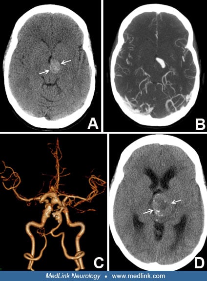
Case 2. A 36-year-old athletic female, without known vascular risk factors, presented during the third trimester of pregnancy with headache, slurred speech, and “twisted mouth.” Head CT scan showed an extra-axial mass compressing the left cerebral peduncle (A), recognized by CT angiography (B) as a partially thrombosed fusiform aneurysm of the left posterior cerebral artery. There was also tortuosity and ectasia of the basilar and left internal carotid arteries (C). Six years later, the vascular mass had expanded (D), producing severe midbrain compression and hydrocephalus, with spastic quadriparesis. She was treated with ventricular drainage and clipping of the left posterior cerebral artery proximal to the aneurysm.
Dilatative arteriopathy in cases like this, with severe progressive multifocal ectasia of cerebral arteries in the absence of a detectable generalized vasculopathy, can produce deficits due to cranial nerve and parenchymal brain compression as well as risks of ischemia or hemorrhage.
|
• Fusiform and dolichoectatic are the final common pathway of vessel wall injury with medial disruption and internal elastic lamina fragmentation. | |
|
• Fusiform and dolichoectatic aneurysm formation may be triggered by hemodynamic, inflammatory, infectious, or genetic factors, although many cases lack an identifiable cause and likely reflect an idiopathic degenerative process. | |
|
• Progression to non-saccular aneurysms involves internal elastic lamina disruption with intimal hyperplasia and neoangiogenesis, followed by intramural hemorrhage, thrombus formation, and eventual hematoma recanalization. |
Fusiform and dolichoectatic aneurysms may arise from a broad range of etiologies. However, they represent the final common pathway of vessel wall injury characterized by disruption of the tunica media and fragmentation of the internal elastic lamina. These findings have been consistently demonstrated in histopathological studies (29; 46; 28). Experimental models support this mechanism, showing that elastase-induced proteolysis can reproduce dolichoectasia and fusiform aneurysm formation, whereas high-flow states can similarly lead to internal elastic lamina breakdown and arterial enlargement (30; 10). The initiating insult to the tunica media may be from hemodynamic stress, inflammatory or infectious injury, or inherited disorders that weaken the vessel wall. In many cases, however, no clear cause is identified, suggesting a spontaneous degenerative process. Associated conditions are summarized in Table 1.
|
• Dissection |
Early reports attributed fusiform aneurysm formation to arteriosclerosis. This hypothesis has been refuted by subsequent pathological and imaging studies. Non-saccular aneurysms and dolichoectasia are characterized by luminal dilatation with disruption of the internal elastic lamina, loss of elastin with increased collagen deposition, medial thinning or degeneration, and typically concentric intimal thickening, resulting in an increased lumen-to-wall ratio. Additional features include upregulation of matrix metalloproteinases, arterial tortuosity, and, in advanced cases, intramural hemorrhage or stagnant flow adjacent to the vessel wall. In contrast, atherosclerosis is defined by the presence of lipid-laden plaques with cholesterol clefts or necrotic cores covered by a fibrous cap, often with eccentric intimal thickening, inflammatory infiltrates, neoangiogenesis, and calcifications, and is typically associated with luminal stenosis. High-resolution vessel wall imaging further supports these distinctions. Accordingly, non-saccular aneurysms and dolichoectasia should be distinguished from atherosclerosis unless true atheromatous pathology is demonstrated, and the term “atherosclerotic aneurysm” is often misleading (28).
Dolichoectasia, although not necessarily synonymous with fusiform aneurysm, is strongly associated with cerebral small vessel disease (71; 61). Across both stroke and non-stroke populations, it is linked to a 2- to 3-fold higher prevalence of imaging markers of small vessel disease, including white matter hyperintensities, silent lacunar infarctions, and cerebral microbleeds (12; 62; 61). Large and small cerebral arteries are structurally and functionally interconnected, and dolichoectasia may promote small vessel disease through hemodynamic alterations leading to distal hypoperfusion and neurovascular dysfunction. Maladaptive arterial remodeling, characterized by reduced pulse wave velocity and pulsatility index, may impair Windkessel function, increasing transmission of pulsatile energy to distal perforating vessels and contributing to microvascular injury (71). Alternatively, dolichoectasia and small vessel disease may arise in parallel from shared pathological mechanisms, including upregulation of matrix metalloproteinases and extracellular matrix rarefaction affecting the arterial media.
Genetics. In some cases, a clear genetic basis for non-saccular aneurysms can be identified. Fabry disease and late onset Pompe disease are hereditary lysosomal storage disorders in which the vacuolization of the vascular smooth muscle leads to the development of dolichoectasia (40; 41). In autosomal dominant polycystic kidney disease, dolichoectasia occurs in about 2% of cases (57). Rarely, in younger patients, fusiform aneurysms are due to congenital defects in the media connective tissue or smooth muscle cells, such as occurs in Marfan syndrome and Ehlers-Danlos syndrome type IV (33). Other genetic conditions associated with intracranial arterial dolichoectasia include pseudoxanthoma elasticum, fibromuscular dysplasia, neurofibromatosis type 1, sickle cell disease, alpha-1-antitrypsin deficiency, moyamoya disease, PHACE syndrome, and arterial tortuosity syndrome (38).
Other causes of non-saccular aneurysms. Fusiform aneurysms may arise from focal injury to the arterial wall caused by spontaneous dissection, trauma, or inflammation. Rarely, they develop secondary to infection of the arterial wall by bacteria or fungi, referred to as infective aneurysms. Additional reported causes include syphilitic arteritis and viral infections, such as HIV and varicella zoster. Autoimmune inflammatory conditions involving the central nervous system, including systemic lupus erythematosus, may also contribute to their formation. In uncommon cases, metastatic neoplastic infiltration of the arterial wall can lead to focal weakening and aneurysm development. Radiation-induced vasculopathy represents another rare mechanism. However, many fusiform aneurysms occur without an identifiable cause, suggesting an underlying idiopathic dilatative arteriopathy, as illustrated in Case 2.
Pathology and pathophysiology. A clinicopathological series of 16 chronic fusiform aneurysms described a characteristic sequence of histological changes (46). The proposed progression begins with disruption of the internal elastic lamina and associated intimal hyperplasia, followed by neoangiogenesis within the thickened intima. With further progression, intramural hemorrhage and thrombus formation occur, followed by hematoma recanalization, typically in aneurysms larger than 20 mm. Intimal neoangiogenesis was observed only in aneurysms greater than 12 mm, whereas intramural hemorrhage was restricted to those exceeding 28 mm.
Following aneurysm formation, lesions may undergo aggressive growth. Expansion of the aneurysm lumen is associated with low wall shear stress, promoting radial enlargement and endothelial dysfunction. This, in turn, facilitates mural thrombus formation, leukocyte transmigration, and matrix metalloproteinase–mediated extracellular matrix degradation. Additional contributors to aneurysm growth include wall neovascularization, intramural hematoma, and microdissections, which can further drive enlargement and mass effect (58).
|
• Fusiform and dolichoectatic aneurysms are uncommon in the general population but are found with increased prevalence in stroke patients. |
Fusiform and dolichoectatic aneurysms are relatively uncommon, accounting for approximately 3% to 13% of all intracranial aneurysms, and are most frequently located within the vertebrobasilar circulation (03). Their rarity is further illustrated in historical large-scale series: in an autopsy cohort exceeding 16,000 cases, only 15 fusiform aneurysms were identified (< 0.1%) (31), whereas in a series of 50,000 cerebral angiograms, 31 cases (0.06%) were detected, distributed across the internal carotid (n=14), vertebrobasilar (n=8), and combined circulations (n=9) (70). Yu and colleagues also reported a strong association with vascular risk factors, particularly hypertension (64%) and tobacco use (74%). Across studies, the mean age at presentation ranges from 40 to 50 years, although cases have been reported from childhood to advanced age (5–85 years) (34; 63). Additionally, males appear to have a higher risk of developing fusiform aneurysms compared with females.
Dolichoectasia, in contrast, is a relatively common vascular condition, particularly among patients with ischemic stroke. Its reported prevalence varies depending on the population studied and the diagnostic criteria applied. In non-stroke populations, vertebrobasilar dolichoectasia is observed in approximately 1% to 4% of individuals, whereas in stroke cohorts the prevalence increases to 8% to 15% (13) and may reach as high as 17% to 19% in series limited to posterior circulation strokes (05; 36). Established risk factors include advanced age, male sex, and hypertension, whereas associations with other traditional vascular risk factors are less consistent. The effect of aging is particularly notable, with arterial diameters increasing incrementally per decade, approximately 0.2 mm in the internal carotid artery and 0.1 mm in the basilar artery. Additionally, these vessels are, on average, larger in men by 0.24 mm and 0.21 mm, respectively (14). No significant differences in prevalence have been identified across racial or ethnic groups.
No specific strategies are known to prevent the development of fusiform and dolichoectatic aneurysms. However, given their association with vascular risk factors, particularly hypertension and tobacco use, strict control of these factors may help slow disease progression.
Giant non-saccular aneurysms can mimic tumors by producing headache, seizures, hydrocephalus, and focal neurologic signs. Pressure on isolated cranial nerves can result in optic atrophy, trigeminal neuralgia, hemifacial spasm, or other cranial neuropathies for which the differential diagnosis is broad. Hypopituitarism and a bitemporal visual field defect can mimic a pituitary neoplasm. There can be increased intracranial pressure with papilledema. Neurovascular imaging usually confirms the diagnosis of aneurysm. Occasionally, a fusiform aneurysm may be misinterpreted as a tumor on non-contrast brain imaging. Unless contrast or vascular imaging is pursued before a biopsy, a disastrous procedure may result.
Transient ischemic attacks and stroke can be caused by large fusiform or dolichoectatic aneurysms as well as by dolichoectasia. The results of a complete work up for other causes of stroke must be considered in reaching this conclusion. In such instances, investigation will frequently demonstrate intraluminal thrombus when the aneurysm is the cause for symptoms.
An abrupt onset of severe headache with or without focal neurologic symptoms suggests intracranial hemorrhage. Hemorrhage has sometimes been considered less likely with fusiform or dolichoectatic aneurysms than with saccular aneurysms. However, without question, hemorrhages can occur. In a long-term prospective study of 156 patients with vertebrobasilar dolichoectasia followed for an average of 9 years, six subarachnoid hemorrhages and 26 intraparenchymal hemorrhages occurred (49).
|
• Vessel imaging to better visualize the fusiform aneurysm can include CTA, MRA, or DSA. | |
|
• MRI/MRA are preferred imaging modalities for dolichoectasia/fusiform aneurysm characterization. | |
|
• CTA has similar advantages to MRA with good resolution for evaluating aneurysmal vascular lumen. | |
|
• High-resolution MRI vessel wall imaging detecting vessel wall enhancement can provide information on the stability of the aneurysm and early signs of aneurysm progression. | |
|
• Vessel imaging to better visualize the fusiform aneurysm can include CTA, MRA, or DSA. | |
|
• MRI/MRA are preferred imaging modalities for dolichoectasia/fusiform aneurysm characterization. |
The diagnostic approach is guided by clinical presentation. Suspected subarachnoid hemorrhage requires urgent non-contrast head CT, followed by lumbar puncture if imaging is negative and suspicion remains high, provided there is no posterior fossa mass effect. CT angiography is typically performed early and can reliably detect non-saccular aneurysms as well as most saccular aneurysms larger than 2 mm. In contrast, presentations with cranial nerve deficits or mass effect should prompt brain MRI with and without contrast and MR angiography.
Fusiform and dolichoectatic aneurysms are identified on imaging as segmental arterial dilatations measuring over 1.5 times the normal vessel diameter without a discrete neck (32). MRI with MRA is the preferred modality for initial detection and characterization, as it better defines true aneurysm size, vessel wall pathology, intraluminal thrombus, and effects on adjacent structures compared with catheter angiography, which primarily outlines the residual lumen. Advanced MRI techniques can detect wall enhancement and intramural hemorrhage, providing early markers of aneurysm progression (46), and may help differentiate primary fusiform aneurysms from dissections by demonstrating features such as an intimal flap or mural hematoma. High-field strength MRI further enhances vessel wall imaging resolution. CT angiography offers comparable spatial resolution for lumen assessment and is particularly sensitive to mural calcifications, with increasing utility in evaluating distal aneurysms. Catheter angiography remains important when detailed vascular anatomy is required to guide endovascular treatment decisions. Emerging imaging approaches may further refine risk stratification, as hemodynamic modeling using CTA and MRA has shown that unstable aneurysms are characterized by lower flow velocities and shear stress, along with increased flow oscillation (06). In addition, high-resolution vessel wall MRI demonstrates more frequent and extensive wall enhancement in fusiform aneurysms, with enhancement correlating with aneurysm size (37; 54).
For dolichoectasia, diagnostic criteria were first formalized in 1986 by Smoker and colleagues using computed tomography (59). Basilar artery ectasia was defined as a diameter greater than 4.5 mm at the level of the mid pons, corresponding to values exceeding two standard deviations above the population mean. Elongation and tortuosity were defined by lateral displacement beyond the clivus or dorsum sellae and by a high bifurcation above the suprasellar cistern. These criteria have since been adapted to MRI and MRA, which offer improved anatomical delineation of vascular abnormalities in relation to adjacent neural structures.
However, existing diagnostic frameworks have important limitations, including exclusion of the anterior circulation, reliance on small and nonrepresentative samples, subjective assessment of tortuosity, and limited clinical applicability. Emerging population-based data suggest that arterial diameter, rather than elongation or tortuosity, is the phenotype independently associated with increased risk of stroke and vascular mortality (15), supporting diameter-based definitions. Using a threshold of two standard deviations above the mean in stroke populations, proposed cutoffs approximate 7 mm for the cavernous internal carotid artery, 4 mm for the middle cerebral artery, and 5 mm for the basilar and vertebral arteries (13). Despite these advances, clinically relevant and standardized diagnostic criteria remain to be established. Furthermore, the risk associated with dolichoectasia appears to be largely driven by hemodynamic alterations, particularly stagnant or disturbed flow. Accordingly, adjunctive techniques that assess cerebral hemodynamics, such as transcranial Doppler, may play an increasingly important role in future evaluation.
There is a lack of prospective data guiding the management of fusiform aneurysms and intracranial arterial dolichoectasia. Treatment aims to reduce the risks of hemorrhage, ischemic stroke, and mass effect, yet the natural history of untreated lesions carries a high risk of complications and mortality (21; 47). Management is particularly challenging due to the absence of a defined aneurysm neck and the need to preserve distal blood flow, limiting the feasibility of conventional surgical clipping. Although a range of surgical and endovascular techniques are available, the optimal timing and selection for intervention remain uncertain and must be individualized. Intervention is generally considered in symptomatic cases, such as those with severe headache, cranial nerve or brainstem compression, hydrocephalus, or hemorrhage, as well as in lesions demonstrating growth on serial imaging, which has been associated with increased risk of rupture and death (42). Overall, treatment decisions require careful consideration of aneurysm location, size, symptoms, and hemodynamic features.
Medical treatment. In patients presenting with acute ischemic stroke, intravenous thrombolysis appears to be safe and effective, and management should follow current acute stroke guidelines (23; 51). Secondary prevention remains more controversial. Although early reports supported anticoagulation to reduce ischemic events (19), the potential for hemorrhagic complications may offset its benefits. In the absence of randomized trials, treatment decisions should be individualized based on patient-specific risk profiles. Conservative management typically includes antithrombotic therapy and aggressive control of vascular risk factors, such as hypertension and hyperlipidemia. In selected cases with luminal thrombus, surgical or endovascular approaches, including aneurysm exclusion or thrombectomy, may reduce embolic risk, though these interventions carry significant risks, including branch occlusion, distal flow compromise, thromboembolism, and arterial dissection.
Surgical treatment. Surgical treatments of non-saccular aneurysms include (1) reinforcement of the aneurysm dome by wrapping, (2) direct clipping, (3) resection and re-anastomosis, (4) trapping and distal bypass, (5) proximal occlusion, (6) transposition, and (7) thrombectomy, endarterectomy, and aneurysmorrhaphy. Reconstructive approaches like wrapping or clipping are generally reserved for lesions that cannot be excluded from the circulation, although wrapping alone is associated with higher rates of rebleeding and progression. Clipping is technically challenging due to the absence of a defined neck, though external remodeling with clips may be feasible in select cases.
Deconstructive strategies aimed at aneurysm exclusion remain the most employed surgical approach, typically achieved through proximal occlusion or trapping, leading to thrombosis and involution of the aneurysm (17). In vertebrobasilar lesions, flow reduction through sacrifice of a single vertebral artery may be effective when collateral circulation is adequate, whereas more extensive occlusion can be considered in select cases. When collateral flow is insufficient, bypass procedures are required to preserve distal perfusion. In patients with significant mass effect, trapping combined with decompression may be indicated. Additionally, in cases of symptomatic vascular tortuosity without thrombus, vessel transposition or microvascular decompression can alleviate brainstem or cranial nerve compression (52).
Endovascular treatment. Advances in catheter technology, coils, balloons, and stents have made endovascular therapy a key alternative to open surgery for fusiform and dolichoectatic aneurysms (72). Deconstructive techniques, such as parent artery occlusion and aneurysm trapping, can be achieved endovascularly using coils or detachable balloons. A key advantage of these approaches over surgical occlusion is the ability to perform a balloon occlusion test, allowing real-time clinical, angiographic, and electrophysiological assessment of collateral circulation and patient tolerance. In cases in which occlusion is not tolerated, combined strategies incorporating microsurgical bypass with endovascular treatment may be necessary. In contrast, reconstructive endovascular approaches aim to exclude the aneurysm while preserving flow through the parent artery. Techniques such as balloon-assisted coiling, stent-assisted coiling, and more complex constructs, including stent-in-stent configurations, have improved the ability to treat aneurysms with wide necks or circumferential involvement (39).
The introduction of flow-diverting stents represents a major advancement, utilizing dense mesh designs to redirect blood flow away from the aneurysm sac, thereby promoting intra-aneurysmal thrombosis and progressive reduction in aneurysm size while maintaining patency of the parent vessel and branch arteries. The Pipeline embolization device has emerged as a widely used tool in this context and has been applied successfully in both anterior and posterior circulation non-saccular aneurysms, including selected ruptured lesions. Furthermore, increasing experience suggests that flow diversion may be feasible even in smaller distal vessels (04; 48).
(A) Pre-procedural anterior and lateral angiographic views of a large (7 x 23 mmm) fusiform basilar artery aneurysm with two daughter sacs, presenting with third cranial nerve compression. (B) Angiographic reconstructed image a...
In a systematic review of 1704 patients with 1737 fusiform aneurysms, endovascular treatment was more commonly used than surgical approaches, particularly in recent years (65% vs. 31%) (63). Surgical management most often involved parent artery occlusion, frequently combined with bypass procedures, whereas reconstructive techniques, such as clipping were less common. Among endovascular strategies, stenting, including flow-diverter devices, and stent-assisted coiling were the predominant approaches. Overall, reconstructive techniques were favored over deconstructive ones (75% vs. 20%) and were associated with lower complication rates, as deconstructive strategies carried approximately double the risk of complications. In this meta-analysis, endovascular therapy emerged as the preferred modality, demonstrating lower mortality and complication rates compared with surgery. Outcomes also varied by location, with aneurysms in the anterior circulation generally associated with better prognosis.
Prevention of aneurysm progression. Finally, the better understanding of the pathophysiological events that lead to the formation and growth of fusiform and dolichoectatic aneurysm has suggested as a future goal the targeting of specific molecular pathways in the progression of these vascular malformations. Similarities in the biological characteristics between aortic aneurysms and non-saccular intracranial aneurysms suggest consideration of medical therapies proposed for prevention of aortic aneurysm growth, such as doxycycline or roxithromycin (53). Several trials have examined the effectiveness of losartan in restricting aortic aneurysm growth in Marfan syndrome with inconclusive results (22). Antiangiogenic drugs such as bevacizumab, rapamycin, or thalidomide, targeting neo-angiogenesis and bleeding within the thickened intima, may prove to have a role in slowing the progression of fusiform aneurysms (58).
There has also been a growing interest in targeting inflammatory pathways to slow progression of fusiform aneurysms. One target of interest is the tumor necrosis factor-alpha (TNF-alpha) inflammatory cascade. A small preliminary study using the TNF-alpha inhibitor infliximab suggested a decreased aneurysmal growth rate and stabilization in two treated patients (07). Other studies have taken a renewed interest in statins and their effect on potentially decreasing vessel wall inflammation and slowing the rate of aneurysm growth.
Surgical and endovascular treatment of fusiform aneurysms is generally reserved for larger, more often symptomatic or progressing aneurysms; therefore, outcomes cannot be directly compared to results observed in aneurysms treated expectantly. In a systematic review, intervention for fusiform and dolichoectatic aneurysms was associated with a substantial risk of complications, with new neurologic deficits occurring in 27% of cases, including stroke in 11.2% and hemorrhage in 3.6% (63). Surgical treatment carried a higher risk profile compared with endovascular approaches, with approximately a 2-fold increased risk of stroke or bleeding after intervention. Outcomes also varied by location, with anterior circulation aneurysms demonstrating a higher risk of complications. At a median follow-up of 3 years, aneurysm occlusion rates were comparable between surgical and endovascular treatments, achieved in 66% and 62% of patients, respectively. Mortality was highest among patients managed conservatively (43.4%), compared with 16.6% in surgically treated patients and 5.5% in those undergoing endovascular therapy. Overall, 45.5% of patients achieved a good functional outcome, highlighting both the risks and potential benefits of intervention in this complex disease.
No clear influence of pregnancy on aneurysm appearance or progression has been described.
Anesthesia techniques similar to those used for other difficult aneurysm surgeries should be used. Electroencephalographic and evoked potential monitoring and barbiturates titrated to burst suppression must be considered. When prolonged interruption of blood flow is likely, hypothermic circulatory arrest may be necessary.
All contributors' financial relationships have been reviewed and mitigated to ensure that this and every other article is free from commercial bias.
Victor J Del Brutto MD MS FAHA
Dr. Del Brutto of the University of Miami Miller School of Medicine and Jackson West Medical Center has no relevant financial relationships to disclose.
See Profile
Steven R Levine MD
Dr. Levine of the SUNY Health Science Center at Brooklyn has no relevant financial relationships to disclose.
See ProfileNearly 3,000 illustrations, including video clips of neurologic disorders.
Every article is reviewed by our esteemed Editorial Board for accuracy and currency.
Full spectrum of neurology in 1,200 comprehensive articles.
Listen to MedLink on the go with Audio versions of each article.
MedLink, LLC
3525 Del Mar Heights Rd, Ste 304
San Diego, CA 92130-2122
Toll Free (U.S. + Canada): 800-452-2400
US Number: +1-619-640-4660
Support: service@medlink.com
Editor: editor@medlink.com
ISSN: 2831-9125
Stroke & Vascular Disorders
May. 03, 2026
Stroke & Vascular Disorders
May. 03, 2026
Stroke & Vascular Disorders
May. 03, 2026
Stroke & Vascular Disorders
Mar. 10, 2026
Stroke & Vascular Disorders
Mar. 10, 2026
Stroke & Vascular Disorders
Mar. 10, 2026
Stroke & Vascular Disorders
Mar. 10, 2026
Stroke & Vascular Disorders
Mar. 10, 2026