Neuropharmacology & Neurotherapeutics
Zonisamide
Apr. 01, 2021
MedLink, LLC
3525 Del Mar Heights Rd, Ste 304
San Diego, CA 92130-2122
Toll Free (U.S. + Canada): 800-452-2400
US Number: +1-619-640-4660
Support: service@medlink.com
Editor: editor@medlink.com
ISSN: 2831-9125
Toll Free (U.S. + Canada): 800-452-2400
US Number: +1-619-640-4660
Support: service@medlink.com
Editor: editor@medlink.com
ISSN: 2831-9125
Nearly 3,000 illustrations, including video clips of neurologic disorders.
Every article is reviewed by our esteemed Editorial Board for accuracy and currency.
Full spectrum of neurology in 1,200 comprehensive articles.
Listen to MedLink on the go with Audio versions of each article.
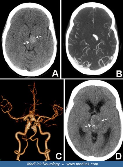
This 59-year-old male had hypertension, diabetes, and dyslipidemia and a history of coronary disease treated with coronary bypass grafting as well as abdominal aortic aneurysm repair. He presented with recurrent posterior circulation ischemic events, improving with intravenous tissue plasminogen activator on one occasion and intravenous heparin therapy on the second and third occasions. He was left with infarction in the territory of the left posterior cerebral artery, with right hemianopsia, and verbal memory deficits. CT scanning of brain revealed an ectatic basilar artery with mural calcifications (A). CT angiography demonstrated basilar artery dolichoectasia (B). He was treated with warfarin and remained stable for 3 years. He then presented with dysarthria, ataxia, and facial droop; MRI scan demonstrated an area of restricted diffusion in the right paramedian pons (C), and CT angiogram showed stable basilar artery ectasia with possible mural thrombus formation (D). (Contributed by Dr. J Brorson.)