
Infectious Disorders
Arboviral encephalitis
May. 15, 2026
MedLink, LLC
3525 Del Mar Heights Rd, Ste 304
San Diego, CA 92130-2122
Toll Free (U.S. + Canada): 800-452-2400
US Number: +1-619-640-4660
Support: service@medlink.com
Editor: editor@medlink.com
ISSN: 2831-9125
Toll Free (U.S. + Canada): 800-452-2400
US Number: +1-619-640-4660
Support: service@medlink.com
Editor: editor@medlink.com
ISSN: 2831-9125
Worddefinition
At vero eos et accusamus et iusto odio dignissimos ducimus qui blanditiis praesentium voluptatum deleniti atque corrupti quos dolores et quas.
Gram-negative meningitis in infants and adults is a severe, life-threatening infection. It requires prompt diagnosis and treatment due to high morbidity and mortality rates in these vulnerable populations.
Neonatal meningitis. Neonatal infections, especially in resource-limited settings, are commonly caused by bacteria such as Escherichia coli, Klebsiella, and Pseudomonas species. Escherichia coli, particularly those with the K1 capsular antigen, is a leading cause of gram-negative meningitis in newborns. Beta-hemolytic streptococci are also significant contributors to neonatal sepsis and meningitis worldwide (32). HIV infection predisposes spontaneous Gram-negative bacillary meningitis in neonates. The mortality and morbidity associated with Gram-negative meningitis remain significant despite advances in antimicrobial chemotherapy. Neonates frequently lack many of the classic features of bacillary meningitis, posing a diagnostic challenge.
Meningitis in older children and adults. Gram-negative bacilli are common organisms in nosocomial meningitis in adults. In patients with Gram-negative bacterial meningitis, Acinetobacter baumannii is emerging as an important causative agent. Nosocomial Gram-negative bacterial meningitis is a complication of various surgical procedures, such as craniotomy, placement of internal or external ventricular catheters, lumbar puncture, intrathecal infusions, or spinal anesthesia; head injury; or at times secondary to metastatic infection in patients with hospital-acquired bacteremia. An outbreak of meningitis and spinal infections with Gram-negative bacteria following local injections for treating chronic back pain was reported in Germany. Gram-negative meningitis may occur following endoscopic endonasal transsphenoidal surgery, but the frequency is quite low. CSF culture is the most important test to establish the diagnosis of Gram-negative meningitis. Metagenomic next-generation sequencing is now a widely used molecular technology that rapidly detects Gram-negative bacteria in CSF specimens of patients with nosocomial meningitis. New beta-lactam antibiotics like cefiderocol, ceftazidime-avibactam, and sulbactam-durlobactam show promising CSF penetration and efficacy against resistant pathogens.
|
Neonatal meningitis | |
|
• Gram-negative bacilli are major causative agents of meningitis in the neonatal period. | |
|
• Escherichia coli carrying the K1 capsular antigen is the most common Gram-negative bacillary organism causing neonatal meningitis. | |
|
• Group B streptococci also represent significant causes of neonatal infection and meningitis. These are not Gram-negative organisms. | |
|
• Gram-negative meningitis in neonates and in the elderly may present without typical signs of meningitis. | |
|
Adult meningitis | |
|
• Nosocomial Gram-negative bacterial meningitis may occur as a complication of neurosurgical procedures, head injury, or secondary to metastatic systemic infection. | |
|
• Gram-negative bacilli can be rare causes of spontaneous meningitis in adults, including individuals without risk factors. | |
|
• Gram-negative meningitis in neonates and in the elderly may present without typical signs of meningitis. | |
|
• Third-generation cephalosporins are highly active against most Gram-negative bacilli and have excellent CSF penetration. | |
|
• Although effective against Gram-negative infections outside the central nervous system, systemically administered aminoglycosides, such as gentamicin, have very poor penetration across the meninges, even in the presence of meningitis. | |
|
• Mortality and long-term sequelae rates are high among adults and children with Gram-negative bacterial meningitis. | |
"Gram-negative meningitis" is a term generally used to encompass those infections of the CSF and meninges due to bacteria such as Enterobacteriaceae and Pseudomonas aeruginosa occurring beyond the neonatal period, exclusive of Neisseria meningitis and Haemophilus influenzae, although some authors use the term to include all of the above. Occasional reports of Gram-negative bacillary meningitis date from as early as the 19th century. Case descriptions of Gram-negative meningitis following abortions, genitourinary procedures, and spinal anesthesia began to appear with some frequency in the 1930s and 1940s. An early literature review of 100 cases of Gram-negative meningitis found that most infections occurred in the neonatal period (04). The first large series of adults with Gram-negative meningitis was described in soldiers who suffered head injuries during World War II (36). Subsequent reports largely characterized Gram-negative meningitis as a nosocomial infection in adults who had undergone neurosurgical procedures (19; 40).
Neonatal Gram-negative meningitis is often diagnosed after the third postnatal day and is associated with higher white blood cell and red blood cell counts in cerebrospinal fluid, whereas adult Gram-negative meningitis typically presents with common meningeal symptoms such as fever, headache, photophobia, and altered mentation, but elderly patients may lack these classic features.
Neonatal meningitis. Neonatal Gram-negative meningitis may present without fever or nuchal rigidity, with atypical seizures, abrupt failure to thrive, bulging fontanelles, necessitating a low threshold for CSF examination. Among neonates, some differences have been noted between Gram-positive meningitis and Gram-negative meningitis. Gram-negative meningitis was more often diagnosed after the third postnatal day and was associated with higher white blood cell and red blood cell counts. Gram-negative meningitis diagnosed in the first 3 days of life was associated with antepartum antibiotic exposure. No difference was noted in either cerebrospinal fluid protein or glucose levels. Additionally, there were no differences in gestational age, birth weight, infant sex, race, or rate of caesarean section (57).
Adult meningitis. Clinical features of Gram-negative meningitis, in adults, often include typical signs and symptoms of any meningitis such as fever, headache, photophobia, neck stiffness, and altered mentation (including signs of cerebral dysfunction such as lethargy, delirium, confusion, or coma), but there is little to distinguish it specifically from other meningeal infections. In severe meningeal inflammation, signs of meningeal irritation may be present. Seizures and focal signs such as cranial nerve palsies may occur in up to 40% of patients (43). Focal neurologic deficits may occur through various mechanisms, including cortical vein or sagittal sinus thrombosis, cerebral artery spasm, subdural empyema, hydrocephalus, septic arteritis or endarteritis obliterans, abscess, or focal cerebral edema (66). In adults, the clinical features of Gram-negative meningitis may be obscured by concurrent surgical or posttraumatic conditions. Head injury, neurosurgical procedures, or underlying systemic illness can mask the classical triad of fever, neck stiffness, and altered sensorium, often delaying recognition and appropriate treatment of the infection.
Elderly patients, especially those with underlying medical illnesses like diabetes mellitus and cardiac disease, may lack many of the classic features of bacillary meningitis. Confusional state may be the only presenting feature in these patients (20).
Spontaneously arising Gram-negative meningitis more commonly occurs abruptly with a relatively fulminant course, whereas post-neurosurgical infection is frequently more insidious with a protracted illness (05).
Prior to the introduction of third-generation cephalosporins, mortality from Gram-negative bacillary meningitis was high, with Pseudomonas meningitis having an 84% mortality rate (15). Mortality rates fell from 34% (pre-1979) to 13% (1980-1988) (16).
Complications occur in up to 64% of patients, including cerebral edema, hydrocephalus, cranial nerve palsies, epidural abscess, subdural empyema, and brain abscess (40).
Factors associated with 30-day mortality or neurologic deterioration include decreased consciousness, blood glucose greater than 180 mg/dL, higher creatinine, and cerebrospinal fluid glucose less than 50 mg/dL (45).
High body temperature, a low CSF glucose, and meropenem-resistant Acinetobacter baumannii infections contribute to poor prognosis in multidrug-resistant cases (13). Advanced cases may involve cerebral edema, hydrocephalus, cranial nerve palsies, and brain abscess (40). Infant survivors of Gram-negative bacillary meningitis frequently experience developmental disabilities and neurologic sequelae, with 61% affected (64).
Neonatal meningitis. In neonates, certain E coli strains with the K1 polysaccharide capsule, which typically inhabit the large intestine of newborns, are the causative agents. The pathogenesis entails the migration of bacteria from the gastrointestinal tract to the bloodstream and subsequently to the central nervous system. The invasion of the central nervous system by E coli begins with the bacteria binding to and penetrating human brain microvascular endothelial cells. For E coli to effectively cross the blood-brain barrier, a significant level of bacteremia is necessary. The outer membrane protein A of E coli K1, which causes meningitis, aids in the invasion of brain microvascular endothelial cells by serving as an adhesion molecule for attachment (55). The penetration of the blood-brain barrier and subsequent CSF infection are facilitated by E coli factors, such as the K1 capsule, flagella, and type S fimbriae through the binding to and invasion of brain microvascular endothelial cells. Host inflammatory responses are crucial in the pathophysiology; these are triggered by bacterial lipopolysaccharides and cell wall components, leading to the release of cytokines and chemokines, activation of immune cells, and neuronal damage. Experimental studies have demonstrated meningeal inflammation and changes in blood-brain barrier permeability following the administration of purified lipopolysaccharide or cell wall products in animal models (60; 68; 09).
Meningitis in older children and adults. Meningitis caused by Gram-negative bacilli commonly occurs in newborns, post-neurosurgical patients, and those with Gram-negative bacteremia. Beyond the first month, Klebsiella species (40%), E coli (15% to 30%), and Pseudomonas aeruginosa (10% to 12%) are frequent causes. Nosocomial infections are increasing, especially in patients over 15 years old.
Gram-negative bacillary meningitis is most common in three settings: newborns, post-head injury, or neurosurgical procedures, and in patients with Gram-negative bacteremia. The causative organisms differ slightly according to the type of patient. In adults beyond the first month of life, the most common causes include Klebsiella species (about 40%), E coli (15% to 30%), and Pseudomonas aeruginosa (10% to 12%). The incidence of nosocomial Gram-negative meningitis is increasing. In a retrospective survey from a developing country, 59% of episodes of acute bacterial meningitis in patients aged 15 years or older were due to nosocomial infections, with Gram-negative bacilli being common (pathogens in 32.1%) of cases (30). Predisposing factors for nosocomial meningitis include previous treatment with broad-spectrum antibiotics, prematurity with low birthweight, and total parenteral nutrition.
Community-acquired Gram-negative bacillary meningitis, often caused by Klebsiella pneumonia, is seen in elderly persons and those who are debilitated, immunosuppressed, or who have alcoholism or diabetes (38; 12). HIV infection is another predisposing factor, with common organisms including Escherichia coli, Klebsiella pneumonia, and non-typhoidal Salmonella in HIV-positive patients (62).
Polymicrobial meningitis, though unusual, appears most commonly due to mixed Gram-negative infection. The frequency of nosocomial Gram-negative meningitis is also increasing, with Acinetobacter baumannii emerging as an important causative agent (52; 13). Gram-negative meningitis in post-neurosurgical patients is most often due to Klebsiella pneumonia, Acinetobacter calcoaceticus var anitratus, E coli, and P aeruginosa (40; 08; 05; 29).
Factors responsible for spontaneous Gram-negative bacilli meningitis include advanced age, presence of cancer, nosocomial exposure, and urinary tract infection (50). A retrospective review of adult patients who had Gram-negative bacilli cultured from CSF following neurosurgical procedures or traumatic head and spinal injury revealed Klebsiella pneumonia, Enterobacter cloacae, and E coli as the most frequent bacterial isolates (07). Acinetobacter meningitis is becoming increasingly common in the post-neurosurgical setting, with mortality exceeding 15% (31). Changes in the epidemiologic trend of acute bacillary meningitis include an increase in patients with a post-neurosurgical state and a rising incidence of Acinetobacter and staphylococcal infections (11).
Disruption of the dura-arachnoid barrier due surgery or trauma, in particular, with depressed skull fractures, can allow entry of skin flora into epidural or subdural spaces, and can provide a direct route for entry of Gram-negative bacilli. High-speed missile injuries like gunshot wounds generally do not produce significant contamination, but the risk of infection increases with delays in debridement. Furthermore, CSF rhinorrhea or otorrhea is associated with a higher incidence of infection, although pneumococcal infections are more common than those caused by Gram-negative bacilli (10). Congenital or anatomical defects, especially related to the neural tube or urogenital system and disseminated strongyloidiasis, can also predispose patients to Gram-negative meningitis.
Pathophysiology involves bacterial penetration of the blood-brain barrier and replication within the CSF, leading to host inflammatory responses triggered by lipopolysaccharides and cell wall components. These responses involve cytokines, chemokines, and immune cells such as granulocytes, macrophages, and microglia, which contribute to neuronal injury and cerebral edema. Meningeal inflammation, as observed in experimental studies with purified lipopolysaccharide and cell wall products from H influenzae, leads to permeability changes in the blood-brain barrier and increased intracranial pressure. The development of cerebral edema is multifactorial, involving vasogenic, cytotoxic, and interstitial mechanisms.
Meningitis causes purulent exudate, mainly composed of neutrophils and bacteria, which can lead to necrosis due to obstruction of meningeal arteries or veins. As the infection progresses, subarachnoid space exudate accumulates, causing noncommunicating hydrocephalus, interstitial cerebral edema, and potential herniation or infarction. Focal neurologic deficits may result from cranial or spinal nerve injury due to exudates, or from cortical and subcortical ischemia and infarction due to inflammation and thrombosis.
Once in the CSF, the low complement and immunoglobulin environment favors bacterial replication. The host inflammatory response to bacterial components, such as lipopolysaccharides, induces cytokine and chemokine release, leading to immune cell activation and neuronal injury. Neuronal damage is caused by hypoxic insult, neurotoxic bacterial products, and immune mediators, resulting in excitatory amino acid production, free oxygen radicals, nitric oxide, and peroxynitrite. This neuronal loss may lead to permanent neurologic sequelae or death.
In severe cases, meningitis leads to the development of a purulent exudate that covers brain surfaces, particularly the cisterns at the base. This exudate composed mainly of neutrophils and bacteria may cause necrosis due to obstruction of meningeal arteries or veins. Subarachnoid space exudate accumulation leads to noncommunicating hydrocephalus interstitial cerebral edema, and potential herniation or infarction. Focal neurologic deficits are often due to exudates injuring cranial or spinal nerves or from cortical and subcortical ischemia and infarction caused by inflammation and thrombosis.
There is no precise epidemiological information regarding Gram-negative meningitis. The overall annual attack rate of bacterial meningitis is roughly 3.0 cases per 100,000 population, although this varies according to age, race, and gender. Gram-negative bacilli account for 1% to 2% of bacterial meningitis in children (ages older than 1 month to 15 years) and approximately 1% to 10% of bacterial meningitis in adults (older than 15 years), although some information suggests the percentage may be as high as 11% to 17% in centers with active neurosurgical services.
Gram-negative meningitis outside of the neonatal period is most often a nosocomial infection. Nosocomial Gram-negative bacterial meningitis is a complication variety of surgical procedures, such as craniotomy, placement of internal or external ventricular catheters, lumbar puncture, intrathecal infusions, or spinal anesthesia; head injury; or at times secondary to metastatic infection in patients with hospital-acquired bacteremia (67). An estimated 50% of nosocomial bacterial meningitis cases occur following neurosurgical procedures and 30% occur after head trauma, especially when CSF rhinorrhea or otorrhea is present. The remaining 20% usually occur in a host of other predisposing conditions that are often found in debilitated patients who would be frequently infected or colonized with Gram-negative bacteria.
Postsurgical central nervous system infections occurred in 41 of 2401 neurosurgical procedures (1.7%) over a 10-year period in a tertiary hospital in Greece. Gram-negative bacteria predominated, with Acinetobacter baumannii isolated in 34.1% and Pseudomonas species in 14.6% of culture-positive cases. External ventricular drain infections were the most frequent source. Empirical therapy was appropriate in 63.4% of cases, yet mortality was high (65.7%). Survivors were significantly younger and had shorter ICU stays. Colistin-tigecycline combinations were the most used regimens. The median onset of infection was 11 days postoperatively. No significant outcome differences were observed based on infection type, pathogen, or comorbidities (41).
A series describing acute bacterial meningitis in 445 adults admitted to the Massachusetts General Hospital (1962 to 1988) identified 40% of the cases as nosocomial, with Gram-negative bacilli as a group accounting for 33% of nosocomial infections but only 3% of community-acquired episodes (16). In this study during the period 1971 to 1988, Gram-negative bacilli were the most common cause (surpassing S pneumoniae), suggesting that for at least some tertiary care centers, this category is now the most common cause of bacterial meningitis.
Of 17,594 CSF cultures analyzed at a Turkish hospital between 2020 and 2024, 1281 (7.3%) were positive. Gram-negative bacteria accounted for 396 (30.9 percent) of isolates. Klebsiella pneumoniae was the most common Gram-negative pathogen, found in 117 (9.1%) cases. Of these, 38 (32.4%) were extended spectrum beta lactamase producers. The study included both neonatal and adult patients. Nearly half of positive cultures were from adults, whereas neonates represented almost one quarter of all samples tested (22).
Communication between the CSF and the environment, often but not always signaled by a CSF leak, is a risk factor for postcraniotomy meningitis, as is perioperative steroid use. In a cohort of 324 patients who underwent craniotomy, almost 40% of the patients developed at least one infection. Meningitis was encountered in 16 procedures (4.8%), and CSF cultures were positive in all. Gram-negative pathogens (Acinetobacter spp, Klebsiella spp, Pseudomonas aeruginosa, Enterobacter cloaceae, Proteus mirabilis) represented 88% of the pathogens responsible for infections in patients undergoing craniotomy (34).
Paul and colleagues reported patients who developed meningitis and spinal infections with Gram-negative bacteria following local injections for the treatment of chronic back pain (47). They reported that 28 of 297 patients who received CT-guided spinal injections developed meningitis or spinal infections. Pseudomonas aeruginosa was responsible for iatrogenic meningitis or spinal infection following therapeutic spinal injections.
A metaanalysis revealed that Escherichia coli, Klebsiella, and Pseudomonas sp cause 20% to 28% of early-onset infant bacteremia and 14% cases of infant meningitis globally, particularly in low- and middle-income countries (24).
A 6-year retrospective study at a Saudi Arabian tertiary hospital analyzed 222 cases of bacterial meningitis, revealing Pseudomonas aeruginosa as the predominant pathogen (43%) (01). Neonates and children were most affected, with nosocomial infections comprising 92% of cases, primarily in intensive care units. Extended-spectrum beta-lactamase resistance was common (12.2%). The findings underscore the burden of bacterial meningitis and the growing issue of antimicrobial resistance.
Neonatal Gram-negative meningitis. Preventive strategies for neonatal Gram-negative meningitis primarily focus on addressing maternal and perinatal factors. Early diagnosis and prompt treatment of maternal infections, the judicious use of intrapartum antibiotics, adherence to aseptic techniques during delivery, and high-quality neonatal care are critical. Particular attention is required for preterm infants and those in neonatal intensive care units, as they are at increased risk for invasive Gram-negative infections. Measures to reduce nosocomial transmission, careful handling of invasive devices, and infection control practices are also key to prevention.
Gram-negative meningitis in older children and adults. Given the limited and sporadic nature of spontaneously arising Gram-negative meningitis in older children and adults, there are no specific community-level preventive measures. However, the question of whether prophylactic antibiotics prior to neurosurgical procedures predispose to Gram-negative infection remains controversial. Most neurosurgeons administer perioperative antibiotics, even for clean procedures, as multiple controlled and uncontrolled studies have shown reduced postoperative infectious complications among recipients (23). Buckwold and colleagues suggested that because most community-acquired or posttraumatic meningitis is caused by Streptococcus pneumoniae or Haemophilus influenzae, perioperative antibiotics may paradoxically increase the risk of Gram-negative infections (08). Other subsequent studies have not confirmed this association (40). A meta-analysis demonstrated that prophylactic antibiotic use significantly reduced postoperative meningitis following craniotomy, supporting their continued use in neurosurgical settings (02).
Gram-negative meningitis often cannot be distinguished on clinical grounds from bacterial meningitis due to other organisms but should be suspected in the hospitalized neurosurgical patient. The diagnosis of meningitis must be considered in any febrile patient with headache and lethargy and not attributed to other processes such as delirium tremens or hepatic encephalopathy unless ruled out by lumbar puncture (48).
Bacterial meningitis, in general, may produce signs and symptoms similar to brain abscess, subdural empyema, and epidural abscess, although these processes more frequently cause focal headache, pain, and neurologic deficits. Once lumbar puncture has been performed, and pleocytosis of the CSF established, the initial differential diagnosis must be broad if the answer is not apparent on Gram stain. Other considerations include viral meningitis, which often causes severe headache, but otherwise, patients are usually alert and awake. Serum C-reactive protein is capable of distinguishing Gram stain-negative bacterial meningitis from viral meningitis on admission with high sensitivity and high specificity (58).
Patients with space occupying lesions (eg, subdural empyema, brain abscess, or necrotic temporal lobe in herpes simplex encephalitis) may present with symptoms that appear to be similar with those of bacterial meningitis. In these patients, lumbar puncture may be complicated by brain herniation (66). Leptospiral, rickettsial, borrelial, and syphilitic infections may mimic an acute bacterial cause. Noninfectious etiologies such as sarcoid, rheumatologic illnesses such as Behçet disease, and malignancies such as lymphoma or metastatic carcinoma must be included, although there may be extra neurologic signs of illness to suggest these diagnoses.
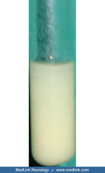
Lumbar puncture is crucial for diagnosing bacterial meningitis, often indicating elevated opening pressures (above 180 mm H2O), CSF pleocytosis, and a predominance of neutrophils. Gram staining can detect Gram-negative bacilli in half of the cases, and CSF culture is vital for definitive confirmation. In cases of bacterial meningitis, CSF analysis typically shows reduced glucose levels (commonly below 40 mg/dL or less than 40% of serum glucose) and increased protein concentrations (exceeding 100 mg/dL). These findings, combined with clinical symptoms and other CSF anomalies, are key to confirming the diagnosis. Normal CSF values differ greatly in neonates compared with older children or adults. Neonates often have higher protein, lower glucose, and different baseline white blood cell counts. These age-specific ranges must be considered when interpreting findings in Gram-negative meningitis.
Once meningitis is suspected, lumbar puncture provides the necessary confirming diagnostic information. Opening pressures are almost always elevated in cases of bacterial meningitis (higher than 180 mm H20). A Gram stain demonstrating Gram-negative bacilli can be found in up to 48% of patients, which is a significantly lower percentage than for the more common meningeal pathogens S pneumoniae (90%), H influenzae (85%), and N meningitidis (75%). Typical CSF leukocyte counts may range from 10 to 10,000, but 1000 to 5000 is the norm, with a neutrophilic predominance in most patients. Hypoglycorrhachia (lower than 50 mg/dL) may be present in up to 60% of patients, and CSF protein concentrations are often elevated due to disruption of the blood-brain barrier; however, the latter is not as specific for bacterial meningitis, and both measurements may be nearly normal in the immunocompromised patient. CSF culture is the most important test to establish the diagnosis of Gram-negative meningitis (63). Community-acquired Gram-negative meningitis is rare in patients without predisposing factors. Genitourinary, gastrointestinal, oral, and sinus sources of meningitis are common and should be looked for in patients with Gram-negative meningitis.
Initial diagnosis is often difficult in the postoperative neurosurgical patient with a fever and CSF pleocytosis, which may be ascribed to either early bacterial infection or to the normal consequence of postsurgical aseptic inflammation. Headache, fever, signs of meningeal irritation, seizures, and abnormal mental status in the setting of recent trauma or neurosurgery are suggestive of central nervous system bacterial infection (63).
There are few rapid diagnostic tests, other than the Gram stain, for identifying the subset of patients with Gram-negative infection. The Limulus lysate assay, prepared from amebocytes of the horseshoe crab Limulus polyphemus, can detect endotoxin present from Gram-negative pathogens (21). This assay can detect Gram-negative bacteria when present in quantities of >103 CFU/mL, with a sensitivity ranging from 71% to 97%. Because of its limited sensitivity and its inability to discriminate amongst the many types of Gram-negative bacteria, this test has not found widespread use as a diagnostic tool.
Metagenomic next-generation sequencing is now an increasingly available molecular diagnostic method that rapidly detects individual pathogens in biological specimens. In a series, metagenomic next-generation sequencing helped in identifying culprit microorganism in CSF specimens in neurosurgical patients with external ventricular and lumbar drainage-associated ventriculitis and meningitis (51). Earlier, in many of these patients, conventional tests had failed to detect any organism in the CSF.
Radiographic procedures have a limited role in the diagnosis of acute bacterial meningitis. Brain imaging (CT or MRI) should precede lumbar puncture if there is clinical evidence of focal neurologic deficit, moderate-to-severe impairment of consciousness, raised intracranial pressure (papilledema), where an intracranial mass lesion is suspected, and in cases with a major convulsive episode. CT scanning may be useful in the subset of patients with basilar skull fractures, as it may detect intracranial air or localize the site of fracture. Site of leaks may also be localized by radionuclide cisternography or by the use of water-soluble contrast dye injected intrathecally prior to CT scanning. Ventriculitis manifests with either intraventricular abscess, ependymal enhancement, or intraventricular loculations on neuroimaging (39).
Gram-negative bacillary meningitis in neonates and infants. Treatment of Gram-negative bacillary meningitis in neonates and infants remains a major clinical challenge because of antibiotic resistance and the variable penetration of drugs into the cerebrospinal fluid. Third-generation cephalosporins, including cefotaxime, ceftriaxone, and ceftazidime, are the preferred first-line agents in community acquired neonatal Gram-negative meningitis. These medicines are chosen because they reliably reach effective levels in the cerebrospinal fluid and show strong activity against Gram-negative bacilli.
When meningitis is caused by Gram-negative organisms that are resistant to cephalosporins, other agents may be required. Colistin, given either intravenously or intrathecally, can be considered in neonates with multidrug resistant organisms (49; 27). Aminoglycosides such as gentamicin are not preferred because they do not reach the cerebrospinal fluid in effective concentrations, even though they are active in other infections outside the blood-brain barrier. Historically, combinations such as intrathecal aminoglycosides with chloramphenicol were used, but these have been replaced by cephalosporins. This change has reduced mortality from very high levels of 40% to 90% down to 6% to 22% percent (14; Kaplan and Patrick 1990; 18).
New medicines are now being studied. Cefiderocol has penetration into the cerebrospinal fluid, ranging from 3% to 70% percent and has shown encouraging results, particularly in neonates. Ceftazidime combined with avibactam and ceftolozane combined with tazobactam also reaches useful levels in the cerebrospinal fluid and has been effective in resistant infections (25). Sulbactam combined with durlobactam and meropenem has worked against carbapenem resistant Acinetobacter infections.
Other medicines such as aztreonam, meropenem, fluoroquinolones, and trimethoprim combined with sulfamethoxazole may also be considered. Meropenem is preferred over imipenem because it is less likely to cause seizures (35; 69; 33; 06). Evidence for fluoroquinolones and trimethoprim combined with sulfamethoxazole in children is limited.
Giving antibiotics directly into the ventricles of the brain in children has not been beneficial and may even be harmful. Shah and colleagues reported that children receiving intraventricular antibiotics had three times higher risk of death compared with children receiving only intravenous treatment (54).
Corticosteroids such as dexamethasone are not recommended in neonatal Gram-negative meningitis because there is no evidence of benefit, and some studies suggest possible harm (03; 12; 59; 66).
Adult Gram-negative bacillary meningitis. In adults, treatment is also based mainly on third-generation cephalosporins such as cefotaxime, ceftriaxone, and ceftazidime. These medicines reach the cerebrospinal fluid well and provide effective coverage against most Gram-negative organisms. Ceftazidime is particularly valuable against Pseudomonas aeruginosa meningitis and has led to marked reductions in mortality compared with older aminoglycoside-based treatments (14; Kaplan and Patrick 1990; 18).
Hospital acquired Gram negative meningitis after neurosurgery or head injury often involves multidrug-resistant bacteria. Resistance to cephalosporins and carbapenems is now common (46; 65). In such cases, colistin given intravenously or intrathecally has shown good results, with rapid clearance of bacteria from the cerebrospinal fluid and acceptable levels of side effects (49; 27). Polymyxins, once avoided due to toxicity, have reappeared as effective options in these infections (17; 42).
Newer medicines such as cefiderocol, ceftazidime combined with avibactam, ceftolozane combined with tazobactam, and sulbactam combined with durlobactam provide additional treatment choices, especially when given together with carbapenems or in extended infusions (25).
Newer medicines such as cefiderocol, ceftazidime combined with avibactam, ceftolozane combined with tazobactuim, and sulbactum combined with durlobactam provide additional treatment choices, especially when given together with carbapenems or in extended infusions.
Intraventricular or intrathecal administration is sometimes used in adults with postsurgical meningitis or device-related infections. Studies have shown lower mortality and relapse rates when these methods are combined with intravenous treatment (61; 53; 56). A large analysis of 602 patients confirmed that giving antibiotics both intravenously and intraventricularly improved survival and cleared the infection without more side effects (37). Other reviews, however, found no clear advantage compared with intravenous treatment alone, so the benefit remains debated (28).
Meropenem is often chosen over imipenem due to lower seizure risk (33; 06). Aztreonam is another useful option because it reaches good cerebrospinal fluid levels. Fluoroquinolones and trimethoprim combined with sulfamethoxazole may be considered in resistant cases, though published experience is limited.
Guidelines from the National Institute for Health and Care Excellence recommend ceftriaxone alone for susceptible Enterobacterales meningitis in adults. Combination with ciprofloxacin did not improve outcomes and was linked with more short-term neurologic complications (44).
Corticosteroids such as dexamethasone are also not recommended in adult Gram-negative meningitis because they do not improve outcomes and may cause harm (12; 03; 66; 59)
All contributors' financial relationships have been reviewed and mitigated to ensure that this and every other article is free from commercial bias.

Ravindra Kumar Garg DM
Dr. Garg of King George's Medical University in Lucknow, India, has no relevant financial relationships to disclose.
See Profile
John E Greenlee MD
Dr. Greenlee of the University of Utah School of Medicine has no relevant financial relationships to disclose.
See ProfileNearly 3,000 illustrations, including video clips of neurologic disorders.
Every article is reviewed by our esteemed Editorial Board for accuracy and currency.
Full spectrum of neurology in 1,200 comprehensive articles.
Listen to MedLink on the go with Audio versions of each article.
MedLink, LLC
3525 Del Mar Heights Rd, Ste 304
San Diego, CA 92130-2122
Toll Free (U.S. + Canada): 800-452-2400
US Number: +1-619-640-4660
Support: service@medlink.com
Editor: editor@medlink.com
ISSN: 2831-9125
Infectious Disorders
May. 15, 2026
General Neurology
May. 13, 2026
Infectious Disorders
May. 12, 2026
Infectious Disorders
May. 12, 2026
Infectious Disorders
May. 05, 2026
Infectious Disorders
May. 01, 2026
Infectious Disorders
Apr. 30, 2026
Headache & Pain
Apr. 10, 2026