


Neurobehavioral & Cognitive Disorders
Mental status examination
Jun. 17, 2026
MedLink, LLC
3525 Del Mar Heights Rd, Ste 304
San Diego, CA 92130-2122
Toll Free (U.S. + Canada): 800-452-2400
US Number: +1-619-640-4660
Support: service@medlink.com
Editor: editor@medlink.com
ISSN: 2831-9125
Toll Free (U.S. + Canada): 800-452-2400
US Number: +1-619-640-4660
Support: service@medlink.com
Editor: editor@medlink.com
ISSN: 2831-9125
Worddefinition
At vero eos et accusamus et iusto odio dignissimos ducimus qui blanditiis praesentium voluptatum deleniti atque corrupti quos dolores et quas.
In this article, the author explains the clinical presentation, pathophysiology, prevention, diagnostic workup, and management of sensorineural hearing loss. Sensorineural hearing loss is most often caused by abnormalities in the hair cells of the organ of Corti in the cochlea. The hair cells may be abnormal at birth or damaged during an individual’s lifetime. There are both external causes of damage (eg, noise, trauma, and infection) and intrinsic abnormalities (eg, some genetic disorders). Less commonly, there may be retrocochlear dysfunction of the auditory nerve as a result of mass lesions, trauma, demyelination, infectious or inflammatory disorders, autoimmune processes, and nutritional disorders. This article does not address sudden sensorineural hearing loss, which is the subject of a separate article on sudden deafness.
|
• With sensorineural hearing loss, hearing is impaired for both air- and bone-conducted sounds. | |
|
• There are different audiogram patterns for different causes of sensorineural hearing loss: presbycusis is typically associated with a downward-sloping, high-frequency loss pattern; noise-induced hearing loss is associated typically with a notched pattern (generally at 4 kHz); and Meniere disease is associated with a low-frequency trough pattern. | |
|
• More than 50% of prelingual deafness is genetic, most often autosomal recessive and nonsyndromic. Genetic bases have also been identified for some postlingual deafness. | |
|
• Functionally significant hearing loss is very common in the elderly, affecting about a third of those aged 70 years or older, adversely impacting quality of life and ability to carry out routine activities and interact socially and, thereby, contributing to isolation, frustration, disappointment, and depression | |
|
• Situations where prevention is most likely to impact hearing outcomes are the prevention of noise-induced hearing loss and the prevention of drug ototoxicity. | |
|
• Cochlear implantation may improve audiologic performance and quality of life in elderly patients, even into their eighties |
In the second century, Galen of Pergamon (c130-c200) believed that hearing loss or deafness could be caused by dysfunction of the ear, acoustic nerve, or brainstem (121). Nevertheless, relatively little was known about the anatomy and physiology of the ear until the discovery of two of the middle ear ossicles (malleus and incus) in the early 16th century by Jacopo Beregario da Carpi (ca. 1460-ca. 1530) in the Provence of Modena, Italy (121).
In 1543, in his De humani corporis fabrica (On the fabric of the human body), Andreas Vesalius (1514-1564) of Brussels later named the two then-known ossicles the “malleus” and the “incus”; he was the first to illustrate these structures (170), and he may have discovered the tensor tympani muscle. Nevertheless, despite the great advancements in anatomy that Vesalius brought about, otology and, particularly, the anatomy of the inner ear were relatively neglected by Vesalius (121; 78).
Gabrielle Falloppio of Modena (Fallopius; 1523-1562) described all three auditory ossicles, the oval and round windows, the chorda tympani (although he was unclear whether this was a nerve), and the eponymous fallopian aqueduct or canal through which the facial nerve passes in the temporal bone (121).
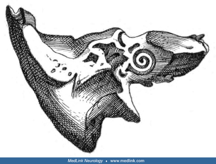
Fallopius recognized the two parts of the inner ear: the “cochlea” and the “labyrinth,” the latter being composed of the vestibule and the semicircular canals (121).
Bartolomeo Eustachi (1524-1574) apparently independently described the stapes, presented cross sections of the temporal bone, and also provided a more complete description of the eponymous Eustachian tube that links the nasopharynx to the middle ear than was known from vague descriptions by Aristotle, Celsus, Vesalius, and others (121).
Little progress was made in understanding the anatomy and physiology of hearing during the 17th century, particularly because after English physician William Harvey (1578-1657) published De Motu Cordis (On the Motion of the Heart and Blood) in 1628, anatomists and physiologists focused heavily on the cardiovascular system and ignored the sensory organs. English physician Thomas Willis (1621-1675) almost a half century after Harvey summarized the contemporary understanding of hearing (177).
He claimed that the auricle collected and channeled sound particles toward the tympanic membrane, which facilitated or prepared the sound for reception in the inner ear. The internal auditory muscles and the ossicles served to sort the sounds. Sounds are then transmitted through the oval window to reverberate in the semicircular canals before reaching the cochlea and then the acoustic nerve. Willis was the first to realize that the cochlea is the auditory sense organ, ie, the site of sensory transduction (121).
The most important otological research in the 18th century was that of Italian physician-scientist Antonio Scarpa (1747-1832). In his Disquisitiones Anatomicae de Audiu et Olfactu (1789), Scarpa was the first to describe the membranous labyrinth (121). Among the later 18th-century otological researchers was Dutch physician Fredrik Ruysch (1638-1731), whose meticulous drawings and skill in the technique of anatomical injection showed details as fine as the injected vessels in the mucous membrane of the complete ossicular chair.
In 1821, French physician Jean-Marc Gaspard Itard (1774-1838) published Traité des Maladies de l'Oreille et de l'Audition, a monograph that laid the foundation of modern otology with the results of Itard's studies of ear diseases in over 170 cases (79). Itard was also later recognized as an educator of deaf-mutes.
French otologist Prosper Ménière (1799-1862) devoted much of his career on diseases of the ear. Meniere studies at the Imperial Institute for Deaf Mutes in Paris (Institut Royal des Sourds et Muets à Paris) helped him recognize a group of patients with episodic and progressive auditory and vestibular symptoms, which he attributed to inner ear pathology (80).
Ménière established that vertigo might be a symptom of labyrinthine disease and specifically recognized a fairly homogenous group of patients with auditory and vestibular dysfunction that he linked to inner ear pathology. Although largely discounted during his lifetime, this work received increased recognition after his death when, in 1874, French neurologist Jean-Martin Charcot (1825-1893) labeled the disease “Maladie de Ménière.” Charcot also noted that episodic symptoms in affected patients ceased when the deafness became complete.
German physician, physiologist, and physicist Hermann Ludwig Ferdinand von Helmholtz (1821-1894) was a major contributor to sensory physiology (81).
While serving as professor of physiology at the University of Heidelberg, Helmholtz wrote Die Lehre von den Tonempfindungen als physiologische Grundlage für die Theorie der Musik (The Doctrine of the Sensations of Tone as a Physiological Basis for the Theory of Music, 1863), in which he introduced his place theory of frequency discrimination (58; 78). Helmholtz suggested that the ear could be understood as a sensory organ whose purpose is to decompose complex sounds (resulting from an admixture of various tones of different frequencies) into their component frequencies--a spectrum analyzer. Helmholtz proposed that each section of the tapering basilar membrane of the cochlea operated as an independent resonator with a natural period of vibration, or resonance, that responded only to sounds that vibrated at that period, like the tensioned strings on a harp. Based on the anatomy of the basilar membrane, which is three to four times wider at the apex than the base, Helmholtz postulated that high-frequency tones are perceived near the base of the cochlea, whereas lower frequencies are perceived toward the apex, just as short harp or piano strings produce high-frequency notes whereas long strings produce low-frequency notes. Helmholtz’s theory of hearing was a place theory, according to which the pitch of a sound is determined by the place where the membrane vibrates, in contrast to temporal or timing theories of hearing in which the frequency of neural firing determines the perception of pitch (78).
As early as 1885, an “auditory chart” was developed that graphically presented auditory acuity as a function of frequency, with responses based on testing with a set of standard tuning forks (172). Later means of graphically presenting auditory acuity as a function of frequency were developed in the early 20th century (172). In 1899, American psychologist Carl Emil Seashore (1866-1949) invented an audiometer, which was marketed initially around 1900 (139; 150; 102; 172).
In the 1920s, Western Electric developed a commercially successful electronic audiometer (172); this went through a series of technological improvements, including the incorporation of bone-conduction testing capability by 1928.
In the 1940s and 1950s, Hungarian-born biophysicist and physiologist Georg (György) von Bekesy (1899-1972) studied experimentally how sound is analyzed and communicated in the cochlea and extended research that had begun in the 19th century in psychoacoustics by Helmholtz (13; 14; 78). Bekesy invented an early type of automated audiometer, which was released in 1946 and continued in use until the 1990s. Bekesy went on to develop a modern place theory of hearing to explain how the cochlea functions as a frequency analyzer, work that overthrew Helmholtz’s earlier resonance theory. Bekesy’s theory was based on his discovery that sound vibrations travel along the basilar membrane in waves, peaking at different places where nerve receptors determine pitch and loudness. Bekesy found that the entire basilar membrane vibrates, although the point of maximum amplitude varies systematically with the fundamental frequency of the vibration because of the tapering shape of the membrane as it stretches along the length of the cochlea. This is the underlying basis for tonotopic or place coding, in which the inner ear hair cells with the greatest response code for the fundamental frequency of the sound. As Helmholtz had postulated for different reasons, Bekesy found that high-frequency tones are perceived near the base of the cochlea, whereas lower frequencies are perceived toward the apex. For his “discoveries of the physical mechanism of stimulation within the cochlea,” Bekesy was awarded the 1961 Nobel Prize in Physiology or Medicine (14; 80).
Other studies, however, have indicated that pitch is coded by a combination of rate information for lower frequencies and place information for higher frequencies; consequently, a number of rate-place schemes have been proposed (24). Moreover, since the 1970s, we have learned that the ear does not act passively, as both Helmholtz and Bekesy had thought, but instead is an active detector (86; 42; 173; 64; 50; 71).
|
• Prelingual hearing loss is present before speech develops, whereas postlingual hearing loss occurs after normal speech development. | |
|
• Children with postlingual deafness experience a much higher linguistic achievement level than those with prelingual deafness. | |
|
• Even children with mild to moderate sensorineural hearing loss have auditory processing deficits that adversely affect language development in children. | |
|
• Patients affected by sensorineural hearing loss have difficulty hearing the speech of others, which sounds mumbled or muffled, and typically have difficulty understanding conversations when there is background noise. | |
|
• Those with high-frequency sensorineural hearing loss, as from presbycusis or noise-induced hearing loss, have particular difficulty perceiving or discriminating high-pitched consonants (eg, ch, f, k, s, t, or th) compared to lower-pitched vowel sounds and also have more difficulty hearing the higher-pitched voices of women and children than they have hearing men’s voices. | |
|
• An individual has sensorineural hearing impairment if the average bone-conducted threshold at 500 Hz, 1000 Hz, and 2000 Hz (ie, the “speech-frequency pure-tone average”) is greater than 25 dB. | |
|
• Sensorineural hearing loss can be considered asymmetric if the interaural difference in pure-tone thresholds is at least 10 dB at two frequencies or at least 15 dB at one frequency. |
Prelingual versus postlingual hearing loss. Prelingual hearing loss is, by definition, present before speech develops, whereas postlingual hearing loss occurs after the development of normal speech. All congenital hearing loss is necessarily prelingual, but not all prelingual hearing loss is congenital. Children with postlingual deafness experience a much higher level of linguistic achievement than those with prelingual deafness. Children who are prelingually deaf and cannot hear sounds below 60 decibels (ie, about the loudness of a vacuum cleaner) will not be able to develop oral language skills comparable to those of their normal-hearing peers. Children born with profound hearing impairment (ie, of 90 decibels or more, or about the loudness of a food blender) generally have delayed or impaired speech, language, and reading development. Even children with mild-moderate unilateral sensorineural hearing loss have a higher frequency of speech and language delay, so such children also need close monitoring of speech, language, and auditory development and functioning (94).
Children with mild to moderate sensorineural hearing loss have auditory processing deficits that adversely affect language development in children (54). Deficits in a general auditory processing are necessary but not sufficient for language difficulties, whereas deficits in slow-rate amplitude modulation (envelope) detection are sufficient but not necessary for language difficulties, and deficits in the discrimination of a single speech contrast (eg, /bɑ/ vs. /dɑ/) are neither necessary nor sufficient for language difficulties (54).
Vestibular dysfunction is a common comorbidity associated with childhood sensorineural hearing loss (57; 45). Early identification of vestibular dysfunction in children with sensorineural hearing loss facilitates early intervention to mitigate the negative impact on motor, behavioral, and neurocognitive development (57; 100).
Sensorineural hearing loss and speech perception. Patients affected by sensorineural hearing loss have difficulty hearing the speech of others, which sounds mumbled or muffled, and typically have difficulty understanding conversations when there is background noise. Those with high-frequency sensorineural hearing loss, as from presbycusis or noise-induced hearing loss, have particular difficulty perceiving or discriminating high-pitched consonants (eg, ch, f, k, s, t, or th) compared to lower-pitched vowel sounds and also have more difficulty hearing the higher-pitched voices of women and children than they have hearing men’s voices. Some sounds may be distorted or perceived as overly loud: when talking at a regular pitch and volume, the patient may have difficulty discriminating speech sounds, whereas compensatory efforts by the speaker to talk in a lower register and more loudly can produce an irritated response from the patient (eg, “You don’t have to yell!”) (82). Talking very loudly or shouting does not necessarily help understanding because it primarily increases the intensity level of vowels rather than consonants; in addition, loud sounds are often uncomfortable for such patients because of recruitment.
Sound distortion and tinnitus. In patients with sensorineural hearing loss, sounds are also frequently distorted so that a pure tone may be heard as a buzz, broadband noise, or a complex mixture of tones. Some patients experience diplacusis (ie, the perception of a single auditory stimulus as two separate sounds, which may differ in pitch or in time).
Sensorineural hearing loss is often associated with subjective tinnitus, which for some patients may be the most troubling symptom as it may impair concentration, preclude adequate sleep, and cause or contribute to anxiety and depression.
Psychiatric associations of sensorineural hearing loss. Sensorineural hearing loss contributes significantly to isolation, stress, and depression and may also cause or contribute to paranoia (30; 190; 69; 148; 188; 189).
A metaanalysis of 20 studies comprising 675,291 individuals found bidirectional associations between sensorineural hearing loss and depression and anxiety (188). The incidence and risk of depression were significantly higher in sensorineural hearing loss subjects than in the general population. The risk of anxiety disorders was also significantly higher in sensorineural hearing loss subjects than in the general population. The prevalence of anxiety disorders among sensorineural hearing loss subjects was about 40%. The bidirectional associations between sensorineural hearing loss, depression, and anxiety were influenced by age.
Sensorineural hearing impairment. An individual has sensorineural hearing impairment if the average bone-conducted threshold at 500 Hz, 1000 Hz, and 2000 Hz (ie, the “speech-frequency pure-tone average”) is greater than 25 dB (10). Sensorineural hearing impairment is graded by the magnitude of the speech-frequency pure-tone average in the better ear, and for clinical purposes, pure-tone averages of 25 dB, 40 dB, and 60 dB can be considered the thresholds of mild, moderate, and severe hearing impairments (28; 51; 10). The World Health Organization grades of hearing impairment are similar but are assessed in the better ear based on a 4-frequency pure tone average of the hearing thresholds at 500, 1000, 2000, and 4000 Hz (Table 1) (179). The degree of hearing loss may also be specified for each frequency or for each frequency region (ie, low, mid, or high frequencies on the audiogram) based on the following broad frequency ranges: low (lower than 500 Hz), middle (501 to 2000 Hz), or high (higher than 2000 Hz) (Table 2) (21).
|
Hearing threshold level* |
Grade |
Impairment |
Description |
|
25 or less |
0 |
No impairment |
No or very slight hearing problems. Able to hear whispers. |
|
26-40 |
1 |
Slight |
Able to hear and repeat words spoken in a normal voice at 1 meter. |
|
41-60 |
2 |
Moderate |
Able to hear and repeat words using a raised voice at 1 meter. |
|
61-80 |
3 |
Severe |
Able to hear some words when shouted into the better ear. |
|
> 80 |
4 |
Profound |
Unable to hear even a shouted voice. |
|
| |||
|
Degree of hearing loss |
Hearing loss range (dB hearing level) |
|
Normal |
–10 to 15 |
Sensorineural hearing loss can be categorized by laterality (ie, unilateral or bilateral, with bilateral sensorineural hearing loss further categorized by symmetry in terms of both degree and configuration), onset (sudden or insidious), and course (static or stable, fluctuating, or progressive).
Asymmetric sensorineural hearing loss. Although a variety of definitions have been proposed in the literature, sensorineural hearing loss can be considered asymmetric if the interaural difference in pure-tone thresholds is at least 10 dB at two frequencies or at least 15 dB at one frequency (162). Asymmetric sensorineural hearing loss is fairly common, especially in those with a history of significant noise exposure (27; 119; 120; 162). Asymmetric sensorineural hearing loss can rarely indicate retrocochlear pathology, such as acoustic neuroma (162). Patients with asymmetric sensorineural hearing loss experience greater impairments in speech discrimination, binaural integration, and interaction compared to patients with symmetric sensorineural hearing loss (36).
In children, prelingual hearing loss can lead to delayed social development, delayed language acquisition, and social isolation from hearing peers. Such problems can be ameliorated by learning and using sign language, attending a school for the deaf, and self-identifying with deaf culture, although this can increase isolation from his or her parents, particularly if they do not know, or make an effort to learn, sign language.
Those with postlingual hearing loss are also more likely to suffer from isolation, and they also experience increased levels of stress and depression compared with their normal-hearing peers; this is particularly true in the elderly, for whom significant hearing loss may also cause or contribute to paranoia (30; 190; 69; 148). Sensorineural hearing loss may also contribute to the development of dementia in the elderly; in animal models, sensorineural hearing loss may lead to dementia-related pathological changes in hippocampal neurons (142).
|
• For sensorineural hearing loss, there is dysfunction of the cochlea (“sensory”) or the auditory nerve (“neural”); although the central auditory pathways could also be considered “neural,” hearing loss resulting from disorders of these pathways is traditionally considered separately as “central” hearing loss. | |
|
• A clinically important distinction in cases of sensorineural hearing loss is whether the condition is “sensory” (ie, cochlear) or whether it is “neural” (ie, “retrocochlear”). | |
|
• Cochlear disorders are typically the purview of audiologists and otolaryngologists whereas retrocochlear disorders are much more likely to reflect neurologic disorders, some of which may require neurosurgical intervention. | |
|
• Sensorineural hearing loss is most often caused by abnormalities in the hair cells of the organ of Corti in the cochlea. |
Sensorineural disorders involve dysfunction of the sensory receptors or conduction of signals from the sensory receptors to the brain. For sensorineural hearing loss, there is dysfunction of the cochlea (“sensory”) or the auditory nerve (“neural”). Although the central auditory pathways could also be considered “neural,” hearing loss resulting from disorders of these pathways is traditionally considered separately as “central” hearing loss. A clinically important distinction in cases of sensorineural hearing loss is whether the condition is “sensory” (ie, cochlear) or whether it is “neural” (ie, “retrocochlear”). Cochlear disorders are typically the purview of audiologists and otolaryngologists whereas retrocochlear disorders are much more likely to reflect neurologic disorders, some of which may require neurosurgical intervention.
Sensorineural hearing loss is most often caused by abnormalities in the hair cells of the organ of Corti in the cochlea, ie, “sensory” rather than neural loss. The hair cells may be abnormal at birth or may be damaged during an individual’s lifetime. There are both external causes of damage (eg, noise, trauma, infection, and radiation) and intrinsic abnormalities (eg, some genetic disorders). Less commonly, there may be dysfunction of the auditory nerve from mass lesions, trauma, demyelination, infectious or inflammatory disorders, autoimmune processes, and nutritional disorders.
Congenital cytomegalovirus is the most common cause of nongenetic sensorineural hearing loss in children (133). In a retrospective chart review of 44 cases of congenital cytomegalovirus infection, 75% had sensorineural hearing loss of varying degree and age at onset: (1) 39% (n=33) passed the universal newborn hearing screen bilaterally, and of these 35% (n=6) ultimately acquired bilateral or unilateral sensorineural hearing loss, detected at a median age of 12 months (mean 20 months); (2) 23% (n=10) did not pass UNHS in one ear and of these 50% (n=5) acquired late-onset hearing loss in the contralateral ear at a median age of 4 months (mean 24 months). Of the 33 children with congenital cytomegalovirus and sensorineural hearing loss, 55% received antiviral medication (ganciclovir and/or valganciclovir). Although both treated and untreated ears experienced progression of hearing loss over time, the group that received antiviral treatment experienced less overall hearing change compared with the untreated group.
|
• Hearing loss is the most common birth defect and the most prevalent sensorineural disorder in developed countries. | |
|
• About 70% of genetic hearing loss is nonsyndromic, and about 30% is syndromic. | |
|
• More than 50% of prelingual deafness is genetic, most often autosomal recessive and nonsyndromic. | |
|
• Half of severe childhood deafness results from simple mendelian inheritance, the vast majority (87%) of which results from autosomal recessive mutations. | |
|
• For individuals 12 years and older in the United States, about 13% (approximately one in eight) have bilateral hearing loss, and about 20% (approximately one in five) have a unilateral or bilateral hearing loss. | |
|
• The prevalence of hearing loss increases with age. |
Hearing loss is the most common birth defect and the most prevalent sensorineural disorder in developed countries (60; 61). Two per 1000 newborns have bilateral permanent sensorineural hearing loss of at least moderate severity (40 dB or greater), but by adolescence, the prevalence increases to 3.5 per 1000 (108).
Etiology of pediatric sensorineural hearing loss. Genetic hearing loss may appear as an isolated finding or as part of a syndrome (68; 125). About 70% of genetic hearing loss is nonsyndromic, and about 30% is syndromic. More than 50% of prelingual deafness is genetic, most often autosomal recessive and nonsyndromic. Most genes associated with genetic forms of deafness are involved in hair bundle morphogenesis, form constituents of the extracellular matrix, function to maintain ion homeostasis in the cochlea, or serve as transcription factors.
Half of severe childhood deafness results from simple mendelian inheritance, the vast majority (87%) of which results from autosomal recessive mutations (44; 125). Consanguineous progeny have a 76% higher risk of developing sensorineural hearing loss than nonconsanguineous progeny (137). Recessive inheritance is responsible for two thirds of congenital deafness (106). At least 16% of the normal population are carriers of a gene for congenital deafness (106; 107). More than 80 genetic loci have been identified among those with autosomal recessive deafness, with responsible loci present on every autosome (ie, a chromosome that is not an allosome or sex chromosome). At least half of autosomal recessive nonsyndromic hearing loss is attributed to autosomal recessive deafness 1A (DFNB1A; OMIM 22090), autosomal recessive deafness 1B (DFNB1B; OMIM 612645), or digenic GJB2/GJB6 deafness (OMIM 22090). DFNB1A is caused by mutations in the GJB2 gene (the “gap junction protein, beta-2” gene) on chromosome 13q12.11, which encodes the gap-junction protein connexin 26, whereas DFNB1B is caused by mutations in the GJB6 gene (the “gap junction protein, beta-6” gene) on chromosome 13q12.11, which encodes the gap-junction protein connexin 30 (116; 05). Genes less frequently implicated in autosomal recessive nonsyndromic hearing loss are SLC26A4, MYO15A, OTOF, (DFNB9, encoding otoferlin), CDH23, TMC1, CLDN14 (DFNB29), TRIOBP, and mtDNA 12S rRNA (175; 74; 93; 122; 67). None of the genes associated with autosomal dominant nonsyndromic hearing loss accounts for a preponderance of cases.
In a study of more than 136,000 children across the United States, elevated blood lead levels greater than 3.5 μg/dL were 1.22 times more likely to have sensorineural hearing loss than those without elevated blood lead levels (129).
Sensorineural hearing loss in older children and adults. For individuals 12 years and older in the United States, 30 million or about 13% (approximately one in eight) have bilateral hearing loss, and 48 million or about 20% (approximately one in five) have a unilateral or bilateral hearing loss (92). The prevalence of hearing loss increases with age; this change reflects the individual impacts of genetic and environmental factors as well as the interactions between specific environmental triggers and an individual's genetic predisposition, as illustrated by aminoglycoside-induced ototoxicity (82). The prevalence of hearing loss is higher in men than women across nearly all age decades and for nearly all regions of the world (03; 92; 180).
Both cross-sectional and prospective analysis indicated that shift work, night shift work, and physically demanding work are associated with increased risk of occurrence, laterality, and severity of sensorineural hearing loss (117).
Sensorineural hearing loss in the elderly. Functionally significant hearing loss is very common in the elderly, affecting about a third of those age 70 years or older (23). This can adversely affect quality of life and compromise an older individual’s ability to carry out routine activities and interact socially, thereby contributing to isolation, frustration, disappointment, and depression (110; 111). The ability to understand spoken speech is often interpreted as an indicator of cognitive abilities, especially in the elderly, so impaired hearing often negatively influences how other people perceive and interact with an elderly person; it also contributes not infrequently to inappropriate diagnoses of dementia by clinicians. In addition, hearing impairment is a significant predictor of postural imbalance and falls in children and older individuals (171; 100).
The underlying causes of sensorineural hearing loss in the elderly are varied and include cumulative effects of prior toxic exposures, noise-induced hearing loss, age-related changes, and genetic susceptibility. Various genes are involved in late-onset hearing loss, and a significant portion of cases of late-onset hearing loss is due to genetic causes (160).
Specific risk factors. A population-based cohort study in Taiwan found that patients with sensorineural hearing loss have a higher incidence of developing glaucoma, but the replicability, potential mechanisms, and significance of this finding are, as yet, unclear (26).
A systematic review and meta-analysis also identified iron-deficiency anemia as a possible risk factor for sensorineural hearing loss (105). The odds of sensorineural hearing loss were 55% higher in individuals with iron-deficiency anemia than those without. The age-specific odds ratios were 1.36 in adults and 3.67 in children.
In the National Health and Nutrition Examination Study (NHANES) database, using a national sample of the U.S. population, the presence of chronic obstructive pulmonary disease (COPD) was associated with elevated hearing thresholds (worse hearing) at each frequency (141). The presence of chronic obstructive pulmonary disease was independently associated with a 3.3 dB increase in the high-frequency pure tone average (3, 4, 6, 8 kHz), and 2.3 dB increase in the low-frequency pure tone average (.5, 1, 2 kHz) after controlling for medical, social, and environmental covariates (141). The presence of chronic obstructive pulmonary disease was independently associated with a 1.85-fold increased odds of isolated low-frequency sensorineural hearing loss.
A systematic review of cross-sectional and case-control studies of the association of type I diabetes with sensorineural hearing loss identified 11 studies of collectively 5792 patients (104). Most of the studies showed that type I diabetes was associated with hearing impairment compared to controls, including differences in pure-tone averages and otoacoustic emissions, increased mean hearing thresholds, altered acoustic reflex thresholds, and problems with the medial olivocochlear reflex inhibitory effect. Significant risk factors for sensorineural hearing loss in type I diabetes included older age, increased disease duration, and higher HbA1C levels.
A retrospective cohort study of approximately 3.6 million adult subjects (age 18 years or older) found that both nicotine and smoke exposure seem to be strongly associated with increased odds for developing sensorineural hearing loss, with chewing tobacco having the strongest association (89); whether this is a causal association or whether tobacco serves only as a proxy for socioeconomic status is unclear.
In a meta-analysis of 18 studies, involving 27,859 cases affected by autoimmune diseases sensorineural hearing loss was significantly related to systemic lupus erythematosus, rheumatoid arthritis, and vitiligo (90); the prevalence of sensorineural hearing loss was 21% in systemic lupus erythematosus, 16% in rheumatoid arthritis, and 39% in vitiligo (90).
|
• Because there is no effective medical or surgical therapy for most instances of sensorineural hearing loss, prevention is essential. | |
|
• Situations where prevention is most likely to impact hearing outcomes are the prevention of noise-induced hearing loss and the prevention of drug ototoxicity. | |
|
• Although at-risk subjects should use hearing protection, a significant proportion of at-risk workers use hearing protection either inconsistently or not at all. | |
|
• Sound does not have to be annoying or painful to cause hearing loss over time. | |
|
• Hearing protection should be worn if individuals find it necessary to raise their voice to carry on a conversation easily. | |
|
• Precautions should be followed to minimize ototoxicity: (1) document preexisting hearing loss or vestibular dysfunction before prescribing vestibulotoxic or ototoxic medications; (2) administer aminoglycosides for no longer than 1 week; (3) avoid aminoglycosides in patients whose calculated creatinine clearance is less than 1.2 L/h; (4) monitor peak and trough aminoglycoside levels and adjust dosing accordingly; (5) use extended dosing intervals (such as once daily instead of multiple daily administration); (6) avoid combinations of aminoglycosides with other nephrotoxic or ototoxic drugs; (7) and discontinue ototoxic agents when clinical vestibulotoxicity or ototoxicity is detected. |
There is no effective medical or surgical therapy for most instances of sensorineural hearing loss. Therefore, prevention is essential. The situations where prevention is most likely to impact hearing outcomes are the prevention of noise-induced hearing loss and the prevention of drug ototoxicity.
Unfortunately, present health promotion initiatives for hearing loss prevention are insufficient and are largely limited to the prevention of noise-induced hearing loss (18; 32; 157). Hearing protection should be used by at-risk subjects (eg, workers in heavy industry, foundry workers, textile industry workers, mill workers, heavy equipment operators, construction workers, machine operators and assemblers, bottling plant workers, soldiers and military aviators and aircrew, professional musicians and sound technicians, miners, farmers, and other agricultural workers, etc.). Unfortunately, a significant proportion of at-risk workers use hearing protection either inconsistently or not at all (112).
There is contradictory evidence that hearing loss prevention programs are effective in the long term (166). Low-quality evidence suggests that stricter legislation can reduce noise levels in workplaces, with substantial reductions demonstrated in some individual circumstances, but there are no controlled studies of the effectiveness of such measures in preserving hearing (166). The limited success of hearing loss prevention programs may be explained by limited use of hearing protection, suboptimal knowledge of noise as a hazard, workplace noisiness, and the benefits of hearing protection devices among some workers (126). Even when full-shift equivalent noise levels are well above the level at which hearing protection devices are required, usage rates are often quite low (35). Improvements are needed in implementing, reinforcing, and evaluating technical interventions and long-term effects. Education is critical because many affected individuals are reluctant to wear hearing protection, and many who wear it do not wear it consistently or correctly. If feasible, environmental controls should be placed to control noise exposure (32; 46). Concurrent with engineering controls, a range of hearing protection devices should be available free of charge, and hearing protection training should be reviewed, particularly for lower-skilled workers (126). Unfortunately, most companies give inadequate attention to environmental noise controls and instead rely primarily on hearing protectors to prevent hearing loss (32; 33). However, without noise controls at the source, exposed workers remain at unnecessary risk (33). Indeed, about two fifths of employees (38%) do not use hearing protectors routinely (32). Hearing protection use correlates with the completeness of company hearing protection programs, suggesting that underuse of hearing protection by employees is “in some substantial part, attributable to incomplete or inadequate company efforts” (32).
In an occupational setting, the Occupational Safety and Health Administration requires baseline and annual audiometric pure-tone air-conduction threshold testing of both ears at 500, 1000, 2000, 3000, 4000, and 6000 Hz for employees exposed to at least 50% of the permissible noise exposure level (OSHA Law and Regulations: 1910.95 Ocupational Noise Exposure). Testing is generally scheduled at least 14 hours after the employee's last noise exposure to minimize the effect of temporary threshold shifts; if this is not possible, employees should wear hearing protection until tested. If an employee's noise exposure exceeds the Occupational Safety and Health Administration’s permissible exposure level, employers may need to establish environmental controls to reduce noise, issue hearing protectors, or rotate employees to less noisy tasks for part of the work day. Some employers issue hearing protection even when the Occupational Safety and Health Administration standards are met because, even at somewhat lower noise levels, some employees may develop permanent noise-induced hearing loss (114). A further complexity is that the Occupational Safety and Health Administration regulations do not cover a variety of workers exposed to excessive noise, including farmers, construction workers, and employees of small businesses where noise levels often exceed recommended levels.
Recreational noise exposure is usually less frequent and of shorter duration than occupational noise exposure, but it may compound occupational noise damage. Many power tools, lawnmowers, firearms, sound speakers, and personal listening devices (eg, MP3 players) exceed safe sound exposure levels. For example, approximate dBA sound levels are 90 for a lawnmower, 100 for a chainsaw, 120 for a rock concert, and 140 for a shotgun blast. The impulsive noise of firearms may be particularly damaging to the cochlea (147; 155). The main hearing loss-related risk factor for teenagers is exposure to recreational noise: frequent attendance at discotheques and pop-music concerts, use of personal stereos, and noise exposure in school workshops (96; 59; 49; 152; 154). Most students are unconcerned about potential effects of loud noise, and the use of hearing protection is minimal (less than 5%) (49).
Sound does not have to be annoying or painful to cause hearing loss over time. Hearing protection should be worn if individuals need to raise their voice to carry on a conversation easily. Hearing protection devices commonly available include earmuffs or earplugs. Ear muffs usually have higher noise-reduction ratings than earplugs; they can be worn in combination in extremely high noise environments, but the maximum noise reduction is approximately 50 dB because of bone conduction through the skull (114), and combination use of earplugs and earmuffs disrupts sound localization (143). Some individuals find ear muffs to be hot, cumbersome, or cosmetically unappealing, any of which can adversely affect usage (08). Training in earplug insertion is important for good attenuation (158). For difficult-to-fit external auditory canals, custom earplugs can be made from an earmold impression. Special earplugs are also available for special needs (eg, for cosmetic considerations, hunting, singing, band members, or specific work environments). In military situations, active noise cancellation earmuffs equipped for communication purposes seem to be the best protection for soldiers during military exercises (115). Misfit earplugs or earmuffs are much less effective and may not adequately protect hearing. Hearing aids (most of which are vented), cotton balls, and earplugs designed for swimming are all inadequate for hearing protection.
Whenever possible, the following precautions should be followed to minimize ototoxicity (22; 97; 159): (1) document preexisting hearing loss or vestibular dysfunction before prescribing vestibulotoxic or ototoxic medications; (2) administer aminoglycosides for no longer than 1 week; (3) avoid aminoglycosides in patients whose calculated creatinine clearance is less than 1.2 L/h; (4) monitor peak and trough aminoglycoside levels and adjust dosing accordingly; (5) use extended dosing intervals (such as once daily instead of multiple daily administration); (6) avoid combinations of aminoglycosides with other nephrotoxic or ototoxic drugs; (7) and discontinue ototoxic agents when clinical vestibulotoxicity or ototoxicity is detected. To avoid permanent impairment, aminoglycoside therapy should cease as soon as symptoms of either auditory or vestibular ototoxicity appear (55). If identified early, much of the symptomatic toxicity is reversible (174; 41; 16).
Specific medications may help prevent sensorineural hearing loss in specific populations. In type 2 diabetics, the use of statins is significantly associated with a 25% reduced risk of sensorineural hearing loss, regardless of age and sex (56). In obese hypertensive men (primarily middle-aged, white men), the use of calcium-channel blockers is associated with a 24% reduced risk of sensorineural hearing loss (153).
Congenital disorders include various genetic factors (including causes of otic capsule dysplasia syndromes and rare chromosomal syndromes), intrauterine infections (rubella, cytomegalovirus, herpes simplex, syphilis), and maternal drug or alcohol abuse (181). Genetic factors cause more than 50% of congenital hearing loss (15). Genetic hearing loss may be autosomal dominant, autosomal recessive, or X-linked.
Multiple conditions cause acquired sensorineural hearing loss after birth, including (1) infectious and postinfectious conditions (eg, COVID-19, suppurative labyrinthitis, meningitis, measles, mumps [ie, epidemic parotitis], herpes zoster oticus [ie, Ramsay Hunt syndrome], zika, and Lyme neuroborreliosis)(144); (2) ototoxic drugs (eg, aminoglycosides [eg, tobramycin], loop diuretics [eg, furosemide], antimetabolites [eg, methotrexate], alkylating agents [eg, cisplatin], salicylates [eg, aspirin], proton pump inhibitors) and drug overdose (eg, methamphetamine) (131); (3) autoimmune factors (including Susac syndrome); (4) traumatic head injury (eg, inner ear concussion, perilymph fistula, shear injury of the eighth nerve, temporal bone fracture); (5) Meniere disease; (6) noise-induced cochlear injury; (7) vascular injury (labyrinthine infarction, superficial siderosis); (8) neoplastic and paraneoplastic conditions (eg, cerebellopontine angle tumors [eg, “acoustic neuroma” or vestibular schwannoma, and meningioma], carcinomatous meningitis) (02), paraneoplastic subacute hearing loss or cochleovestibulopathy); (9) cranial radiation therapy (66); (10) nutritional disorders (eg, profound B-vitamin deficiencies); (11) intracranial hypertension; (12) sickle-cell disease; and (13) age-related changes (ie, presbycusis) (12; 113; 62; 99; 151; 167; 184). Diabetes and chronic obstructive pulmonary disease are also associated with an increased risk of sensorineural hearing loss, but the pathogenetic mechanisms are unclear (136; 141). Vitiligo is associated with an increased risk of sensorineural hearing loss, presumably indicating an autoimmune mechanism (88; 91).
Multiple conditions cause acquired sensorineural hearing loss after birth, including infectious and postinfectious conditions (eg, suppurative labyrinthitis, meningitis, measles, mumps [ie, epidemic parotitis], herpes zoster oticus [ie, Ramsay Hunt syndrome]), ototoxic drugs (eg, aminoglycosides [eg, tobramycin], loop diuretics [eg, furosemide], antimetabolites [eg, methotrexate], and salicylates [eg, aspirin]), autoimmune factors (including Susac syndrome), traumatic head injury (eg, inner ear concussion, perilymph fistula, shear injury of the eighth nerve, temporal bone fracture), Meniere disease, noise-induced cochlear injury, vascular injury (labyrinthine infarction, superficial siderosis), neoplastic and paraneoplastic conditions (eg, cerebellopontine angle tumors [eg, “acoustic neuroma” or vestibular schwannoma, and meningioma], carcinomatous meningitis, paraneoplastic subacute hearing loss or cochleovestibulopathy), cranial radiation therapy, nutritional disorders (eg, profound B-vitamin deficiencies, as seen in prisoners of war), and age-related changes (ie, presbycusis) (12; 113).
Subacute hearing loss with or without associated vestibular dysfunction can occur as a paraneoplastic condition in association with small-cell lung cancer, thymoma, neuroblastoma, and B-cell follicular lymphoma (98; 53; 43; 168; 132; 40; 63). In patients with paraneoplastic subacute hearing loss, the damage is generally cochlear or cochleovestibular. Some cases of subacute hearing loss may have anti-Hu antibodies (43; 63; 132) or glutamic acid decarboxylase autoantibody (168), but the antigens and antibodies involved in paraneoplastic cochleovestibulopathy remain unknown (53), and, consequently, the mechanism of paraneoplastic cochleovestibulopathy is not well understood.
Common causes of sensorineural hearing loss in the elderly include aging (ie, presbycusis), noise exposure, and vascular occlusive disease (47). Distinguishing among these various causes is facilitated by attention to history (eg, noise or toxin exposure, trauma, etc.), timing of onset and course of hearing loss, determination of whether the hearing loss is unilateral or bilateral, the distribution of hearing loss as a function of sound frequency, associated manifestations (eg, vertigo), and family history. Subjective tinnitus is often associated with sensorineural hearing loss, although the perceived abnormal sounds are generated centrally, probably at least in part by a “release” mechanism akin to that proposed for Bonnet visual hallucinations. Genetic factors contribute to many late-onset cases of bilateral sensorineural hearing loss. In a Japanese study, potentially causative genetic variants were identified in 60% of the cases when genetic analysis of 63 known deafness genes was performed using massively parallel DNA sequencing (160).
Conditions that commonly produce cochlear sensorineural hearing loss include genetic disorders, intrauterine infections, infectious or postinfectious disorders, vascular damage or ischemia, noise exposure, ototoxicity, autoimmune disorders, Ménière disease, age-related changes (ie, presbycusis), and trauma. Causes of retrocochlear sensorineural hearing loss include superficial siderosis, some viral infections (eg, measles), meningitis, carcinomatous meningitis, internal auditory canal and cerebellopontine angle tumors, and head injury.
|
• The U.S. Preventive Services Task Force has concluded that current evidence is insufficient to assess the balance of benefits and harms of screening for hearing loss in asymptomatic adults aged 50 years or older without diagnosed hearing loss in the primary care setting. | |
|
• Elderly individuals who complain of or acknowledge hearing impairment require audiometry, whereas those who deny hearing impairment should be screened with the whispered-voice test. | |
|
• The Weber and Rinne tests should not be used for general screening. | |
|
• Pure-tone audiometry (air and bone conduction) involves the determination of the lowest intensity at which an individual hears a sequence of pure tones as a function of frequency (or pitch); specific frequencies from 250 (close to middle C) to 8000 Hz are tested using earphones and oscillators. | |
|
• Patients with asymmetric sensorineural hearing loss may be followed with serial audiograms (every 6 months) if they have known significant asymmetric noise exposure, no recent changes in hearing or word recognition, and no associated symptoms or signs (eg, vertigo, hemiataxia). | |
|
• Patients without a history of significant asymmetric noise exposure or other known cause of asymmetric sensorineural hearing loss, as well as those with progression of asymmetric sensorineural hearing loss, should undergo brainstem auditory-evoked potential testing. | |
|
• MRI imaging is not a cost-effective screening technique for acoustic neuroma among all patients with asymmetric sensorineural hearing loss. | |
|
• MRI should be reserved for cases with a high clinical suspicion or cases with abnormal or inconclusive results from brainstem auditory-evoked potential testing. | |
|
• The “rule 3000” is a clinical decision rule that can help guide a more cost-effective approach to utilization of MRI in patients with asymmetric sensorineural hearing loss: if asymmetric sensorineural hearing loss of at least 15 dB is present at 3000 Hz, MRI should be performed; in contrast, if there is less than 15 dB of asymmetry at this frequency, patients can be followed biannually with audiometry. | |
|
• MRI is also recommended to evaluate congenital total hearing loss, as this may aid in identifying and classifying inner ear abnormalities, determining contraindications to cochlear implantation, and predicting intraoperative difficulties in those considered candidates for cochlear implantation. | |
|
• A stepwise diagnostic approach, including imaging, genetic testing, and pediatric evaluation, can identify a cause for sensorineural hearing loss in two thirds of affected children. |
Screening for hearing loss. The U.S. Preventive Services Task Force has concluded that current evidence is insufficient to assess the balance of benefits and harms of screening for hearing loss in asymptomatic adults aged 50 years or older without diagnosed hearing loss in the primary care setting (109). Elderly individuals who complain of or acknowledge hearing impairment require audiometry, whereas those who deny hearing impairment should be screened with the whispered-voice test (10). With the whispered voice test, the examiner whispers a set of three random numbers and letters while gently using the end of his or her finger to occlude and rub the external auditory canal of the patient’s nontested ear (rubbing is necessary because occlusion of the external auditory canal does not provide sufficient masking) (10). The whispered voice test should be performed with the examiner either standing behind the patient or with the patient’s eyes closed to prevent lip-reading. To ensure that the patient understands the instructions, a trial run using a loud voice is recommended. Individuals who perceive the whispered voice do not require further testing, whereas those unable to perceive the whispered voice require audiometry (10). The Weber and Rinne tests should not be used for general screening.
Distinguishing causes of sensorineural hearing loss. Distinguishing among the various causes of sensorineural hearing loss is facilitated by attention to history (eg, family history of hearing impairment, exposures during pregnancy for congenital cases, age of onset, noise or toxin exposure, trauma, etc.), onset (eg, sudden, subacute, or chronic), course (eg, static, fluctuating, or progressive), determination of whether the hearing loss is unilateral or bilateral, the pattern or profile of hearing loss (ie, distribution of hearing loss as a function of sound frequency), and associated manifestations (eg, vertigo, ataxia, etc.).
Because there is significant variability between providers regarding the management of patients with asymmetric sensorineural hearing loss, evidence-based guidelines would be useful in guiding imaging practices (138). In general, otolaryngologists are more likely to observe asymmetric findings with serial audiograms alone, whereas otology, neurotology, and neurology specialists are more likely to obtain neuroimaging (138). However, in the absence of very careful patient selection, MRI imaging is not a cost-effective screening technique for identifying retrocochlear pathology. See recommendations below regarding the use of serial audiograms, brainstem auditory evoked potential studies, and MRI.
Audiometry. Audiometry subjectively determines how the individual processes auditory information; consequently, audiometry requires patient cooperation and the ability to communicate effectively concerning auditory perceptions of presented acoustic stimuli. Pure-tone audiometry (air and bone conduction) involves the determination of the lowest intensity at which an individual hears a sequence of pure tones as a function of frequency (or pitch). Specific frequencies from 250 (close to middle C) to 8000 Hz are tested using earphones and oscillators. Intensity or loudness is measured in decibels (dB), which is a logarithmic unit used to describe the ratio of two sound pressures, where the reference threshold is the average threshold for a normal-hearing adult. The audiogram is a graphic profile of hearing thresholds (in dB hearing level) as a function of frequency (in Hz). The pattern of auditory thresholds as a function of frequency shown on an audiogram is helpful diagnostically; for example, a low-frequency pattern of sensorineural hearing loss is typical of Ménière disease, Wolfram syndrome, and Susac syndrome; a sloping high-frequency hearing loss pattern is typical of presbycusis, and a notched pattern (with the notch at 4 kHz) is typical of noise-induced hearing loss. Audiologists use special behavioral techniques to test hearing in infants and young children.
Speech audiometry tests include the assessment of speech reception thresholds, speech detection thresholds, and word recognition (21). The speech reception threshold is the lowest intensity level at which a patient can correctly repeat half of a series of common bisyllabic words from a closed set that are spoken with equal emphasis on both syllables (so-called “spondee” words, like “baseball,” “airplane,” and “playground”), preferably using recorded materials rather than monitored live voice (ie, spoken by the audiologist, processed, and presented at a specific intensity). The speech reception threshold should correspond closely to the pure-tone average: assuming that a patient fully understands the audiologist’s directions, a speech reception threshold substantially better than the pure-tone average suggests possible malingering or exaggeration of hearing loss whereas a speech reception threshold that is substantially worse than the pure-tone average may indicate hearing loss with severe distortion of speech sounds (21). In certain circumstances (eg, when a patient has difficulty understanding speech or is hesitant to guess when uncertain), the audiologist may use a limited set of test words that are first practiced with the patient; in this situation, the speech reception threshold more closely approximates the threshold of the best component frequency used in the computation of the pure-tone average rather than the pure-tone average itself (21). When the speech reception threshold cannot be measured (as in infants), speech detection threshold may be used instead: speech detection threshold is the lowest intensity level at which a patient can detect the presence of speech, and it generally corresponds to the best test frequency threshold (21). Word recognition (or “speech discrimination”) measures a patient’s ability to repeat a list of monosyllabic words at a suprathreshold level (approximately 40 dB above the speech reception threshold or at a comfortable listening level if this is too loud for the patient) (21).
Physiologic tests of the auditory system can objectively determine the functional status of the auditory system and can be performed at any age. Such tests include immittance testing, evoked otoacoustic emissions, and brainstem auditory evoked response testing (also known as auditory brainstem response testing). Immittance testing (ie, tympanometry, acoustic reflex thresholds, and acoustic reflex decay) is used to assess middle ear pressure, tympanic membrane mobility, Eustachian tube function, mobility of the middle ear ossicles, and the function of the acoustic reflex pathway. Evoked otoacoustic emissions are sounds originating within the cochlea as a manifestation of the activity of the cochlear outer hair cells; evoked otoacoustic emissions are present in ears with hearing sensitivity better than 40 to 50 dB hearing level and are measured using an external-auditory-canal probe with a microphone and transducer. Brainstem auditory evoked response or auditory brainstem response testing uses a series of clicks to evoke electrophysiologic responses in the eighth cranial nerve and auditory brainstem, which are recorded with surface electrodes. The auditory brainstem response wave V detection threshold can be used to estimate auditory thresholds in patients who cannot cooperate (eg, young children and aphasic or demented patients) or will not cooperate (eg, malingering patients) with behavioral tests such as pure-tone audiometry. Because broadband click or tone burst stimuli are needed to elicit sufficient neural synchrony for an observable response with auditory brainstem response threshold estimation, this technique cannot be used to provide a frequency profile of hearing as in an audiogram (21). The auditory brainstem response wave V detection threshold does not assess low frequency (lower than 1500 Hz) auditory sensitivity but instead correlates roughly with hearing sensitivity in the range from 1500 to 4000 Hz. Nevertheless, auditory brainstem response threshold estimation does not measure hearing per se, and such thresholds may be normal even in the absence of the auditory cortex when sound perception is obviously impossible (21).
Patients with asymmetric sensorineural hearing loss may be followed with serial audiograms (every 6 months) if they have known significant asymmetric noise exposure, no recent changes in hearing or word recognition, and no associated symptoms or signs (eg, vertigo, hemiataxia) (162). Patients without a history of significant asymmetric noise exposure or other known cause of asymmetric sensorineural hearing loss as well as those with progression of asymmetric sensorineural hearing loss should undergo brainstem auditory-evoked potential testing (162).
Ancillary testing (neuro-otologic tests, neuroimaging, and genetic testing). Ancillary testing (eg, videonystagmography or electronystagmography, vestibular-evoked myogenic potentials, magnetic resonance imaging, temporal bone CT, and molecular genetic testing) is helpful in selected cases, including some cases with congenital hearing loss or acquired asymmetric sensorineural hearing loss. In general, cases with acquired symmetric sensorineural hearing loss, in the absence of other manifestations (eg, vestibular or central nervous system signs or symptoms), do not require ancillary testing.
Neuroimaging. MRI imaging is not a cost-effective screening technique for acoustic neuroma among all patients with asymmetric sensorineural hearing loss (72). Among young adult men receiving screening audiometry as military recruits, only 0.3% had asymmetric sensorineural hearing loss, and only 0.3% of these had a causative intracranial lesion, none of whom had a vestibular schwannoma (72); one patient had an enlarged vestibular aqueduct, and the other patient had asymmetric prominence of the cisternal portion of the eighth cranial nerve without contrast enhancement of unclear basis.
In a retrospective cohort study of MRI for asymmetric hearing loss at a single academic institution in the United States from 2012 to 2022, 600 patients met the inclusion criteria (85). Eighteen (3.0%) had abnormal findings likely responsible for asymmetric hearing loss, whereas 40 (6.7%) had abnormal findings with "follow-up needed," three of whom had interventions to manage their incidental findings. The authors concluded that "Incidentalomas occur over twice as often as retrocochlear pathologies responsible for ASNHL (asymmetric sensorineural hearing loss)."
MRI should be reserved for cases with a high clinical suspicion or cases with abnormal or inconclusive results from brainstem auditory-evoked potential testing (162). Various prediction rules have been proposed (135; 04; 29; 124). The “rule 3000” is a clinical decision rule that can help guide a more cost-effective approach to utilization of MRI in patients with asymmetric sensorineural hearing loss; if asymmetric sensorineural hearing loss of at least 15 dB is present at 3000 Hz, MRI should be performed. In contrast, if there is less than 15 dB of asymmetry at this frequency, patients can be followed biannually with audiometry (135; 04). Another proposed rule could be called the "rule 1000": a hearing asymmetry of at least 15 dB at 1000 Hz is an important clinical predictor of abnormal MRI findings in patients with asymmetric sensorineural hearing loss (124).
MRI has also been recommended in the evaluation of congenital total hearing loss, as this may aid in identifying and classifying inner ear abnormalities, determining contraindications to cochlear implantation, and predicting intraoperative difficulties in those considered candidates for cochlear implantation (31; 165; 70). Inner ear abnormalities were found by MRI in 20% to 40% of imaged ears among patients with congenital profound or total deafness (31; 165).
However, although both CT and MRI have important and often complementary diagnostic utility, in a multi-institutional retrospective chart review of patients under 18 years of age, CT had superior diagnostic yield in identifying abnormalities associated with bilateral sensorineural hearing loss (187). In this study, approximately a quarter of the 313 patients had an abnormal finding on CT or MRI. The concordance rate was 92%. The diagnostic yields of CT and MRI were 25% and 18%, respectively. CT was significantly more likely to yield an abnormal finding versus MRI. The most common findings on CT were cochlear and semicircular canal abnormalities, whereas the most common findings on MRI were cochlear nerve aplasia/hypoplasia and semicircular canal abnormalities.
Higher rates of abnormalities are identified by CT and MR imaging in children with profound hearing loss (approximately 40%) compared to milder hearing loss approximately 10% to 30%), and in asymmetric sensorineural hearing loss (approximately 50%) compared to symmetric sensorineural hearing loss (approximately 30%) (165). In one study of pediatric unilateral sensorineural hearing loss patients who underwent CT of the temporal bone, MRI, or both as part of their clinical work-up, uniform criteria for pursuing imaging were not employed, but even in this grab sample (obtained from a multiinstitutional retrospective chart review at tertiary medical centers) there was a significant yield of abnormalities detected by CT or MRI—40% of 2019 had abnormalities identified by CT of the temporal bone, MRI, or both (140); almost all of these were results of optic capsule dysplasia, most commonly enlarged vestibular aqueduct and cochlear abnormalities (eg, cochlear nerve aplasia or hypoplasia) (140).
In 476 children with unilateral sensorineural hearing loss (156 congenital, 252 postnatal, and 68 of unknown onset), MRI was more sensitive than CT in detecting an anatomic cause of sensorineural hearing loss (70); an absent or hypoplastic cochlear nerve was the most common finding.
When superficial siderosis is suspected, a thorough evaluation is necessary to localize the source of bleeding. MRI is the most important diagnostic study in conjunction with lumbar puncture and selective use of angiography. Although CT may suggest the diagnosis of superficial siderosis, MRI is more sensitive and specific (149; 19; 76). Although used in the past (176), there is little role for brain biopsy any longer to diagnose this condition. Nevertheless, even with thorough diagnostic evaluation, the source of bleeding remains unidentified in many cases (103; 76). When superficial siderosis is diagnosed on brain imaging and no source is identified, spinal imaging is essential (87; 52).
Stepwise diagnostic approach in children. A stepwise diagnostic approach, including imaging, genetic testing, and pediatric evaluation, can identify a cause for sensorineural hearing loss in two thirds of affected children (164). If sensorineural hearing loss is diagnosed within the first 3 weeks after birth, cytomegalovirus (CMV) testing should be performed to identify patients that may benefit from antiviral treatment (73). If sensorineural hearing loss is diagnosed in an infant after 3 weeks, testing can be done using dried blood spot samples if testing capability is available. In children with bilateral sensorineural hearing loss, genetic testing should be performed, ideally with next-generation sequencing techniques. Ophthalmological evaluation must be done on all children with sensorineural hearing loss. High-resolution CT and/or MRI should be performed to assess for anatomic causes of hearing loss and to determine candidacy for cochlear implantation when indicated. In a study of 423 children with sensorineural hearing loss, the most commonly identified cause among those with bilateral sensorineural hearing loss was a genetic mutation (26%), whereas among those with unilateral sensorineural hearing loss, the most commonly identified cause was a structural anomaly of the temporal bone (27%) (164). The probability of finding an etiologic diagnosis is significantly higher in children under 1 year of age and in children with profound sensorineural hearing loss (164).
Genetic testing. Genetic factors can contribute to approximately 20% of childhood unilateral sensorineural hearing loss (84) and at least half of severe childhood bilateral sensorineural hearing loss (125; 130). Genetic tests are available for only a subset of the recognized causes of genetic hearing loss, including some with a high frequency as a cause of deafness (ie, mutations in GJB2 with autosomal recessive deafness 1B or DFNB1B [OMIM 612645]), USH2A with Usher syndrome type IIA (OMIM 276901), and STRC with autosomal recessive deafness 16 or DFNB16 (OMIM 603720), some associated with another recognizable clinical or radiological feature (ie, SLC26A4 with Pendred syndrome [OMIM 274600] and enlarged vestibular aqueduct, and some associated with a recognizable audiogram profile (eg, WFS1 with low-frequency sensorineural hearing loss in Wolfram syndrome I [OMIM 222300]) (128; 130). One retrospective chart review of 430 pediatric patients with nonsyndromic sensorineural hearing loss at a tertiary pediatric hospital found that some genetic testing was ordered in 28% of cases, and comprehensive genetic testing was ordered in 25% of cases (127). Of those with comprehensive genetic testing, a definitive genetic etiology was identified in only 12%.
Sensorineural hearing loss associated with nonsyndromic large vestibular aqueduct syndrome has been localized to 7q31 and results from recessive mutations in the anion transporter gene SLC26A4 responsible for many cases of Pendred syndrome (01; 163; 20; 17; 182; 09; 183; 118; 123; 25; 145). SLC26A4 codes for pendrin, a membrane transporter that can exchange anions between the cytosol and the extracellular fluid (118). Both Pendred syndrome and nonsyndromic hearing loss associated with an enlarged vestibular aqueduct can also result from disruption of the transcriptional control of SLC26A4, either by interfering with a key transcriptional element in the SLC26A4 promoter that binds FOX11 (a transcriptional activator of SCL26A4) or by mutations in FOX11 itself (183). Considerable phenotypic variability exists among patients with biallelic SLC26A4 mutations, but the basis for this variability is unknown and may relate to modifier genes or environmental factors (145). Wolfram syndrome (WFS1; OMIM 222300) is an autosomal-recessive allelic disorder characterized by optic atrophy, diabetes mellitus, hearing loss, and diabetes insipidus; it is caused by homozygous or compound heterozygous mutations in the WFS1 gene on chromosome 4p16.1.
In mitochondrial disease testing in young adults with new-onset sensorineural hearing loss that does not have some other explanation, one third of individuals with molecularly confirmed mitochondrial disease present with sensorineural hearing loss (75). The audiometric profile of such cases includes mild to moderate sensorineural hearing loss at lower frequencies and moderate sensorineural hearing loss at 2 kHz and higher frequencies (75).
|
• Healthcare professionals and family members can improve communication with affected patients by facing the person (so that the impaired person can easily see face and lip movements), speaking in a lower register with short, simple sentences, and eliminating extraneous background sounds when possible. | |
|
• Hearing aids are the principal form of rehabilitation for sensorineural hearing loss and can significantly improve communication and reduce hearing handicap. | |
|
• Hearing aids are effective for treating mild to moderate hearing loss when the device is appropriately selected and fit for the patient and when the patient is motivated and able to use the device. | |
|
• Even in demented patients, hearing aids are generally well tolerated and reduce disability caused by hearing impairment, so dementia per se is not a reason to preclude evaluation for a hearing aid; however, hearing aids do not reduce cognitive dysfunction, behavioral problems, or psychiatric manifestations in such patients. | |
|
• Assistive devices and training in “speech-reading” are helpful for many elderly patients with presbycusis and other forms of sensorineural hearing loss. | |
|
• Instruction in sign language should be considered for those with severe hearing loss not corrected by a hearing aid. | |
|
• Cochlear implantation may improve audiologic performance and quality of life, including among elderly patients, even into their eighties. |
Healthcare professionals and family members can improve communication with affected patients by facing the person (so that the impaired person can easily see face and lip movements), speaking in a lower register with short, simple sentences, and eliminating extraneous background sounds when possible. Hearing aids are the principal form of rehabilitation for sensorineural hearing loss and can significantly improve communication and reduce hearing handicap (178; 48).
Hearing aids are effective for treating mild to moderate hearing loss when the device is appropriately selected and fit for the patient and when the patient is motivated and able to use the device. Hearing aids are remarkably effective for patients with unilateral tinnitus with acquired ipsilateral sensorineural hearing loss with approximately 80% to 90% of patients reporting improvements in tinnitus annoyance and loudness (161). Early-stage use of hearing aids preserves auditory cortical structure in children with sensorineural hearing loss (185). Even in demented patients, hearing aids are generally well tolerated and reduce disability caused by hearing impairment, so dementia per se is not a reason to preclude evaluation for a hearing aid; however, hearing aids do not reduce cognitive dysfunction, behavioral problems, or psychiatric manifestations in such patients (06). Patients generally require a period of acclimatization to hearing aid use, which is associated with an apparent gain in the signal-to-noise ratio and lower cognitive load required for use (101). Sociodemographic factors are significantly associated with hearing amplification referral rates and time until amplification for children with identified congenital sensorineural hearing loss (34).
In addition, assistive devices (eg, a built-in telephone amplifier, low-frequency doorbells, amplified ringers, close-captioned television decoders, flashing alarm clocks, flashing smoke detectors, and alarm bed vibrators) and training in “speech-reading” (ie, using visual cues to help determine what is being spoken) are helpful for many elderly patients with presbycusis and other forms of sensorineural hearing loss (48; 178).
Instruction in sign language should be considered for those with severe hearing loss not corrected by a hearing aid. For selected patients with profound hearing loss that is not improved by a simple hearing aid, a cochlear implant device may provide functional hearing (113); there is no absolute age threshold beyond when this procedure cannot be done for appropriate patients.
Cochlear implantation may improve audiologic performance and quality of life, including among elderly patients, even into their eighties (77; 37; 146; 65; 156). Elderly people (greater than 75 years) who are acceptable cochlear implantation candidates should be offered the same opportunity to benefit from cochlear implantation as younger adult candidates (156). Pain and transient vertigo are the most common complications reported in elderly patients following cochlear implantation (37). Bilateral cochlear implants may provide audiological benefit over unilateral cochlear implants and hearing-specific quality-of-life improvements (11).
In children, guidelines for cochlear implantation require the following:
|
“(1) bilateral profound bilateral sensorineural hearing loss in children aged 9 to 24 months and bilateral severe to profound sensorineural hearing loss in children older than 2 years; (2) use of appropriately fitted hearing aids for 3 months (this can be waived if there is evidence of ossification); and (3) demonstration of limited progress with auditory, speech, and language development" (07). |
Speech and language measures and health-related quality of life can improve in children undergoing unilateral cochlear implantation for asymmetric sensorineural hearing loss (95) and also apparently for cochlear implantation in children with unilateral sensorineural hearing loss (186), although a wider experience will be necessary before this should influence management guidelines. Insurance type and cytomegalovirus status are significantly associated with referrals for and timing of cochlear implantation for children with congenital sensorineural hearing loss (34).
Targeted management of the underlying etiology may be necessary in a small fraction of patients with sensorineural hearing loss, including some patients with barotrauma, traumatic brain injury, acoustic trauma (ie, exposure to excessively loud noise), sudden sensorineural hearing loss, progressive asymmetric sensorineural hearing loss (which may represent the effects of a tumor of the eighth cranial nerve or some other cerebellopontine angle mass), subacute bilateral progressive sensorineural hearing loss (which may represent autoimmune inner ear disease), and fluctuating sensorineural hearing loss (which may represent Meniere disease, especially in association with subjective tinnitus, aural fullness, and vertigo). Meniere disease may be treated medically with a low-sodium diet, diuretics, and corticosteroids, but if the vertigo is not medically controlled, various surgical procedures may be considered. Sensorineural hearing loss either from tumors of the eighth nerve or compressing the eighth nerve generally is not reversed with surgical removal or irradiation; if the hearing loss is mild and the tumor is very small, hearing may be saved in 50% of those undergoing hearing preservation surgery for tumor removal.
All contributors' financial relationships have been reviewed and mitigated to ensure that this and every other article is free from commercial bias.

Douglas J Lanska MD MS MSPH
Dr. Lanska of the University of Wisconsin School of Medicine and Public Health has no relevant financial relationships to disclose.
See ProfileNearly 3,000 illustrations, including video clips of neurologic disorders.
Every article is reviewed by our esteemed Editorial Board for accuracy and currency.
Full spectrum of neurology in 1,200 comprehensive articles.
Listen to MedLink on the go with Audio versions of each article.
MedLink, LLC
3525 Del Mar Heights Rd, Ste 304
San Diego, CA 92130-2122
Toll Free (U.S. + Canada): 800-452-2400
US Number: +1-619-640-4660
Support: service@medlink.com
Editor: editor@medlink.com
ISSN: 2831-9125
Neurobehavioral & Cognitive Disorders
Jun. 17, 2026
Neuro-Oncology
May. 27, 2026
Neuro-Oncology
May. 27, 2026
Neuropharmacology & Neurotherapeutics
May. 14, 2026
General Neurology
May. 13, 2026
General Child Neurology
May. 12, 2026
Neuro-Oncology
Apr. 30, 2026
Neuropharmacology & Neurotherapeutics
Apr. 23, 2026