Neuro-Oncology
Dermoid and epidermoid tumors
Feb. 12, 2024
MedLink®, LLC
3525 Del Mar Heights Rd, Ste 304
San Diego, CA 92130-2122
Toll Free (U.S. + Canada): 800-452-2400
US Number: +1-619-640-4660
Support: service@medlink.com
Editor: editor@medlink.com
ISSN: 2831-9125
Toll Free (U.S. + Canada): 800-452-2400
US Number: +1-619-640-4660
Support: service@medlink.com
Editor: editor@medlink.com
ISSN: 2831-9125
Nearly 3,000 illustrations, including video clips of neurologic disorders.
Every article is reviewed by our esteemed Editorial Board for accuracy and currency.
Full spectrum of neurology in 1,200 comprehensive articles.
Listen to MedLink on the go with Audio versions of each article.
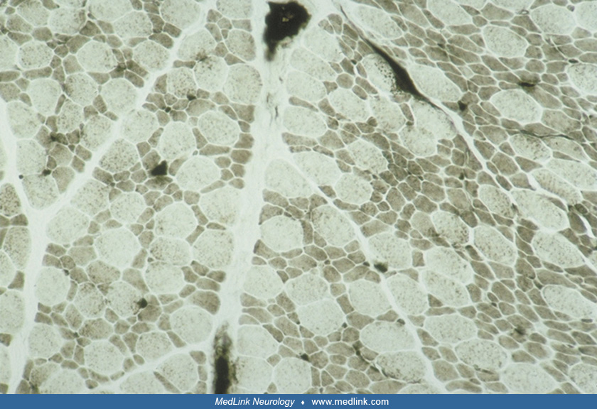
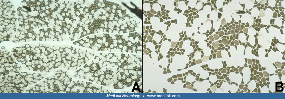
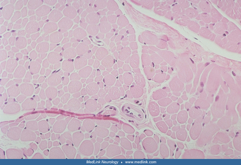
(A to C) NADH-TR of several different fascicles shows that all small fibers are type I and all large fibers are type II, but some fascicles are composed nearly entirely of small type I fibers. ATPase stains (not shown) confirmed a similar histochemical pattern of fiber types. Serial muscle biopsies of children with congenital muscle fiber-type disproportion frequently show a greater number of type I fibers at older ages than in infancy, as in this case, presumably due to continued conversion over time of some type II fibers to type I. Magnification x 250. (Contributed by Dr. Harvey Sarnat.)