Neuro-Oncology
Brainstem gliomas in childhood
Dec. 28, 2023
MedLink®, LLC
3525 Del Mar Heights Rd, Ste 304
San Diego, CA 92130-2122
Toll Free (U.S. + Canada): 800-452-2400
US Number: +1-619-640-4660
Support: service@medlink.com
Editor: editor@medlink.com
ISSN: 2831-9125
Toll Free (U.S. + Canada): 800-452-2400
US Number: +1-619-640-4660
Support: service@medlink.com
Editor: editor@medlink.com
ISSN: 2831-9125
Nearly 3,000 illustrations, including video clips of neurologic disorders.
Every article is reviewed by our esteemed Editorial Board for accuracy and currency.
Full spectrum of neurology in 1,200 comprehensive articles.
Listen to MedLink on the go with Audio versions of each article.
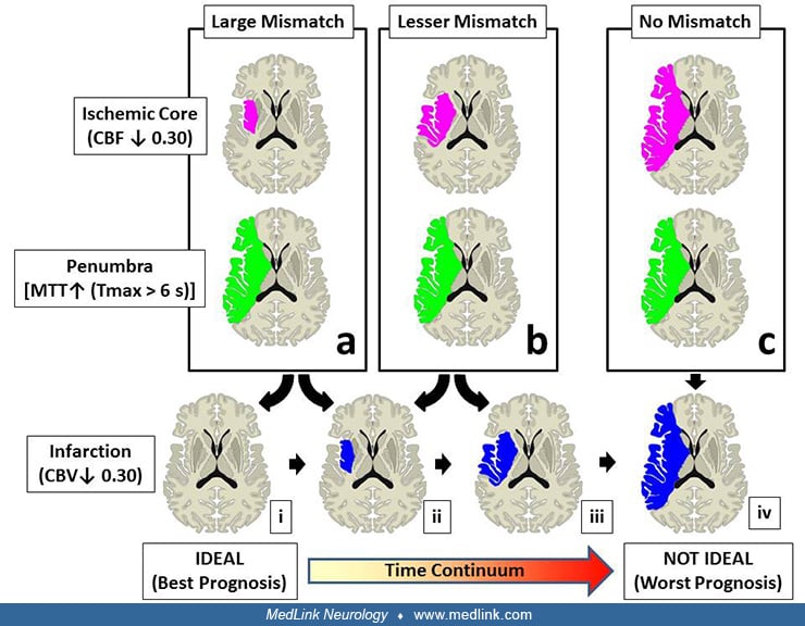
(a) The "ideal" patient is one with a large mismatch between the ischemic core and the penumbra volume, particularly if the cerebral blood volume is maintained normal (i). Although this is the picture of the typical onset, as time elapses, cerebral blood volume will begin to decrease in the core, spreading centrifugally (ii) to span the entire area of ischemia. (b) A lesser degree of mismatch is seen later, as the ischemic core also expands, indicating a less than "ideal" situation for a beneficial reperfusion effect. The abnormally low cerebral blood volume will also spread (from ii to iii), suggesting the progressive loss of tissue to infarction. (c) Finally, unless the tissue is revascularized, all evidence of parameters mismatch is lost, suggesting irreparable damage and futility of intervention. NOTE: As a practical step, we have used the same color scheme typically applied by the RAPID software (iSchemaView) to display cerebral blood flow and mean transit time. (Contributed by Dr. Camilo Gomez.)


