Developmental Malformations
Malformations of the brain
Sep. 23, 2023
MedLink®, LLC
3525 Del Mar Heights Rd, Ste 304
San Diego, CA 92130-2122
Toll Free (U.S. + Canada): 800-452-2400
US Number: +1-619-640-4660
Support: service@medlink.com
Editor: editor@medlink.com
ISSN: 2831-9125
Toll Free (U.S. + Canada): 800-452-2400
US Number: +1-619-640-4660
Support: service@medlink.com
Editor: editor@medlink.com
ISSN: 2831-9125
Nearly 3,000 illustrations, including video clips of neurologic disorders.
Every article is reviewed by our esteemed Editorial Board for accuracy and currency.
Full spectrum of neurology in 1,200 comprehensive articles.
Listen to MedLink on the go with Audio versions of each article.
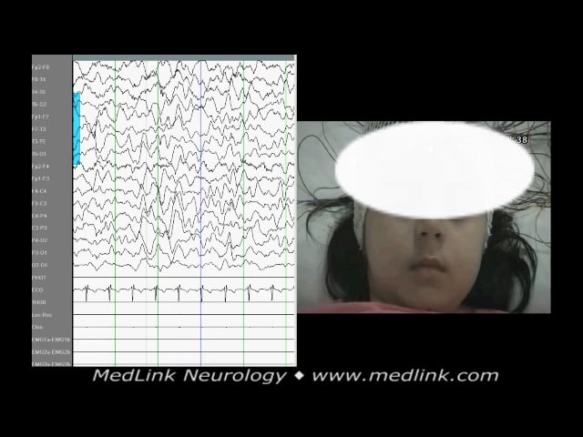
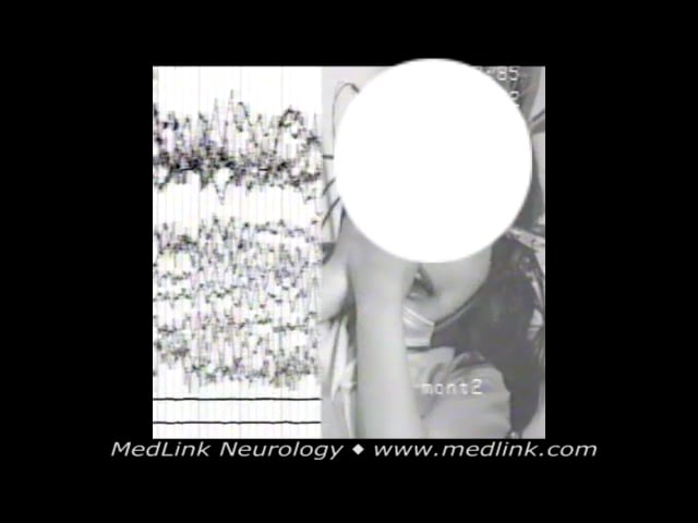
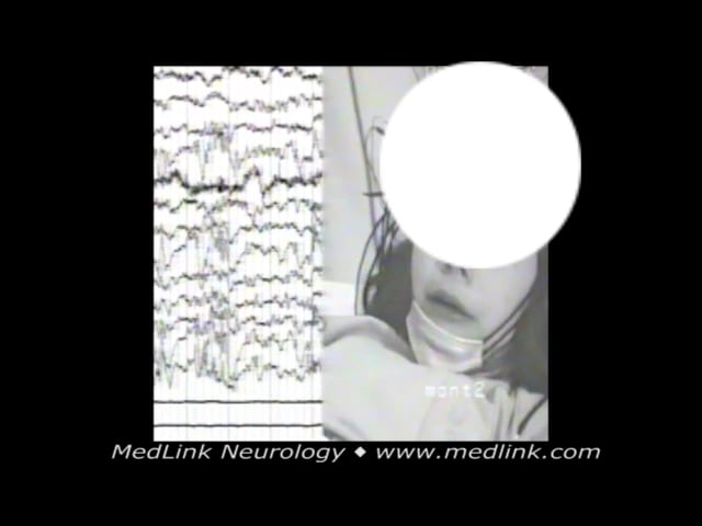
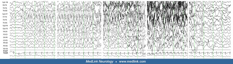
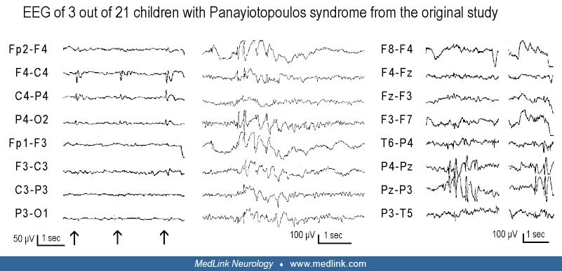
Left top and bottom: EEG of two girls with Panayiotopoulos syndrome. Right top: EEG of a boy with frequent, brief visual seizures of elementary visual hallucinations and, occasionally, blindness. Right bottom: EEG a 15-year-old boy with symptomatic occipital lobe epilepsy. In routine EEG, high-amplitude, continuous occipital sharp and slow wave complexes (occipital paroxysms) occurred immediately after closing of the eyes, lasting as long as the eyes were closed. The EEG normalized immediately after opening the eyes and continued as long as the eyes were open, though some breaks in occipital spikes occurred. The activation of the occipital paroxysms was due to the elimination of central vision and fixation (left of the vertical bar, symbol of eyes with glasses). Occipital spikes were inhibited by fixation (right of the vertical bar, symbol of eyes without glasses). (Used with permission: Panayiotopoulos CP. Inhibitory effect of central vision on occipital lobe seizures. Neurology 1981;31:330-3.)