Single enzyme defects of peroxisomal beta-oxidation
Mar. 15, 2023
MedLink®, LLC
3525 Del Mar Heights Rd, Ste 304
San Diego, CA 92130-2122
Toll Free (U.S. + Canada): 800-452-2400
US Number: +1-619-640-4660
Support: service@medlink.com
Editor: editor@medlink.com
ISSN: 2831-9125
Toll Free (U.S. + Canada): 800-452-2400
US Number: +1-619-640-4660
Support: service@medlink.com
Editor: editor@medlink.com
ISSN: 2831-9125
Nearly 3,000 illustrations, including video clips of neurologic disorders.
Every article is reviewed by our esteemed Editorial Board for accuracy and currency.
Full spectrum of neurology in 1,200 comprehensive articles.
Listen to MedLink on the go with Audio versions of each article.
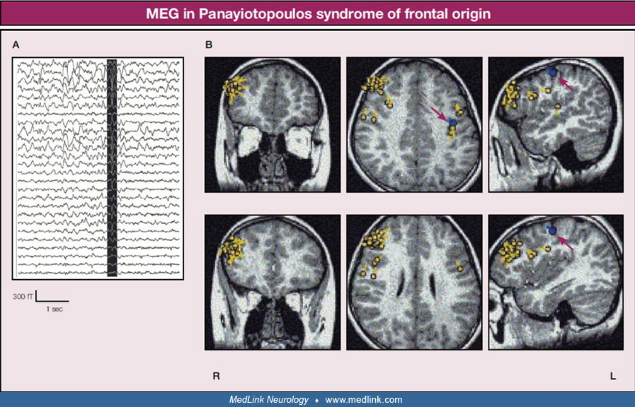
In rolandic epilepsy, equivalent current dipoles of spikes are located and concentrated in the rolandic regions and have regular directions. In self-limited epilepsy with autonomic seizures, equivalent current dipoles of spikes are located and concentrated bilaterally in the rolandic regions and right occipital area. The directions of each equivalent current dipole in each area are regular as if three small round toothbrushes are placed in each of the three areas. Small yellow circles represent locations, and yellow arrows represent directions of equivalent current dipoles. Blue circles and arrows represent bilateral somatosensory evoked magnetic field. (Reproduced with permission from: Kanazawa O. Benign Rolandic epilepsy and related epileptic syndromes: electrophysiological studies including magnetoencephalography in ictal and interictal phenomena. In: Benjamin SM, editor. Trends in epilepsy research. New York: Nova Science Publishers, 2005b:19-54. Kanazawa O, Tohyama J, Akasaka N, Kamimura T. A magnetoencephalographic study of patients with Panayiotopoulos syndrome. Epilepsia 2005a;46(7):1106-13.)