Epilepsy & Seizures
Aphasic seizures
Oct. 02, 2023
MedLink®, LLC
3525 Del Mar Heights Rd, Ste 304
San Diego, CA 92130-2122
Toll Free (U.S. + Canada): 800-452-2400
US Number: +1-619-640-4660
Support: service@medlink.com
Editor: editor@medlink.com
ISSN: 2831-9125
Toll Free (U.S. + Canada): 800-452-2400
US Number: +1-619-640-4660
Support: service@medlink.com
Editor: editor@medlink.com
ISSN: 2831-9125
Nearly 3,000 illustrations, including video clips of neurologic disorders.
Every article is reviewed by our esteemed Editorial Board for accuracy and currency.
Full spectrum of neurology in 1,200 comprehensive articles.
Listen to MedLink on the go with Audio versions of each article.
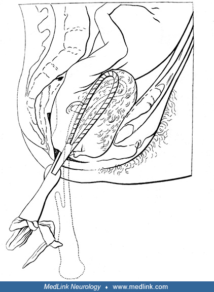
This uterus was removed in the course of excising an ovarian mucinous cystadenoma. The photo shows the uterus and adnexa (less the initially excised left ovarian tumor) from the posterior aspect. The pale-yellow, right ovary is clearly seen on the upper right of the image. Both oviducts, visible at the top right and left, adjoin their respective separate cornua of this malformed uterus. (Photograph by Dr. Ed Uthman on May 26, 2000. Public domain.)