Neuro-Ophthalmology & Neuro-Otology
Meniere syndrome
May. 23, 2023
MedLink®, LLC
3525 Del Mar Heights Rd, Ste 304
San Diego, CA 92130-2122
Toll Free (U.S. + Canada): 800-452-2400
US Number: +1-619-640-4660
Support: service@medlink.com
Editor: editor@medlink.com
ISSN: 2831-9125
Toll Free (U.S. + Canada): 800-452-2400
US Number: +1-619-640-4660
Support: service@medlink.com
Editor: editor@medlink.com
ISSN: 2831-9125
Nearly 3,000 illustrations, including video clips of neurologic disorders.
Every article is reviewed by our esteemed Editorial Board for accuracy and currency.
Full spectrum of neurology in 1,200 comprehensive articles.
Listen to MedLink on the go with Audio versions of each article.
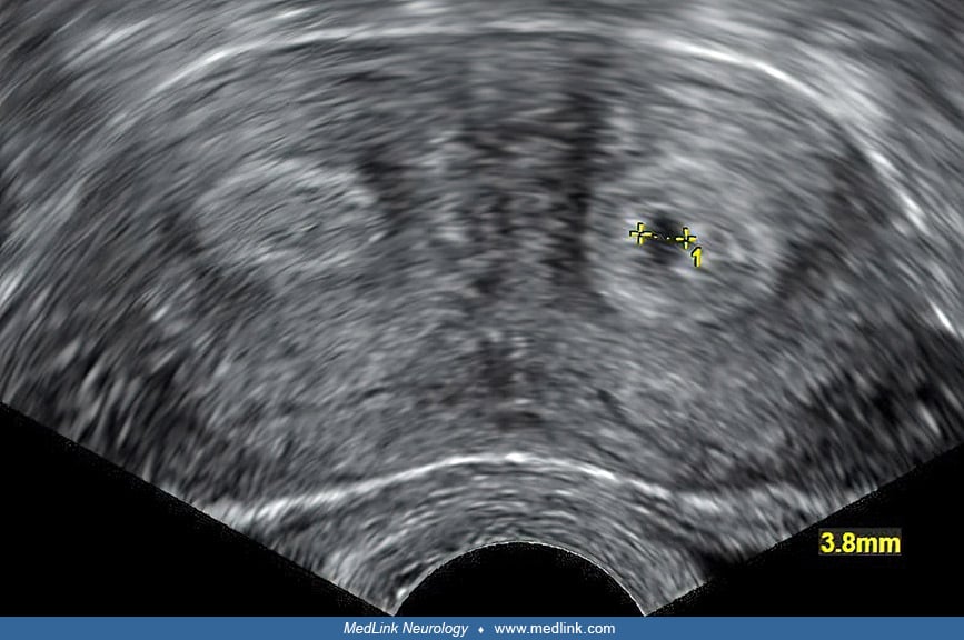
Transvaginal ultrasound showing a cross-section of a bicornuate uterus, with two cavities or "horns" visible at the left and right, respectively. In the "horn" to the right, a gestational sac is visible, with a diameter of 5.7 mm, corresponding to a gestational age of between 4 and 5 weeks. This projection did not transect the maximal diameter, thus, giving a bit smaller diameter in the image. The patient sought medical care because of a positive pregnancy test despite having an intrauterine device (IUD) with copper, presumably because the IUD was located in the other "horn." (Courtesy of Mikael Häggström on July 14, 2014. Public domain.)