Infectious Disorders
Epstein-Barr virus infections of the nervous system
Nov. 11, 2023
MedLink®, LLC
3525 Del Mar Heights Rd, Ste 304
San Diego, CA 92130-2122
Toll Free (U.S. + Canada): 800-452-2400
US Number: +1-619-640-4660
Support: service@medlink.com
Editor: editor@medlink.com
ISSN: 2831-9125
Toll Free (U.S. + Canada): 800-452-2400
US Number: +1-619-640-4660
Support: service@medlink.com
Editor: editor@medlink.com
ISSN: 2831-9125
Nearly 3,000 illustrations, including video clips of neurologic disorders.
Every article is reviewed by our esteemed Editorial Board for accuracy and currency.
Full spectrum of neurology in 1,200 comprehensive articles.
Listen to MedLink on the go with Audio versions of each article.
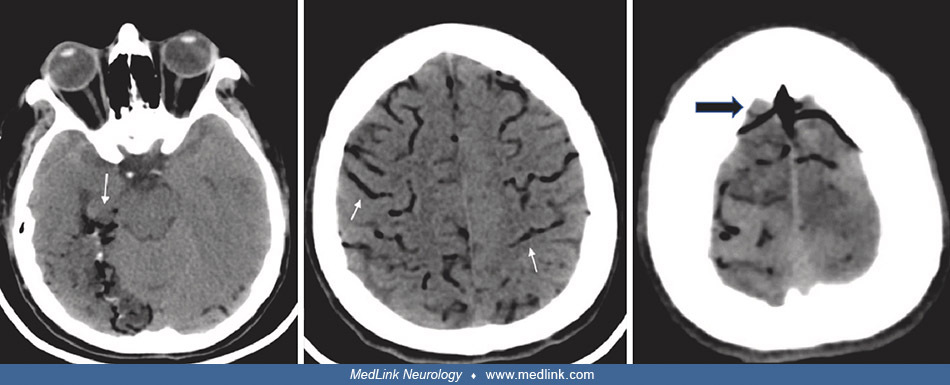
Findings on susceptibility-weighted imaging from a head MRI in a patient with lung cancer and spontaneous cerebral gas emboli. Small dot-like low signal (circles) in the right frontal cortex (left image) and the left parietal subcortex (right image) on susceptibility-weighted imaging. (TR/TE). (Source: Inatomi S, Izumi T, Eura N, et al. Electroencephalographic findings after convulsive seizures due to cerebral arterial air embolism secondary to lung cancer: a case report. J Med Case Rep 2022;16[1]:137. Creative Commons Attribution 4.0 International License [CC BY 4.0], https://creativecommons.org/licenses/by/4.0.)