Infectious Disorders
Tick borne rickettsial diseases of the CNS
Mar. 05, 2023
MedLink®, LLC
3525 Del Mar Heights Rd, Ste 304
San Diego, CA 92130-2122
Toll Free (U.S. + Canada): 800-452-2400
US Number: +1-619-640-4660
Support: service@medlink.com
Editor: editor@medlink.com
ISSN: 2831-9125
Toll Free (U.S. + Canada): 800-452-2400
US Number: +1-619-640-4660
Support: service@medlink.com
Editor: editor@medlink.com
ISSN: 2831-9125
Nearly 3,000 illustrations, including video clips of neurologic disorders.
Every article is reviewed by our esteemed Editorial Board for accuracy and currency.
Full spectrum of neurology in 1,200 comprehensive articles.
Listen to MedLink on the go with Audio versions of each article.
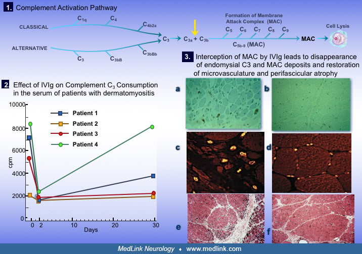
(A) Cross-section of an H&E-stained muscle biopsy demonstrates the classic perifascicular atrophy (layers of atrophic fibers at the periphery of the fascicle) with necrosis and some inflammatory infiltrates (H&E stain ×100.) (B) Deposition of complement (membranolytic attack complex in green) on the endothelial cell wall of endomysial vessels (stained in red with a lectin Ulex Europaeus) leads to destruction of endothelial cells (confirmed in orange based on the superimposition of red on green). Consequently, in the muscles of patients with dermatomyositis, (C) the density of the endomysial capillaries (in red) is reduced, especially at the periphery of the fascicle, with dilatation of the lumen of the remaining capillaries in an effort to compensate for the ischemic process (x560). (Dalakas MC. Polymyositis, dermatomyositis and inclusion body myositis. In: Kasper DL, Fauci AS, Hauser SL, Longo DL, Jameson JL, Loscalzo J, editors. Harrison’s principles of internal medicine. 19th edition. New York: McGraw-Hill, 2015b:2195-202.)