Neuromuscular Disorders
Neurogenetics and genetic and genomic testing
Dec. 09, 2024
MedLink, LLC
3525 Del Mar Heights Rd, Ste 304
San Diego, CA 92130-2122
Toll Free (U.S. + Canada): 800-452-2400
US Number: +1-619-640-4660
Support: service@medlink.com
Editor: editor@medlink.com
ISSN: 2831-9125
Toll Free (U.S. + Canada): 800-452-2400
US Number: +1-619-640-4660
Support: service@medlink.com
Editor: editor@medlink.com
ISSN: 2831-9125
Nearly 3,000 illustrations, including video clips of neurologic disorders.
Every article is reviewed by our esteemed Editorial Board for accuracy and currency.
Full spectrum of neurology in 1,200 comprehensive articles.
Listen to MedLink on the go with Audio versions of each article.
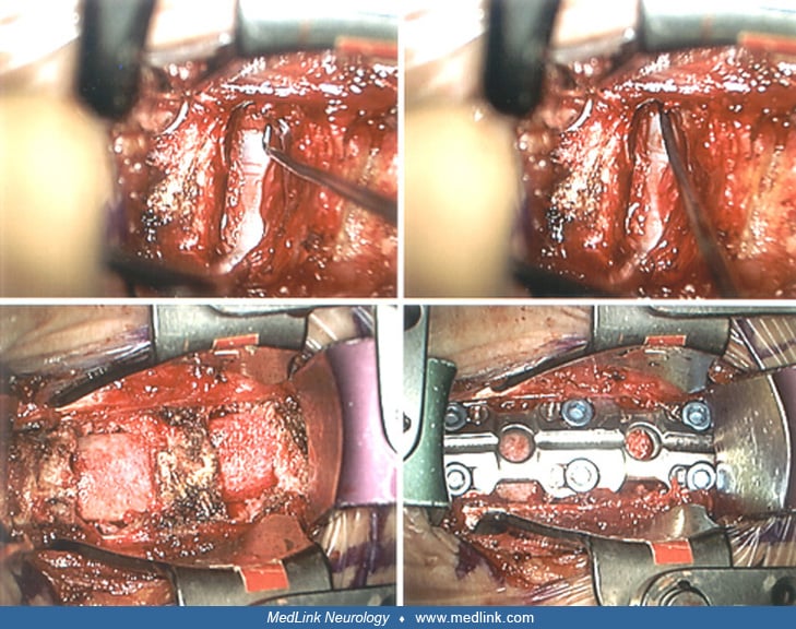
This panel is a sequence of surgical steps oriented with the patient’s head to the left, and feet to the right, as the surgeon views the field. The upper left photograph illustrates the C5-6 interspace after the disc and calcified posterior longitudinal ligament have been removed, exposing the dura covering the spinal cord. The nerve hook indicates an epidural vein over the left C6 root sleeve. The upper right photograph illustrates passage of the nerve hook out the left C6 foramen, confirming thorough decompression of the root after removal of disc fragments. Allograft tricortical iliac crest bone grafts are in position at each of the interspaces, C5-6 and C6-7, in the lower left photograph. In the lower right photograph, the titanium plate spanning C5-7 is held by a pair of screws in each vertebral body, allowing earlier mobilization of the patient while maintaining satisfactory alignment with retained disc height. (Contributed by Dr. Saul Schwarz.)