Peripheral Neuropathies
Autonomic neuropathy: treatment
Jul. 14, 2026
MedLink, LLC
3525 Del Mar Heights Rd, Ste 304
San Diego, CA 92130-2122
Toll Free (U.S. + Canada): 800-452-2400
US Number: +1-619-640-4660
Support: service@medlink.com
Editor: editor@medlink.com
ISSN: 2831-9125
Toll Free (U.S. + Canada): 800-452-2400
US Number: +1-619-640-4660
Support: service@medlink.com
Editor: editor@medlink.com
ISSN: 2831-9125
Nearly 3,000 illustrations, including video clips of neurologic disorders.
Every article is reviewed by our esteemed Editorial Board for accuracy and currency.
Full spectrum of neurology in 1,200 comprehensive articles.
Listen to MedLink on the go with Audio versions of each article.
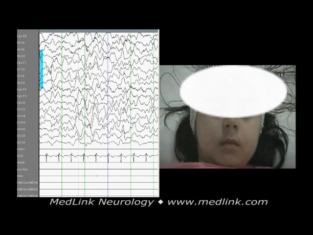
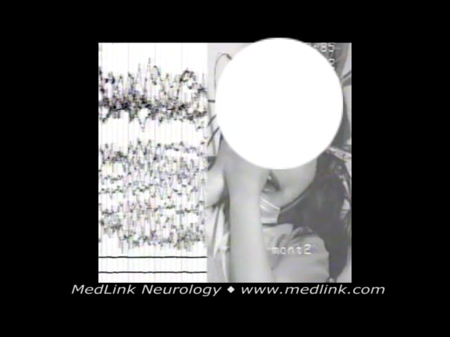
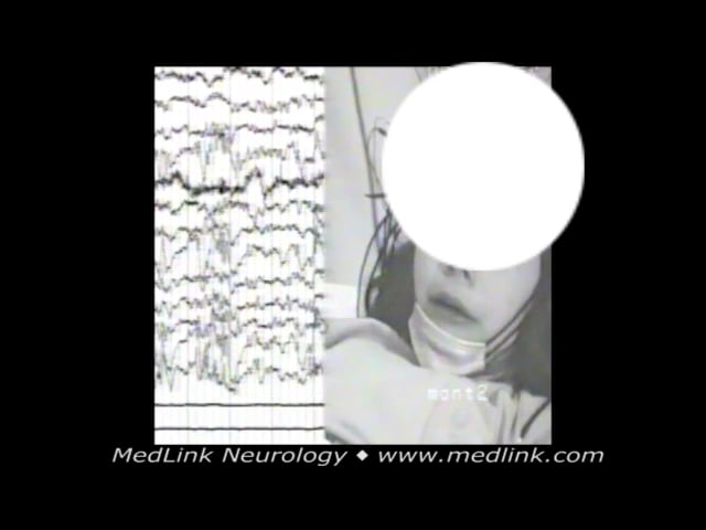
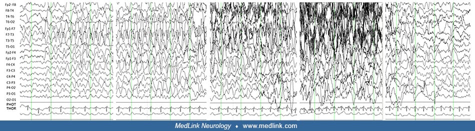
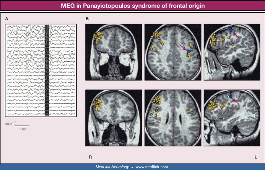
The patient had seizures typical of self-limited epilepsy with autonomic seizures from the age of 4 years. EEGs initially showed occipital spikes, but at the age of 13, EEG had bifrontal spikes, and MEG was performed. The patient’s younger brother also had self-limited epilepsy with autonomic seizures preceded by febrile seizures and followed by rolandic seizures. (A) MEG waveforms. The reversed colored MEG waveforms, in white in the vertical dark zone, were analyzed. (B) Magnetic source images revealed clustering equivalent current dipoles of spike discharges alongside the right inferior frontal sulcus, but the orientations were not so regular. All MRIs are T1-weighted. The pale-colored solid circles and tails represent the locations and orientations of equivalent current dipoles of the spike discharges. The early somatosensory evoked field was modeled using a single equivalent current dipole approach to estimate the spatial source of response, whereas the dark-colored solid circles and tails indicated by red arrows represent the locations and orientations of somatosensory evoked fields (N20). (Reproduced with permission from (Saitoh N, Kanazawa O, Toyama J, Akasaka N, Kamimura T. Magnetoencephalographic findings of self-limited epilepsy with autonomic seizures with frontal epileptic discharges. Pediatr Neurol 2007;36:190-4.)