Sleep Disorders
NonREM parasomnias
Dec. 13, 2025
MedLink, LLC
3525 Del Mar Heights Rd, Ste 304
San Diego, CA 92130-2122
Toll Free (U.S. + Canada): 800-452-2400
US Number: +1-619-640-4660
Support: service@medlink.com
Editor: editor@medlink.com
ISSN: 2831-9125
Toll Free (U.S. + Canada): 800-452-2400
US Number: +1-619-640-4660
Support: service@medlink.com
Editor: editor@medlink.com
ISSN: 2831-9125
Nearly 3,000 illustrations, including video clips of neurologic disorders.
Every article is reviewed by our esteemed Editorial Board for accuracy and currency.
Full spectrum of neurology in 1,200 comprehensive articles.
Listen to MedLink on the go with Audio versions of each article.
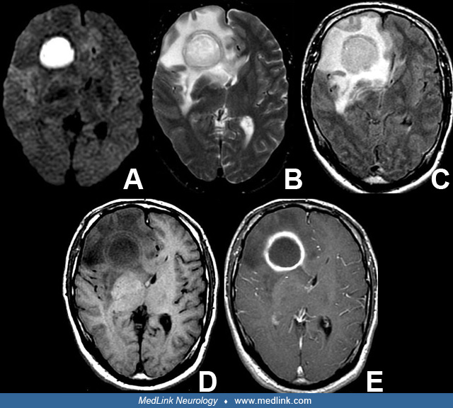
This 49-year-old male was told that he no longer needed SBE prophylaxis for dental procedures despite the presence of his patent foramen ovale. Three weeks after undergoing routine teeth cleaning, this patient developed a seizure. Axial MR imaging of a pathologically proven brain abscess in this patient reveals (A) marked restricted diffusion on diffusion-weighted images, (B) high T2 signal, and (C) FLAIR signal due to edema and a well circumscribed mass in the right frontal lobe. Once the capsule has formed (usually within 2 to 3 weeks), the walls of the mass are often (D) clearly delineated even without on T1 weighted imaging, and (E) the walls show brisk enhancement with contrast and demarcates the wall from the necrotic center. (Contributed by Dr. Diana Gomez-Hassan.)