Epilepsy & Seizures
Focal clonic seizures
Mar. 19, 2025
MedLink, LLC
3525 Del Mar Heights Rd, Ste 304
San Diego, CA 92130-2122
Toll Free (U.S. + Canada): 800-452-2400
US Number: +1-619-640-4660
Support: service@medlink.com
Editor: editor@medlink.com
ISSN: 2831-9125
Toll Free (U.S. + Canada): 800-452-2400
US Number: +1-619-640-4660
Support: service@medlink.com
Editor: editor@medlink.com
ISSN: 2831-9125
Nearly 3,000 illustrations, including video clips of neurologic disorders.
Every article is reviewed by our esteemed Editorial Board for accuracy and currency.
Full spectrum of neurology in 1,200 comprehensive articles.
Listen to MedLink on the go with Audio versions of each article.
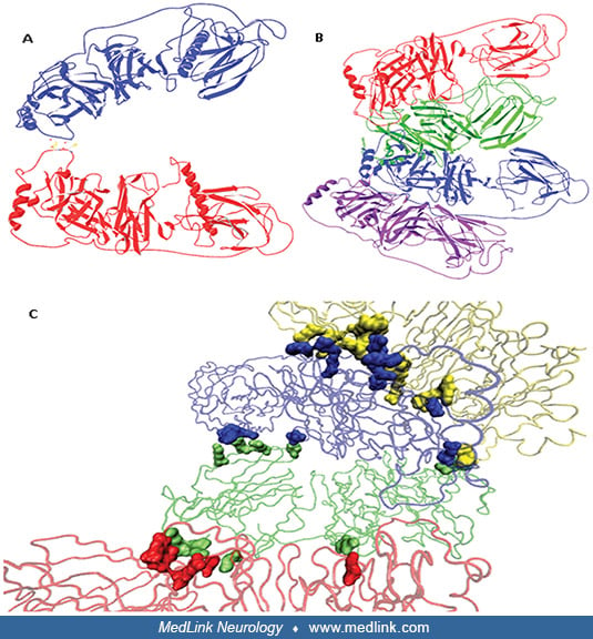
Ribbon diagram (Richardson diagram) of dopamine beta-hydroxylase. (A) Model for the dimeric form of dopamine beta-hydroxylase. Chains A and B are shown in blue and red, respectively. (B) Model for the tetrameric form of the enzyme. Chains A, B, C, and D are depicted in blue, red, green and purple, respectively. (C) Residues forming a tetramer interface. Subunits are highlighted in tube conformation with chains A, B, C, and D highlighted in blue, red, green and yellow, respectively. Residues are represented with the same colors as that of the corresponding chain color. (Source: Kapoor A, Shandilya M, Kundu S. Structural insight of dopamine β-hydroxylase, a drug target for complex traits, and functional significance of exonic single nucleotide polymorphisms. PLoS One 2011;6(10):e26509. Creative Commons Attribution-Share Alike 3.0 Unported license.)