Neuropharmacology & Neurotherapeutics
Nusinersen
Apr. 23, 2026
MedLink, LLC
3525 Del Mar Heights Rd, Ste 304
San Diego, CA 92130-2122
Toll Free (U.S. + Canada): 800-452-2400
US Number: +1-619-640-4660
Support: service@medlink.com
Editor: editor@medlink.com
ISSN: 2831-9125
Toll Free (U.S. + Canada): 800-452-2400
US Number: +1-619-640-4660
Support: service@medlink.com
Editor: editor@medlink.com
ISSN: 2831-9125
Nearly 3,000 illustrations, including video clips of neurologic disorders.
Every article is reviewed by our esteemed Editorial Board for accuracy and currency.
Full spectrum of neurology in 1,200 comprehensive articles.
Listen to MedLink on the go with Audio versions of each article.
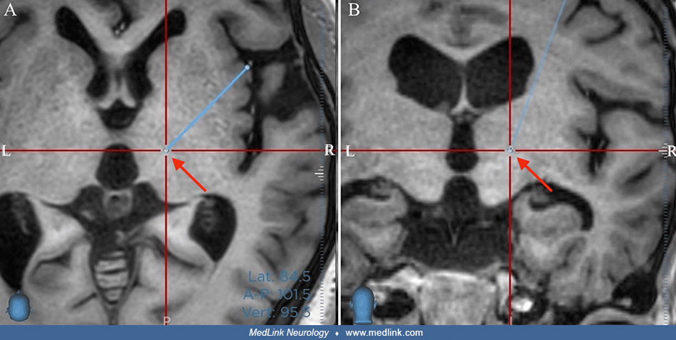
Postoperative coronal T2-weighted MRI following unilateral ventro-oral (Vo) thalamotomy in a middle-aged woman with peripheral posttraumatic dystonia with complex regional pain syndrome. As in a normal MRI, the left side of the screen is the patient's right. The coagulation area is almost identical to the previous target. The red arrow points to the lesioned area. (From: Kuramoto Y, Taira T, Tsuji S, Kubo T, Yoshimura S. Successful unilateral ventro-oral (Vo) thalamotomy for peripheral post-traumatic dystonia with complex regional pain syndrome: a case report. Cureus 2025;17[5]:e83536. Creative Commons Attribution 4.0 International [CC BY 4.0] license, creativecommons.org/licenses/by/4.0.)