Sleep Disorders
Fatal familial insomnia
Sep. 25, 2024
MedLink, LLC
3525 Del Mar Heights Rd, Ste 304
San Diego, CA 92130-2122
Toll Free (U.S. + Canada): 800-452-2400
US Number: +1-619-640-4660
Support: service@medlink.com
Editor: editor@medlink.com
ISSN: 2831-9125
Toll Free (U.S. + Canada): 800-452-2400
US Number: +1-619-640-4660
Support: service@medlink.com
Editor: editor@medlink.com
ISSN: 2831-9125
Nearly 3,000 illustrations, including video clips of neurologic disorders.
Every article is reviewed by our esteemed Editorial Board for accuracy and currency.
Full spectrum of neurology in 1,200 comprehensive articles.
Listen to MedLink on the go with Audio versions of each article.
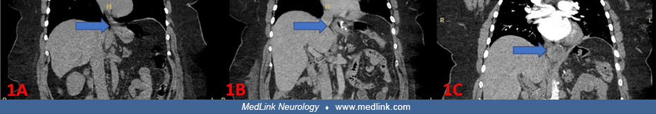
CT imaging showing progressive hiatal hernia in a 54-year-old woman with swallow syncope as a late complication of sleeve gastrectomy. (A) CT of the abdomen and pelvis showing absence of a hiatal hernia (blue arrow) 13 months before the patient's laparoscopic sleeve gastrectomy. (B) CT of the abdomen and pelvis showing the presence of a hiatal hernia (blue arrow) 8 days after the patient's laparoscopic sleeve gastrectomy. (C) CT angiogram of the chest showing progressive enlargement of a hiatal hernia (blue arrow) 2 months after the patient's laparoscopic sleeve gastrectomy. (From: Rudensky F, Khan SN, Chalasani P. Swallow syncope as a late complication of sleeve gastrectomy. Cureus 2025;17[3]:e81434. Creative Commons Attribution 4.0 International [CC BY 4.0] license, creativecommons.org/licenses/by/4.0.)